BIOLOGICAL BASIS OF BEHAVIOR 8 10 THE NERVOUS

BIOLOGICAL BASIS OF BEHAVIOR 8 -10%

THE NERVOUS SYSTEM
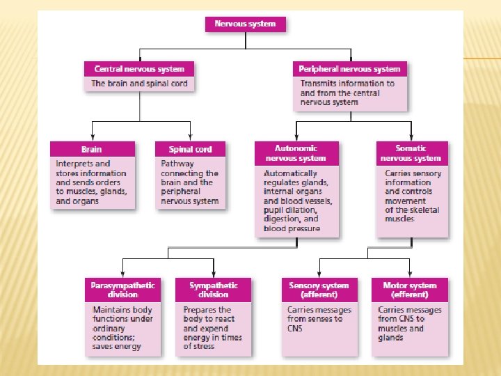

CENTRAL NERVOUS SYSTEM Central Nervous System Brain Spinal Cord

THE SPINAL CORD Complex cable of nerves that connects brain to rest of the body Carries motor impulses from the brain to internal organs and muscles Carries sensory information from extremities and internal organs to the brain

PERIPHERAL NERVOUS SYSTEM Peripheral Nervous System Somatic Nervous System Autonomic Nervous System Sympathetic Division Parasympathetic Division

Peripheral Nervous System – nerves that lie outside the brain and spinal cord. PERIPHERAL NERVOUS SYSTEM

THE SOMATIC NERVOUS SYSTEM Somatic Nervous System - handles voluntary movement (raising hand, walking, jump, etc. )
- HANDLES AUTOMATIC PROCESSES (HEART RATE, DIGESTION, PERSPIRATION, ETC. ,")
THE AUTONOMIC NERVOUS SYSTEM (ANS)- HANDLES AUTOMATIC PROCESSES (HEART RATE, DIGESTION, PERSPIRATION, ETC. , ) Sympathetic division mobilizes the body’s resources for emergencies; creates the fight-or-flight Most active when you are angry, afraid, or aroused Fight-or-flight response Increases heart rate and breathing Stops digestion

THE AUTONOMIC NERVOUS SYSTEM Parasympathetic division Brings you back to normal Calms body Produces effects opposite to those of the sympathetic division Reduces heart rate and breathing Restores

TYPES OF NEURONS Afferent/Sensory neurons Carry Efferent/Motor neurons Carry information from sensory systems to the brain information from the brain to muscles and glands Interneurons Carry information between other neurons
 in the human brain Neurons")
NEURONS: THE MESSENGERS About 100 billion neurons (nerve cells) in the human brain Neurons have many of the same features as other cells Nucleus Cytoplasm Cell What membrane makes neurons unique is their shape and function
 Contains nucleus Axon Carry information to the")
STRUCTURE OF NEURONS Dendrites Cell Body (Soma) Contains nucleus Axon Carry information to the cell body from other neurons Can be altered as a result of neuroplasticity Carries information to the next cell Myelin Sheath Insulates the axon and speeds up the neural impulse
 gap between neurons Terminal button Enlarged The")
THE SYNAPSE Synaptic Tiny space (synaptic cleft) gap between neurons Terminal button Enlarged The area at the end of an axon synapse Composed of the terminal button of one neuron, the synaptic space, and the dendrites or cell body of the receiving neuron

TRANSMISSION BETWEEN NEURONS Synaptic vesicles Neurotransmitters Chemicals released by synaptic vesicles Receptor sites Sacs in terminal button that release chemicals into synaptic space Location on receptor neuron for specific neurotransmitter Reuptake When the sending neuron reabsorbs the chemicals for reuse.

THE SYNAPSE Synaptic Vesicles Terminal Button/ Axon Terminal Neurotrans mitter Synaptic Space Receptor Sites

THE NEURAL IMPULSE Ions Charged molecules Resting Potential When more negative ions are inside the neuron than outside Charge is approximately -70 m. V Neuron is not transmitting information

THE NEURAL IMPULSE Action Potential Sudden, massive change in charge in the neuron Occurs when depolarization reaches the threshold of excitation Ions flow across cell membrane Neuron is now transmitting information
 When the electrical charge of a cell moves away")
THE NEURAL IMPULSE Polarization (off) When the electrical charge of a cell moves away from zero Depolarization When (on) the electrical charge of a cell moves toward zero

THE NEURAL IMPULSE Polarization Resting Potential Depolarization Action Potential

THE NEURAL IMPULSE

THE NEURAL IMPULSE All-or-None A Law neuron either fires or it does not When it does fire, it will always produce an impulse of the same strength Intensity of a stimulus is coded by the frequency of action potentials

THE NEURAL IMPULSE Absolute refractory period Period immediately after an action potential when another action potential cannot occur Relative refractory period Period following absolute refractory period when a neuron will only respond to a stronger than normal impulse

NEUROTRANSMITTERS AND BEHAVIOR Neurotransmitters are the chemicals released by neurons. They are usually classified as excitatory neurotransmitters or inhibitory neurotransmitters. Excitatory neurotransmitter: they excite connecting neurons and cause them to fire. Inhibitory neurotransmitters: inhibit or prevent the next neuron from firing. Some chemicals can function as neurotransmitters but aren’t actually neurotransmitters (think drugs). Agonist – mimics action of a neurotransmitter Antagonist – opposes action of a neurotransmitter
 for the")
Some Well-Known Neurotransmitters Take a look at the word document (web chart) for the neurotransmitters you need to know! Must have an index card for each neurotransmitter.

THE ENDOCRINE SYSTEM
 into the bloodstream in order to")
Endocrine System – glands that secrete chemicals (hormones) into the bloodstream in order to help control bodily functioning ENDOCRINE SYSTEM
 that control metabolism Parathyroid glands")
THE ENDOCRINE SYSTEM Thyroid gland Secretes hormones (primarily thyroxin) that control metabolism Parathyroid glands Control levels of calcium and phosphate which in turn controls levels of excitability

THE ENDOCRINE SYSTEM Pineal gland Secretes melatonin which regulates the sleep-wake cycle Pancreas Regulates blood-sugar levels and helps with digestion Secretes insulin and glucagon

THE ENDOCRINE SYSTEM Pituitary gland Referred to as the “master gland” because it regulates many other glands – tells the other glands when to release hormones Gonads Ovaries and testes secrete estrogens androgens (which include testosterone) Adrenal glands Secretes hormones in reaction to stress (adrenaline)

STRUCTURE OF HUMAN BRAIN Brain: The three primary divisions forebrain Cortex Basal ganglia Limbic system The midbrain Involved The in sensory and motor functions hindbrain Medulla Pons Reticular formation Cerebellum

FIGURE 5. 3: MAJOR STRUCTURES OF THE HUMAN BRAIN Copyright © 2016, 2012 Pearson Education, Inc. All Rights Reserved
 The Hindbrain Medulla: marrow” means “the deep inside region")
THE HINDBRAIN (1 OF 2) The Hindbrain Medulla: marrow” means “the deep inside region or First large swelling at the top of the spinal cord, forming the lowest part of the brain Point where sensory nerves from the left and right sides of the body crossover Controls life-sustaining functions such as breathing, swallowing, and heart rate Pons: means “bridge” Larger swelling above the medulla that connects the lower sections of the brain to the upper sections Plays a part in sleep, dreaming, left–right body coordination, and arousal
 Reticular formation (RF): area of neurons running through the")
THE HINDBRAIN (2 OF 2) Reticular formation (RF): area of neurons running through the middle of the medulla and the pons and slightly beyond Responsible for selective attention and ignoring constant unchanging information Reticular activating system (RAS): stimulates the upper part of the brain, keeping people awake and alert Cerebellum: Part means “little brain” of the lower brain located behind the pons Controls involuntary, rapid, fine motor movement and coordinates voluntary movements that have to happen in rapid succession
 Limbic system: subcortical structures located in")
THE FOREBRAIN The Limbic System (1 of 3) Limbic system: subcortical structures located in the inner margin of the upper brain; involved in learning, emotion, and motivation It includes: Thalamus Hypothalamus Hippocampus Amygdala
 Thalamus: Somewhat round structure in the center of")
The Limbic System (2 of 3) Thalamus: Somewhat round structure in the center of the brain Receives sensory information from the lower part of the brain, processes it, relays it to the proper areas of the cortex Damage to the thalamus might result in the loss or partial loss of sensations Hypothalamus: Small structure in the brain located below the thalamus and directly above the pituitary gland Responsible for motivational behavior such as sleep, hunger, thirst, and sex
 Hippocampus: meaning “seahorse” Curved structure located within each")
The Limbic System (3 of 3) Hippocampus: meaning “seahorse” Curved structure located within each temporal lobe Responsible for the formation of long-term declarative memories Amygdala: Located meaning “almond” near the hippocampus Responsible for fear responses and the memory of fear Information from the senses goes to the amygdala before the upper part of the brain stimulating quicker response

FIGURE 5. 4: THE LIMBIC SYSTEM Copyright © 2016, 2012 Pearson Education, Inc. All Rights Reserved

Cortex Outermost covering of the brain consisting of densely packed neurons One-tenth of an inch thick on average Wrinkled surface allows for more surface area for neurons to fit in the small space inside the skull Responsible for higher thought processes and interpretation of sensory input
 Two hemispherical sections of the brain cortex Corpus callosum:")
CEREBRAL HEMISPHERES (1 OF 2) Two hemispherical sections of the brain cortex Corpus callosum: meaning “hard bodies” Tough, thick band of neural fibers that connects the right and left cerebral hemispheres Contralateral organization: For specific regions, each hemisphere is responsible for the opposite side of the body, for control or for receiving information Plays a role in information coming from many of the sense organs to the brain, and in the motor commands originating in the brain going to the rest of the body
 • Each hemisphere can be divided into four lobes:")
CEREBRAL HEMISPHERES (2 OF 2) • Each hemisphere can be divided into four lobes: – – Occipital Parietal Temporal Frontal

FIGURE 5. 5: THE LOBES OF THE BRAIN Copyright © 2016, 2012 Pearson Education, Inc. All Rights Reserved
 Occipital lobe: Section of the brain")
FOUR LOBES OF THE BRAIN (1 OF 5) Occipital lobe: Section of the brain located at the rear and bottom of each cerebral hemisphere Contains the visual centers of the brain Primary visual cortex: processes visual information from the eyes Visual association cortex: helps identify and make sense of the visual information from the eyes
 Parietal Lobes Sections of the brain")
FOUR LOBES OF THE BRAIN (2 OF 5) Parietal Lobes Sections of the brain located at the top and back of each cerebral hemisphere just under the parietal bone Comprises the somatosensory cortex: Area of cortex at the front of the parietal lobes The cells at the top of the brain receive information from the bottom of the body and vice versa Responsible for processing information from the skin and internal body receptors for touch, temperature, and body position
 Temporal lobes: Areas of the cortex")
FOUR LOBES OF THE BRAIN (3 OF 5) Temporal lobes: Areas of the cortex located just behind the temples Responsible for the sense of hearing and meaningful speech Comprises: Primary auditory cortex Auditory association area An area involved in language in the left temporal lobe
 Learning Objective 5. 2: Understand the")
FOUR LOBES OF THE BRAIN (4 OF 5) Learning Objective 5. 2: Understand the structures and functions of the various parts of the central nervous system. Frontal Areas lobes: of the brain located in the front and top Responsible for higher mental functions of the brain –planning, personality, memory storage, impulse control, complex decision making and the production of fluent speech Helps control emotions through its connection to the limbic system
 Frontal lobes (continued): It comprises Prefrontal")
FOUR LOBES OF THE BRAIN (5 OF 5) Frontal lobes (continued): It comprises Prefrontal cortex: most forward part of the frontal lobes; responsible for performing mental and motor tasks Motor cortex: Rear section of the frontal lobe; resembles the somatosensory cortex Controls the movements of the body’s voluntary muscles by sending commands out to the somatic division of the peripheral nervous system Comprises mirror neurons that fire when an animal performs an action or when an animal observes that same action being performed by another

FIGURE 5. 6: THE MOTOR AND SOMATOSENSORY CORTEX • The motor cortex controls the voluntary muscles of the body. • Body parts are drawn larger or smaller according to the number of cortical cells devoted to that body part. • The somatosensory cortex receives information about the sense of touch and body position. Copyright © 2016, 2012 Pearson Education, Inc. All Rights Reserved
 Areas within each lobe of the cortex")
ASSOCIATION AREAS OF CORTEX (1 OF 3) Areas within each lobe of the cortex responsible for the coordination and interpretation of information, as well as higher mental processing Make connections between the sensory information coming into the brain and stored memories, images, and knowledge Majority of these areas present in the frontal lobe
 Broca’s area: Present in the left frontal")
ASSOCIATION AREAS OF CORTEX (2 OF 3) Broca’s area: Present in the left frontal lobe of most people; allows smooth and fluent speech Broca’s aphasia: Condition resulting from damage to the area causes broken speech – halting with mispronounced words https: //www. youtube. com/watch? v=f 2 Ii. MEb. M n. PM
 Wernicke’s Part area: of the left temporal")
ASSOCIATION AREAS OF CORTEX (3 OF 3) Wernicke’s Part area: of the left temporal lobe; involved in understanding the meaning of words Wernicke’s aphasia: Damage to the area leads to a condition where the affected person is able to speak fluently and pronounce entirely wrong words correctly https: //www. youtube. com/watch? v=3 oef 68 Ya b. D 0
 Hemispheric Roger specialization experiment: Sperry cut through the corpus")
SPLIT-BRAIN RESEARCH (1 OF 2) Hemispheric Roger specialization experiment: Sperry cut through the corpus callosum, creating a split brain, to cure epilepsy; initial success in animal and human subjects Special testing conducted by sending messages to only one side of the brain in patients with a split brain
 Results: Left hemisphere specializes in language, speech, handwriting, calculation")
SPLIT-BRAIN RESEARCH (2 OF 2) Results: Left hemisphere specializes in language, speech, handwriting, calculation (math), sense of time and rhythm, and thought analysis Right hemisphere specializes in processing involving perception, visualization, spatial perception, recognition of patterns, faces, emotions, melodies, expression of emotions, and comprehension of simple language

TABLE 5. 1: SPECIALIZATION OF THE TWO HEMISPHERES Copyright © 2016, 2012 Pearson Education, Inc. All Rights Reserved

FIGURE 5. 7: THE SPLIT-BRAIN EXPERIMENT Copyright © 2016, 2012 Pearson Education, Inc. All Rights Reserved

ADAPTABILITY Neuroplasticity – the brain can change in response to the environment and to genes i. e. , growing new dendritic spins

PEEKING INSIDE THE LIVING BRAIN Studying humans and animals with brain damage or manipulated brain tissue Three methods: Lesioning: sending an electrical current strong enough to destroy the target neurons through the tip of the wire surgically inserted into the brain Brain stimulation: temporarily disrupting or enhancing the normal functioning of specific brain areas through electrical stimulation, resulting in changes in behavior or cognition Neuroimaging techniques: directly imaging the brain’s structure or its function
 Lesioning: Anesthetize animals. Purposefully damage part of animal's brain")
MANIPULATION TECHNIQUES (1 OF 2) Lesioning: Anesthetize animals. Purposefully damage part of animal's brain through electrical current to study that area scientifically. Test the animal to see what has happened to its abilities.
 Brain Stimulation: Invasive techniques: Stimulating from the inside Deep")
MANIPULATION TECHNIQUES (2 OF 2) Brain Stimulation: Invasive techniques: Stimulating from the inside Deep brain stimulation (DBS): electrodes placed in specific deep-brain areas and electrode wires are then routed to an impulse generator that sends impulses to specific brain areas of interest Noninvasive techniques: Stimulating from the outside Transcranial magnetic stimulation (TMS): magnetic pulses are applied to the cortex using special copper wire coils positioned over the head
: brainimaging using computer-aided series of Xrays; shows stroke")
MAPPING BRAIN STRUCTURE Computed tomography (CT): brainimaging using computer-aided series of Xrays; shows stroke damage, tumors, injuries, and abnormal brain structure Magnetic resonance imaging (MRI): more detailed than CT; shows effects of very small strokes
 Electroencephalogram Records (EEG): electric activity of the cortex")
Mapping Brain Function (1 of 2) Electroencephalogram Records (EEG): electric activity of the cortex below specific areas of the skull using an electroencephalograph Small metal-disk or sponge-like electrodes placed directly on the scalp with a special solution to help conduct electrical signals Electrodes are connected to an amplifier in turn connected to a computer to view the information Resulting electrical output forms waves indicates stages of sleep, seizures, and even the presence of tumors; determination of active areas of brain during mental tasks
 Positron emission tomography (PET): brain-imaging through injection of")
Mapping Brain Function (2 of 2) Positron emission tomography (PET): brain-imaging through injection of a radioactive glucose into the subject; computer detects activity of brain cells consuming the glucose a color-coded image of the brain with lighter colors indicates greater activity Functional (f. MRI): Computer magnetic resonance imaging tracks changes in the oxygen levels of the blood Active brain areas are identified by superimposing the scan over the picture of the brain’s structure

FIGURE 5. 1: MAPPING BRAIN STRUCTURE a. CT scan from a 5 -year-old girl with a head injury and skull fracture b. In an adult individual Copyright © 2016, 2012 Pearson Education, Inc. All Rights Reserved

FIGURE 5. 2: MAPPING BRAIN FUNCTION a. EEG record b. PET scan image Copyright © 2016, 2012 Pearson Education, Inc. All Rights Reserved c. f. MRI image
 Genetics: the science of heredity DNA")
ROLE OF CHROMOSOMES AND GENES (1 OF 2) Genetics: the science of heredity DNA (deoxyribonucleic acid) Special molecule that contains the genetic material of an organism Consists of two very long sugar–phosphate strands, each linked together by amines or bases arranged in a particular pattern Amines contain the genetic code for building proteins
 Gene: section of DNA having the")
ROLE OF CHROMOSOMES AND GENES (2 OF 2) Gene: section of DNA having the same arrangement of chemical elements Chromosome: tightly wound strand of genetic material or DNA; found in the nucleus of a cell Humans have 46 chromosomes in each cell (except the egg and the sperm) – 23 pairs Most characteristics are determined by 22 such pairs, called autosomes The two chromosomes of the 23 rd pair are called the sex chromosomes: XX being female and XY being male

DOMINANT AND RECESSIVE GENES Dominant gene: a gene that actively controls the expression of a trait Recessive gene: a gene that influences the expression of a trait only when paired with an identical gene https: //www. youtube. com/watch? v=prk. HKjf. U m. Ms

GENETIC PROBLEMS Diseases carried by recessive genes are inherited when a child inherits two recessive genes, one from each parent Examples: cystic fibrosis, sickle-cell anemia, Tay-Sachs disorder (leads to brain cells being destroyed), phenylketonuria (PKU), etc. Phenylketonuria: condition in which an infant is born without the ability to break down phenylalanine (an amino acid) that controls coloring of skin and hair Inappropriate levels could damage brain and hamper intellectual

Figure 6. 1: Dominant and Recessive Genes and PKU The variation of parents carrying zero, one, or two recessive genes and the result of this in their offspring is shown. a) If only one parent carries the PKU gene, the children might be carriers but will not have PKU. b) Only if both parents are carriers of PKU will a child have the one in four possibility of having PKU. Copyright © 2016, 2012 Pearson Education, Inc. All Rights Reserved

CHROMOSOME PROBLEMS Problem with the number of chromosomes Chromosome can end up in the wrong cell, creating imbalance: one cell with only 22 and the other with 24 Can cause mild to severe problems in development Example: Down syndrome Developmental delay caused by an extra chromosome in the 21 st pair Symptoms include the physical characteristics of almond-shaped, wide-set eyes; intellectual disability

Down Syndrome Copyright © 2016, 2012 Pearson Education, Inc. All Rights Reserved

CHARLES DARWIN AND NATURAL SELECTION Common belief that human behavior is shaped by biology, environment, and evolutionary forces Charles Darwin 19 th-century British scientist Theory of evolution: principle stating that species of plants and animals change gradually over the course of many generations Natural selection: traits that contribute to survival are more likely to be passed on

HUMAN BEHAVIOR GENETICS Family studies - Assess Hereditary influence; Nature Assume that close family members share more of a trait than non-relatives Used to assess the heritability of psychological disorders or traits

Human Behavior Genetics Twin studies - Assess Hereditary influence by comparing identical and fraternal twins Used to determine how heritable a trait or disorder may be Identical twins would have highest heritability

Human Behavior Genetics Family Studies & Twin Studies LIMITATIONS – Raised in the same environment so difficult to rule out nurture influence

HUMAN BEHAVIOR GENETICS Adoption Assess studies hereditary and environmental factors by examining adopted children and both their biological and their adoptive parents

STATES OF CONSCIOUSNESS – AWARENESS OF INTERNAL AND EXTERNAL STIMULI Waking consciousness Thoughts, feelings, and perceptions that occur when we are awake and alert Altered States of Consciousness A mental state that differs noticeably from normal waking consciousness, including sleep, dreaming, meditation, or drug-induced states

BIOLOGICAL RHYTHMS AND SLEEP BIOLOGICAL RHYTHMS – INTERNAL BIOLOGICAL CLOCK CIRCADIAN RHYTHMS – 24 HOUR BIOLOGICAL CYCLE JET LAG – Fly across time zones, your biological clock keeps time as usual, but the official clock changes -Experience difficulty falling asleep and poor quality sleep -If for several days – may feel fatigued, sluggish, and irritable during the day time Chronic Jet Lag – deficits in cognitive ability MELATONIN – regulate the human biological clock
 Monitors electrical activity of the brain over time by means")
BRAIN ACTIVITY Electroencephalograph (EEG) Monitors electrical activity of the brain over time by means of recording electrodes attached to the surface of the scalp Activity in the brain – brain waves Different patterns of EEG activity are associated with different states of consciousness Beta, Alpha, Theta, Delta

BIOLOGICAL RHYTHMS AND SLEEP CON’T Beta Waves – Normal waking thought, alert problem solving Alpha Waves – Deep relaxation, blank mind, meditation Theta Waves – Light sleep Delta Waves – Deep Sleep B. A. T. D.
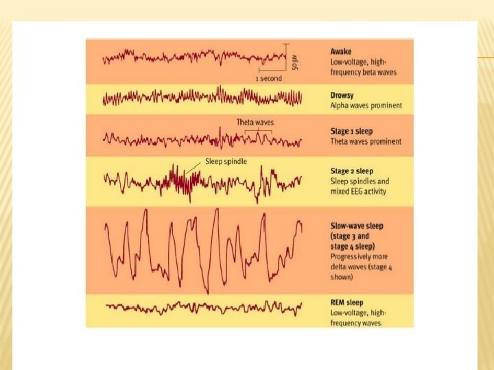

THE RHYTHMS OF SLEEP Brain waves and sleep stages Stage 1 Stage 2 Stage 3 Stage 4 REM sleep

CIRCADIAN CYCLES: THE BIOLOGICAL CLOCK Circadian cycles are those that last “about a day” Circadian rhythms are governed by an area of the hypothalamus called the suprachiasmatic nucleus (SCN) Controls body temperature, metabolism, blood pressure, hormone levels, and hunger Jet lag is the result of desynchronization of the circadian rhythm

http: //www. youtube. com/watch? feature=play er_detailpage&v=rt. CQ 9 jz. C-Ek http: //www. youtube. com/watch? feature=play er_detailpage&v=Wts. SI-uyr. QI
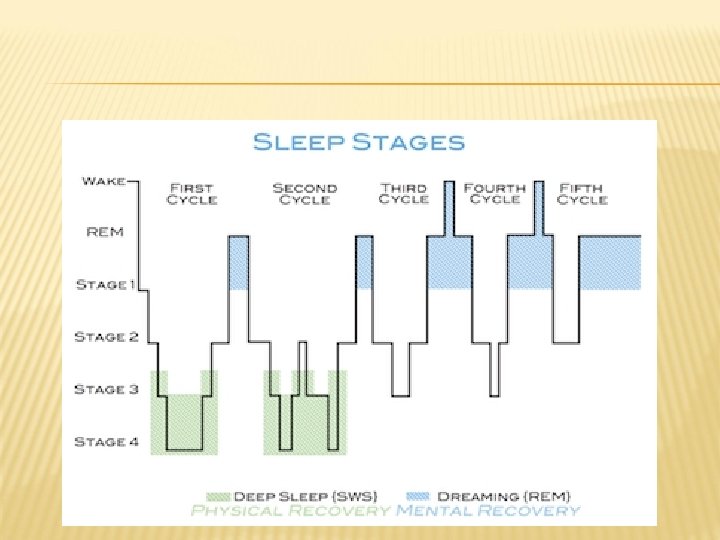

SLEEP CYCLE The sleep cycle is every 90 minutes REM – when we dream/do not move Nightmares REM takes over for stage 1 – there is no stage 5 REM rebound – don’t get enough sleep Crash for 12 hours on a Saturday, brain is confused and it makes us dream a lot and not get deep sleep Not a good thing

Sleep paralysis – while you are dreaming your body can’t move Only happens in REM Theory of why this happens – so we don’t act our dreams NREM (stage 4) – usually where Night Terrors occur

AGE TRENDS – INFANTS VS. ADULTHOOD Infants spend much more of their sleep time in the REM stage than adults do. In the first few months, REM accounts for about 50% of babies sleep, as compared to 20% of adults sleep Adulthood – Percentage of time spent in stage 1 increases slightly This shift toward lighter sleep may contribute to the increased frequency of nighttime awakenings seen among the elderly

CHANGES IN REM AND NREM

SLEEP DISORDERS Sleeptalking and sleepwalking Usually occurs during Stage 4 sleep More common in children Sleepwalking more common in boys Night terrors Episodes of fright that occur during stages 3 or 4 of NREM sleep Person may sit up or scream, but likely will not recall the episode in the morning

SLEEP DISORDERS Insomnia Difficulty falling asleep or remaining asleep Affects about 35 million Americans May be related to stress, depression, medication Can also be caused by noise, temperature, or trying to sleep in a new environment

SLEEP DISORDERS Apnea Person stops breathing momentarily during sleep Affects about 10 to 12 million Americans

SLEEP DISORDERS Narcolepsy Suddenly falling asleep without warning during waking hours Narcoleptics often experience loss of muscle tone as well May also drop into REM sleep immediately, causing hallucinations Likely caused by a central nervous system defect

SLEEP DISORDERS Sleep apnea video – Shaq https: //www. youtube. com/watch? v=4 Jki. Wv. Wn 2 a U Dog with narcolepsy video https: //www. youtube. com/watch? v=X 0 h 2 nle. WT w. I https: //www. youtube. com/watch? v=-z. VCYdrw-1 o

DREAMS

WHY DO WE DREAM? Dreams as unconscious wishes Freud thought dreams were the “royal road to the unconscious” Manifest content What the dreamer remembers about the dream Latent content The hidden, unconscious meaning of the dream

WHY DO WE DREAM? Dreams and information processing Information gathered during the day is reprocessed to strengthen memory Dreams and neural activity Activation-synthesis hypothesis Random outbursts of nerve-cell activity are interpreted as stories by higher brain centers Dreams and waking life Extension of concerns in daily life

http: //www. youtube. com/watch? v=7 GGzc 3 x 9 WJU&edufilter=l. Xc. DYxo. YBxh. Py 6 qm. NJ 3 K KQ&safe=active

DRUG-ALTERED CONSCIOUSNESS

SUBSTANCE USE AND ABUSE Substance Use Using a substance but it does not yet interfere with a person’s life Substance Pattern Abuse of drug use that diminishes one’s ability to fulfill responsibilities May result in repeated use in dangerous situations May lead to legal difficulties related to drug use

SUBSTANCE USE AND ABUSE Tolerance PROGRESSIVE DECREASE IN A PERSON’S RESPONSIVENESS TO A DRUG More substance is required to obtain the original effect Withdrawal Physical discomfort when the substance is stopped

SUBSTANCE USE AND ABUSE Dependence Compulsive use of a substance Also known as addiction PHYSICAL DEPENDENCE – A PERSON MUST CONTINUE TO TAKE A DRUG TO AVOID WITHDRAWAL ILLNESS PSYCHOLOGICAL DEPENDENCE – A PERSON MUST CONTINUE TO TAKE A DRUG TO SATISFY INTENSE MENTAL AND EMOTIONAL CRAVING FOR THE DRUG
 Developing a tolerance Experiencing withdrawal Using substance")
DEPENDENCE (4 OF THE FOLLOWING 7 SYMPTOMS) Developing a tolerance Experiencing withdrawal Using substance for a longer period or in greater quantities than intended Presence of a desire or repeated attempts to Spending a lot of time using/obtaining the substance Reduction or cessation of usual activities Continued use despite awareness of drug’s harmful effects

DEPRESSANTS Depressant drugs slow behavior by either speeding up or slowing down nerve impulses Common depressants are Alcohol Barbiturates Opiates

ALCOHOL Most used psychoactive drug in Western societies Although most often used in moderation, about 14 million Americans have problems with alcohol Men are three times more likely to be problem drinkers

ALCOHOL Highly addictive Even moderate amounts can affect Perception Motor processes Memory Judgment Visual acuity Depth perception Cognitive functioning

ALCOHOL Overall effect is to calm the nervous system Sometimes perceived as a stimulant because it relaxes inhibitions

BARBITURATES “Downers” or “Bars” Often Used to treat insomnia Can interfere with sleep patterns and cause dependence Effects are similar to alcohol

OPIATES Derived from the opium poppy Includes opium, morphine, and heroin Opiates resemble endorphins, the body’s natural painkillers Causes euphoria followed by clouded mental functioning

STIMULANTS Substances that excite the central nervous system Includes drugs such as Caffeine Nicotine Amphetamines Cocaine

CAFFEINE Naturally occurring substance found in coffee, tea, cocoa, and chocolate Also added to soft drinks and pain medications Increases alertness In high doses, caffeine can cause anxiety, headaches, heart palpitations, insomnia, and diarrhea

NICOTINE Found in tobacco Considered by many to be the most addictive stimulant in use today Affects levels of several neurotransmitters Depending on amount and time smoked, can have either sedative or stimulating effects Can lead to numerous withdrawal symptoms, including nervousness, headaches, and irritability

AMPHETAMINES Chemically similar to epinepherine, a hormone that activates the sympathetic nervous system Increase alertness as well as feelings of wellbeing Can cause euphoria followed by a crash, including severe depression Leads to cycle of addiction
 Ecstasy acts as both a stimulant")
AMPHETAMINES Forms can include methamphetamine and ecstasy (MDMA) Ecstasy acts as both a stimulant and hallucinogen Even short-term use of ecstasy may have long-term consequences

COCAINE Blocks reabsorption of dopamine Produces increased alertness, motivation, and euphoria Crash leads to anxiety, depression, and strong cravings

HALLUCINOGENS Substances perception LSD Produces that distort visual and auditory hallucinations and delusions similar to a psychotic state Can result in psychosis, memory loss, paranoia, panic attacks, nightmares and aggression

MARIJUANA THC, the active ingredient in marijuana, produces symptoms such as Mild hallucinations Euphoria Enhanced sense of well-being Relaxation Distortion of time Some users may experience anxiety and paranoia

EXPLAINING ABUSE AND ADDICTION Biological factors Some people may be genetically predisposed to addiction Psychological, Expectations, social, and cultural factors social setting, and cultural beliefs and values can affect usage patterns Attitudes and beliefs about drug use may come from family environment

MEDITATION Meditation Techniques which improve the ability to focus and relax Suppresses activity of the sympathetic nervous system

MINDFULNESS- MEDITATION Jon Kabat- Zinn created Mindfulness. Meditation. He recommends devoting 45 minutes each day to quieting the mind, something we all need to balance increasingly busy and technological lives.

MINDFULNESS- MEDITATION Many chronic illnesses, including heart disease, cancer, and diabetes are caused or worsened because of lifestyle choices. Mindfulness meditation programs teach people to take more control over their lives, whether they need to reduce stress in general or take an active role in managing chronic conditions, such as coping with pain.

MINDFULNESS- MEDITATION https: //www. youtube. com/watch? v=4 Ea. MJO o 1 jks

KEY TERMS CHECKLIST Central nervous system Peripheral nervous system Somatic nervous system Autonomic nervous system Fight or flight Parasympathetic division afferent/sensory neurons Efferent/motor neurons Parts of a neuron The synapse Neurotransmitters The neural impulse Endocrine system Sympathetic division Thyroids, pituitary gland, gonads, etc. Midbrain Hindbrain Forebrain Four lobes of the brain Broca’s area/aphasia Wernicke’s area/aphasia Roger sperry Neuroplasticity Lesioning Imaging techniques Plus motor and somatosensory cortex MRI, CT, EEG, etc. Charles Darwin Circadian rhythm Brain waves Depressants Stimulants hallucinogens
- Slides: 123