BIOLOGI MOLEKULER SIKLUS SEL Anwari Adi Nugroho S

BIOLOGI MOLEKULER SIKLUS SEL Anwari Adi Nugroho, S. Pd. , M. Pd.

Fungsi sel yang paling mendasar berupa duplikasi akurat sejumlah besar DNA di dalam kromosom, dan kemudian memisahkan hasil duplikasi tersebut hingga terjadi dua sel baru yang identik.
 phase")
Phases of the Cell Cycle 1. Siklus sel terdiri dari 1. Mitotic (M) phase (mitosis and cytokinesis) 2. Interphase (cell growth and copying of chromosomes in preparation for cell division) 2. Interphase (terdiri dari sekitar 90% dari siklus sel) yang dapat dibagi dalam sub fase: 1. G 1 phase (“first gap”) 2. S phase (“synthesis”) 3. G 2 phase (“second gap”)

Pada sel prokariota yang tidak memiliki inti sel, siklus sel terjadi melalui suatu proses yang disebut pembelahan biner, sedang pada sel eukariota yang memiliki inti sel, siklus sel terbagi menjadi dua fase fungsional, fase S dan M, dan fase persiapan, G 1 dan G 2:
 1. Terjadi replikasi DNA. 2. Pada umumnya, sel tubuh manusia membutuhkan")
Fase S (Sintesis) 1. Terjadi replikasi DNA. 2. Pada umumnya, sel tubuh manusia membutuhkan waktu sekitar 8 jam untuk menyelesaikan tahap ini. 3. Hasil replikasi kromosom yang telah utuh, segera dipilah bersama dengan dua nuklei masing-masing guna proses mitosis pada fase M.

0. 5 µm A eukaryotic cell has multiple chromosomes, one of which is represented here. Before duplication, each chromosome has a single DNA molecule. Once duplicated, a chromosome consists of two sister chromatids connected at the centromere. Each chromatid contains a copy of the DNA molecule. Mechanical processes separate the sister chromatids into two chromosomes and distribute them to two daughter cells. Figure 12. 4 Chromosome duplication (including DNA synthesis) Centromere Sister Separation chromatids of sister chromatids Centromeres Sister chromatids
 PEMBELAHAN SEL Mitosis (tidak terjadi reduksi jumlah kromosom) Meiosis (terjadi reduksi")
Amitosis (Pembelahan biner) PEMBELAHAN SEL Mitosis (tidak terjadi reduksi jumlah kromosom) Meiosis (terjadi reduksi jumlah kromosom)
 1. Pada tahun 1882, ahli anatomi Jerman Walther Flemming mengembangkan pewarna")
Fase M (mitosis) 1. Pada tahun 1882, ahli anatomi Jerman Walther Flemming mengembangkan pewarna untuk mengamati kromosom selama mitosis dan sitokinesis 2. Bagi Flemming, terlihat sel tumbuh membesar 3. Sekarang dapat diketahui banyak peristiwa kritis terjadi selama tahapan siklus sel
 1. Terjadi pada sel tubuh (somatis) dan menghasilkan sel anak dengan")
Fase M (mitosis) 1. Terjadi pada sel tubuh (somatis) dan menghasilkan sel anak dengan jumlah kromosom sama dengan sel induk. 2. Kromosom hasil pembelahan mitosis berpasangan sehingga disebut diploid (2 n). 3. Hasil akhir pembelahan ini adalah 2 sel anak yang masing-masing memiliki sifat dan jumlah kromosom yang sama dengan induknya.
")
Fase M (mitosis)
 Nucleolus Nuclear envelope Chromatin (duplicated) Plasma")
G 2 OF INTERPHASE Centrosomes (with centriole pairs) Nucleolus Nuclear envelope Chromatin (duplicated) Plasma membrane PROMETAPHASE PROPHASE Early mitotic spindle Aster Centromere Chromosome, consisting of two sister chromatids Fragments of nuclear envelope Kinetochore Nonkinetochore microtubules Kinetochore microtubule

METAPHASE ANAPHASE Metaphase plate Spindle Centrosome at one spindle pole TELOPHASE AND CYTOKINESIS Cleavage furrow Daughter chromosomes Nuclear envelope forming Nucleolus forming

THE SPINDLE 1. The mitotic spindle mikrotubul yang mengontrol pergerakan kromosom selama mitosis 2. Spindle muncul dari sentromer a. spindle microtubules b. Asters 3. Perakitan spindle microtubules dimulai dari sentrosom – microtubule organizing center 4. Sentrosom bereplikasi membentuk dua sentrosom yang bermigrasi ke kutub yang berlawanan, dan spindle microtubules tumbuh dari sentrosom 5. Aster (a radial array of short microtubules) muncul dari tiap sentrosom

THE SPINDLE Spindle memiliki struktur seperti web terbuat dari microtubule. Sangat penting pada mitosis karena mengatur kromosom untuk berada posisi yang benar A cell at metaphase Mitotic center Microtubule a spindle

Some spindle microtubules : Berikatan dengan kinetochores chromosomes Aster Sister chromatids Centrosome Metaphase Plate Kinetochores Overlapping nonkinetochore microtubules Kinetochores microtubules Microtubules 0. 5 µm Chromosomes Centrosome 1 µm

Chromosomes attached to spindle during nuclear division
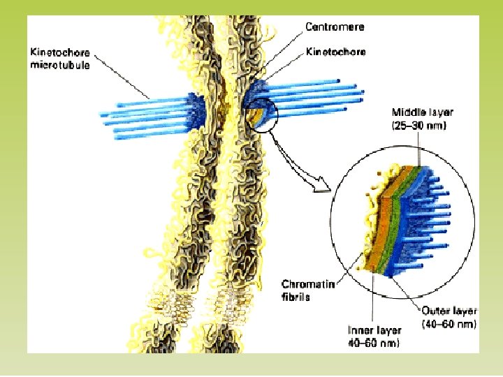

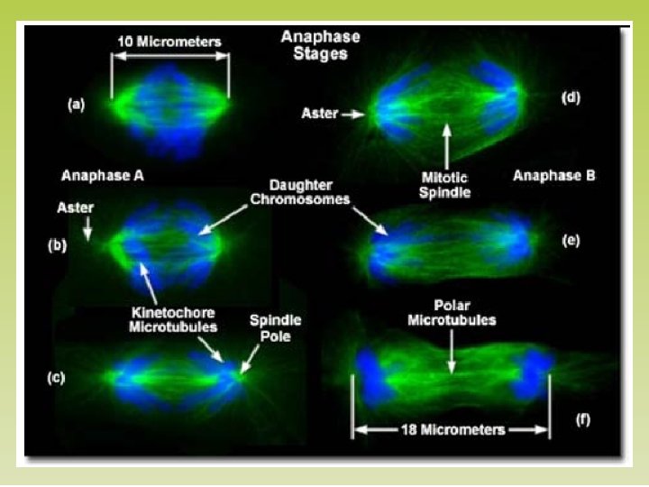
Pada anafase, sister chromatid berpisah dan bergerak sepanjang kinetochore microtubules menuju arah berlawanan ujung sel Kinetochor e Spindle pole

CYTOKINESIS Pada sel hewan Cytokinesis terjadi oleh proses yang disebut cleavage, membentuk sebuah a cleavage furrow Cleavage furrow 100 µm Contractile ring of microfilaments Daughter cells (a) Cleavage of an animal cell (SEM)
 Vesicles forming cell plate")
Pada sel tumbuhan selama cytokinesis terbentuk plat sel (cell plate) Vesicles forming cell plate Wall of patent cell 1 µm Cell plate New cell wall Daughter cells (b) Cell plate formation in a plant cell (SEM)

Mitosis In A Plant Cell Nucleus Chromatine Nucleolus condensing 1 Prophase. The chromatin is condensing. The nucleolus is beginning to disappear. Although not yet visible in the micrograph, the mitotic spindle is staring to from. Figure 12. 10 Chromosome 2 Prometaphase. We now see discrete chromosomes; each consists of two identical sister chromatids. Later in prometaphase, the nuclear envelop will fragment. 3 Metaphase. The 4 spindle is complete, and the chromosomes, attached to microtubules at their kinetochores, are all at the metaphase plate. Anaphase. The 5 chromatids of each chromosome have separated, and the daughter chromosomes are moving to the ends of cell as their kinetochore microtubles shorten. Telophase. Daughter nuclei are forming. Meanwhile, cytokinesis has started: The cell plate, which will divided the cytoplasm in two, is growing toward the perimeter of the parent cell.

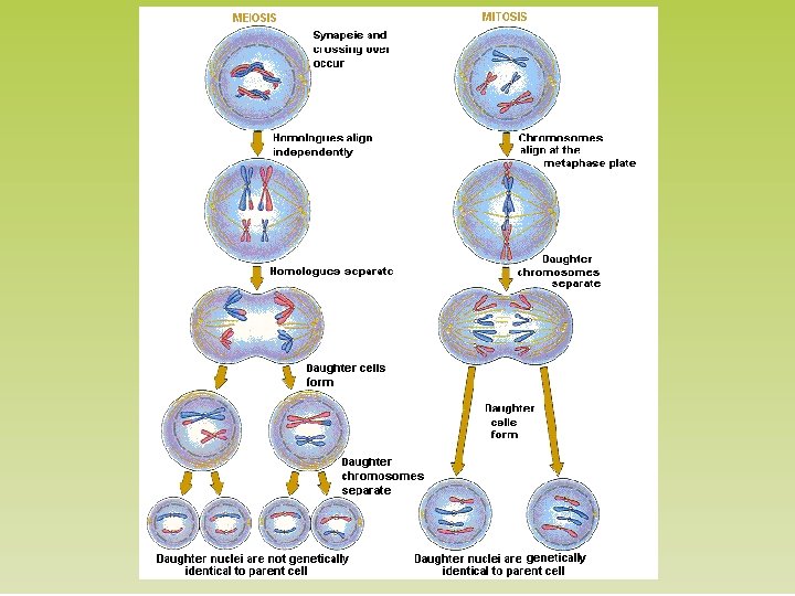
Meiosis 1. Meiosis dan mitosis hampir sama caranya hanya yg berbeda perilaku pembelahan kromosom pada meiosis I 2. Pembelahan sel gamet

Meiosis I Profase I Prometafase I Anafase I Telofase I Metafase I Sitokinesis I
 kromosom berkelompok dan memendek 2. Zygoten (berpasangan) Kromosom homolog")
Profase I 1. Leptoten (ramping) kromosom berkelompok dan memendek 2. Zygoten (berpasangan) Kromosom homolog membentuk pasanganya yg disebut bivalen dan membentuk komplek sinoptik , gen mengalami rekombinasi melalui pindah silang 3. Pakiten : bivalen memendek dan tebal 4. Diploten : kromosom homolog sedikit tertarik , kromatid dan kiasmata terpisah 5. Diakinesis: Sentromer menjauh kromatid terus memendek

Lima Tahapan Profase I Leptoten Zigoten Diploten Pakiten Diakinesis

Meiosis II Profase II Telofase I Anafase II Telofase II Metafase II

SPERMATOGENESIS

OOGENESIS
 1. Fasa G yang terdiri dari G 1 dan G 2")
Fase G (gap) 1. Fasa G yang terdiri dari G 1 dan G 2 adalah fase sintesis zat yang diperlukan pada fase berikutnya. 2. Pada sel mamalia, interval fase G 2 sekitar 2 jam, sedangkan interval fase G 1 sangat bervariasi antara 6 jam hingga beberapa hari. 3. Sel yang berada pada fase G 1 terlalu lama, dikatakan berada pada fase G 0 atau “quiescent”. Pada fase ini, sel tetap menjalankan fungsi metabolisnya dengan aktif, tetapi tidak lagi melakukan proliferasi secara aktif. 4. Sebuah sel yang berada pada fase G 0 dapat memasuki siklus sel kembali, atau tetap pada fase tersebut hingga terjadi apoptosis. 5. Pada umumnya, sel pada orang dewasa berada pada fase G 0. Sel tersebut dapat masuk kembali ke fase G 1 oleh stimulasi antara lain berupa: perubahan kepadatan sel, mitogen atau faktor pertumbuhan, atau asupan nutrisi.
- Slides: 32