BIOL 3340 Chapter 3 Chapter 3 Microbial Cell

BIOL 3340 Chapter 3

Chapter 3 Microbial Cell Structure

Types of Cells Two major classes: eukaryotes & prokaryotes. Differences: the materials making up the nucleus of eukaryotic cells are separated from the rest of the cell by the nuclear membrane, whereas in prokaryotic cells these materials are not separated. • All animals and plant cells are eukaryotic including fungi. Bacteria, cyanobacteria and the mycoplasmas are prokaryotic.
 – spheres ldiplococci")
Size, Shape, and Arrangement of Bacterial cells Cocci (s. , coccus) – spheres ldiplococci (s. , diplococcus) – pairs lstreptococci – chains lstaphylococci – grape-like clusters ltetrads – 4 cocci in a square lsarcinae – cubic configuration of 8 cocci

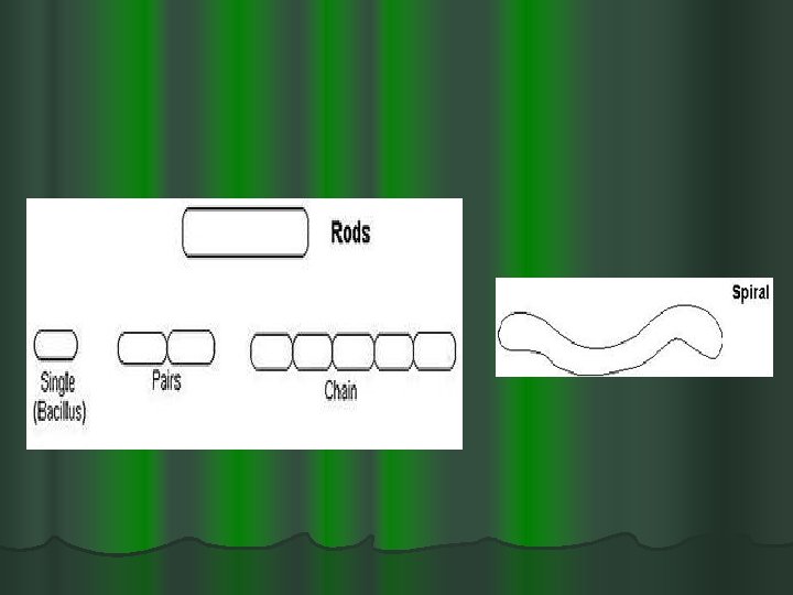
: – rods l coccobacilli – very")
……Size, Shape, and Arrangement Bacilli (s. , bacillus): – rods l coccobacilli – very short rods l vibrios – resemble rods, comma shaped l spirilla (s. , spirillum) – rigid helices l spirochetes – flexible helices l mycelium – network of long, multinucleate filaments Check on line lab Manual for Bacterial shapes)

……Size, Shape, and Arrangement l Sizes: l Typically ~ 0. 1 - 20 m (with some exceptions) l Typical coccus: ~ 1 m (e. g. Staphylococcus) l Typical short rod: ~ 1 x 5 m (e. g. E. coli) l Barely within the best resolution of a good compound light microscope

Bacterial Shapes

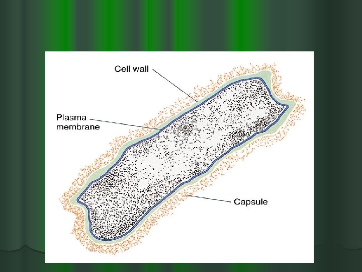
Cell Structure of Procaryotes Prokaryotic cells The constituents of a typical bacterium are as follows: Bacterial Cell Wall and Capsule – bacteria are surrounded by a cell wall, which not only contains polysaccharide but also contains protein and lipid. l In some bacteria, the cell wall is surrounded by the capsule. l The cell wall and capsule provide shape and form to the bacterium and also acts as a physical barrier between the bacterium and its environment. l Nucleoids – in bacteria the nuclear material is concentrated in a region called the nucleoid within the cytoplasm. l

A typical Bacterial Cell

…. Cell Structure There is no membrane-bound nucleus in prokaryotes. l Instead the DNA is located within a specialized region of the cytoplasm of the cell called the nucleoid region. l There is no nuclear membrane surrounding the nucleoid. l l Bacterial flagella – many bacteria possess one or more flagella for locomotion.

Gram-negative Cell Walls and Acid Fast cell wall in Chapter 3

Procaryotic Cell Membranes: l membranes are an absolute requirement for all living organisms. l plasma membrane encompasses the cytoplasm l some procaryotes also have internal membrane systems

Functions of the Plasma Membrane l separation of cell from its environment l selectively permeable barrier l some molecules are allowed to pass into or out of the cell l transport systems aid in movement of molecules l detection of and response to chemicals in surroundings with the aid of special receptor molecules in the membrane

Fluid Mosaic Model of Membrane Structure

…. . Plasma membrane

…. . Plasma membrane

…Phospholipid layer l polar ends l interact with water l hydrophillic l nonpolar ends l insoluble in water l hydrophobic

Membrane Proteins Peripheral proteins: l loosely associated with the membrane and easily removed Integral proteins l embedded within the membrane and not easily removed

Procaryotic Cytoplasm Cytoplamic Matrix: l Cytoplasm contains the nucleoid, ribosomes and inclusion bodies l lacks organelles bound by unit membranes l composed largely of water l is a major part of the protoplasm (the plasma membrane and everything within)

. . Cytoplasmic Matrix l Viscous aqueous suspension of proteins, nucleic acid, dissolved organic compounds, mineral salts l Network of protein fibers similar to the eukaryotic cytoskeleton. Cytoplasmic Inclusion Bodies: l granules of organic or inorganic material that are stockpiled by the cell for future use. l some are enclosed by a single-layered membrane

…. Cytoplasmic inclusions: l Glycogen Granules l Poly- -hydroxybutyrate granules l Lipid droplets l Gas vacuoles l Metachromatic granules (Phosphate crystals or volutin granules) l Sulfur Granules

Ribosomes: l complex structures consisting of protein and RNA l sites of protein synthesis l smaller than eucaryotic ribosomes 70 S l eucaryotic ribosomes 80 S l procaryotic

The Nucleoid: l irregularly shaped region l location of chromosome l usually l not 1/cell membrane-bound

The Procaryotic Chromosome The Chromosomes: l usually a closed circular, double-stranded DNA molecule l looped and coiled extensively

Plasmids: l usually small, closed circular DNA molecules l exist and replicate independently of chromosome l have relatively few genes present

Procaryotic Cell Walls Prokaryotic Cell Wall: l rigid structure that lies just outside the plasma membrane (detail to continue)

Functions of Cell Wall l provides characteristic shape to cell l protects the cell from osmotic lysis l may also contribute to pathogenicity l very few procaryotes lack cell walls

Cell Walls of Bacteria Gram Staining developed by Gram in 1888: l bacteria are divided into two major groups based on the response to gram-stain procedure l gram-positive bacteria stain purple l gram-negative bacteria stain pink l staining reaction due to cell wall structure


Gram Positive and Gram negative

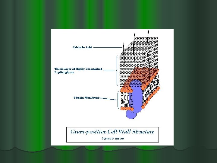
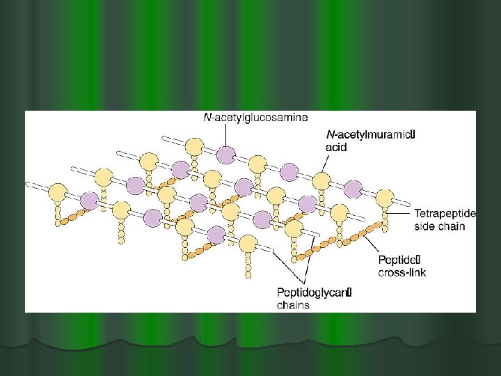
Gram-Positive Cell Walls l Gram positive bacteria composed primarily of peptidoglycan l Peptidoglycan are polymers which contains N-acetylglucosamine and N-acetylmuramic acid and several different amino acids • Walls contain teichoic acid ( polymers of glycerol or ribitol joined by phosphate groups)

. . Gram-Positive Cell Walls l The periplasmic space lies between plasma membrane and cell wall and is smaller than that of gram-negative bacteria l periplasm has relatively few proteins l enzymes secreted by gram-positive bacteria are called exoenzymes

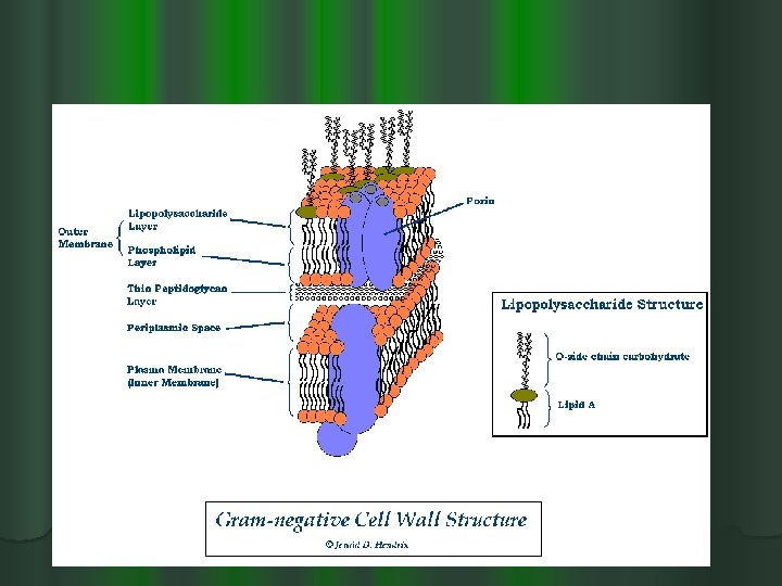
Gram-Negative Cell Walls l consist of a thin layer of peptidoglycan surrounded by an outer membrane l outer membrane composed of lipids, lipoproteins, and lipopolysaccharide (LPS) l no teichoic acids

…. . Gram Negative l more complex than gram-positive walls l periplasmic space differs from that in gram -positive cells l may constitute 20 -40% of cell volume l many enzymes present in periplasm

Gram Positive and Negative cell Wall

Assignments l Features of a prokaryotic cell l List the differences between a gram positive and gram negative cell wall.

Variations on Cell Wall Architecture Acid-fast Cell Walls: l Many genera in the “High GC gram-positive” bacterial group contain mycolic acids, embedded in the peptidoglycan. l Mycolic acids are a class of waxy, extremely hydrophobic lipids. l Certain genera contain very large amounts of this lipid, and are difficult to gram stain. l These genera may be identified by the “acidfast” staining technique. l Includes Mycobacterium and Nocardia.

. . Variations on Cell Wall Architecture Mycoplasmas: l Bacteria that are naturally have no cell walls l Includes Mycoplasma and Ureaplasma Archaea : l Have archaea cell walls with no peptidoglycan l Many have cell walls containing pseudomurein, a polysaccharide similar to peptidoglycan but containing Nacetylglucosamine and Nacetyltalosaminuronic acid.

Capsules, Slime Layers, and S-Layers of material lying outside the cell wall l capsules l usually composed of polysaccharides l well organized and not easily removed from cell l slime layers l similar to capsules except diffuse, unorganized and easily removed. l a capsule or slime layer composed of polysaccharides can also be referred to as a glycocalyx

Glycocalyx

S-layers: l regularly structured layers of protein or glycoprotein. l in bacteria the S-layer is external to the cell wall. l Regular “floor tile” pattern. l Function not clear -- Stability?

Functions of Capsules, Slime Layers, and S-layers l protection from host defenses (e. g. , phagocytosis) l protection from harsh environmental conditions (e. g. , desiccation) , chemicals or osmotic stress l attachment to surfaces l facilitate motility l nutrient Storage
 l short, thin, hairlike, proteinaceous appendages up to")
Pili and Fimbriae (s. , fimbria) l short, thin, hairlike, proteinaceous appendages up to 1, 000/cell l mediate attachment to surfaces sex pili (s. , pilus): l similar to fimbriae except longer, thicker, and less numerous (1 -10/cell) l required for mating

Fimbriae

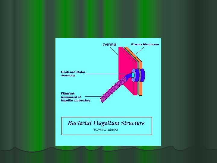
…Fimbriae Function s: l Mobility l Almost all motile bacteria are motile by means of flagella l Motile vs. non motile bacteria. l Different species have different flagella arrangements Structure: Filament composed of the protein flagellin l Hook & Rotor Assembly & Permits rotational "spinning" movement l

…Fimbriae

Chemotaxis l movement towards a chemical attractant or away from a chemical repellent l concentrations of chemical attractants and chemical repellents detected by chemoreceptors on surfaces of cells

Bacterial Endospores Bacterial Spores l are formed by some bacteria as dormant structures. l resistant to numerous environmental conditions e. g heat, radiation, chemicals, nutrient depletion, desiccation, and waste buildup. l Bacterial spores are NOT a reproductive structure, like plant or fungal spores. l Produced by very few genera of bacteria l Major examples Bacillus & Clostridium

…endospores Sporogenesis

Sporogenesis: l l l Also called endospore formation or sporulation normally commences when growth ceases because of lack of nutrients A copy of the bacterial chromosome is surrounded by a thick, durable spore coat. When the vegetative cell dies and ruptures, the free spore is released. When spore encounters favorable growth conditions, spore coat ruptures and a new vegetative cell is formed.

. . Sporogenesis Complex multistage process

Spore Germination

Bibliography l l l http: //en. wikipedia. org/wiki/Scientific_method https: //files. kennesaw. edu/faculty/jhendrix/bio 33 40/home. html Lecture Power. Points Prescott’s Principles of Microbiology-Mc Graw Hill Co. http: //www. bio. mtu. edu/campbell/prokaryo. htm http: //molecularbiology. suite 101. com/article. cfm/cell_structure http: //water. me. vccs. edu/courses/ENV 108/lesso n 5_2. htm
- Slides: 60