Biochemistry of Vitamins and Minerals Biochemistry of Vitamins

Biochemistry of Vitamins and Minerals

Biochemistry of Vitamins characters: • They are essential organic compounds for normal health and growth since they are not synthesized in human body. • They must be supplied in the diet in the required very small amounts and deficiency of any vitamin in the body results in a disease. • They are present in normal food in small amounts. • They act as coenzymes. • They do not enter in tissue structure as carbohydrates, fats and proteins. • They are not oxidized for energy production as carbohydrates, fats and proteins. • Provitamins: These are precursors of vitamins that converted into vitamins inside the body e. g. Carotenes are provitamin A. • Vitaimers: These are different forms of one vitamin e. g. Vitamin D has 2 vitamers; D 2, & D 3.

• • • Classification: Vitamins can be classified according to their solubility into two main categories: A. Fat soluble vitamins: they include A, K. E&D vitamins. Characters: 1. They are soluble in fat solvents. 2. They need bile salts for absorption. 3. They can be stored in the body. B. Water soluble vitamins: they include vitamin C and B complex group. Characters: 1. They are soluble in water 2. Most of them are not stored in the body.
 • (Anti-night blindness, Anti-xerophthalmia vitamin)")
• Fat soluble vitamins • Vitamin A (Retenoids) • (Anti-night blindness, Anti-xerophthalmia vitamin) • Provitamin: • - the provitamin is called carotenoids or carotenes
 a- Liver, eggs and")
• • • Sources: 1. Animal sources: (RETINYL ESTER) a- Liver, eggs and milk fat. b- Fish liver oils e. g. shark liver oil. 2. Plant sources: a- Vitamin A is present in plants as provitamin A; Carotenes. b- Carotenes ( , and ): Present in carrots, potato and tomatoes.

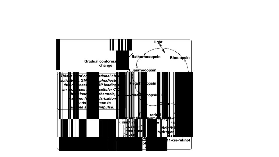
Functions of vitamin A: • Vitamin A acts as a hormone. It act on DNA. It has the following functions: • 1. Role of vitamin A in Vision = Visual cycle =Rhodopsin cycle:

• 2. Role of vitamin A in Reproduction: • 3. Role of vitamin A in Growth: • Retinol is essential for normal growth and bone & teeth formation. • 4. Maintenance of epithelial cells: • 5. Antioxidant (anticancer) action

• • Deficiency of vitamin A 1. Eye: a- Night blindness: impaired dark adaptation. b- Xero-ophthalmla: dryness and roughness of cornea. • 2. Growth retardation. • 3. Skin and mucus membranes: Roughness of skin (goose skin) and mucus membranes e. g. urinary system. This leads to infection .
 have three vitamers: K 1 K 2 and K 3")
Vitamin K (Anti-haemorrhagic vitamin) have three vitamers: K 1 K 2 and K 3 1. Naturally occurring vitamin K (fat soluble): Vitamin K 1 (phylloquinone): is the major form of vitamin K in plants e. g. spinach, cauliflower and cabbage. Vitamin K 2 (menaquinone): is formed inside the intestine by intestinal bacteria. 2. Synthetic (e. g. K-viton drug): ) is water-soluble Vitamin K 3 (Menadione) and it is most potent member.

Functions of vitamin K: 1. Synthesis of some blood clotting factors in liver: prothrombin (factor II), and factors VII, IX and X. 2. in bones. Synthesis of osteocalcin (calcium binding protein)

• Deficlency of vitamin K: • -. Deficiency of vitamin K is rare because intestinal bacteria synthesize it. • -. Deficiency leads to impairment of blood clotting.
 (Antioxidant & anti-sterility vitamin) Structure: - Vitamin")
• • • Vitamin E (tocopherols) (Antioxidant & anti-sterility vitamin) Structure: - Vitamin E in the diet is called Tocopherols. There are four types of tocopherols ; 1 - -tocopherol , 2 - β- tocopherol , 3 - γ- tocopherol, 4 - δ-tocopherol.

• Sources: • Vegetables and seed oils e. g. cotton , olive and coconut oils.
 Vitamin E prevents oxidation of cell")
• Functions: • A. Antioxidant: • 1) Vitamin E prevents oxidation of cell components (e. g. polyunsaturated fatty acids, DNA and cell membranes) by free radicals. • • 2) Vitamin E is natural antioxidant that added to oils to prevent the rancidity. • • 3) vitamin E protects against heart diseases: • As it acts as antioxidant, vitamin E prevent oxidation of LDL. Oxidized LDL causes heart disease. • • 5) It prevents hemolytic anemia through keeping of the integrity of RBCs membrane preventing their heamolysis. • B. It has antisterility effect: •
: • • • Vitamin D have two vitamers a-Vitamin D 2(")
Vitamin D (Calciferol): • • • Vitamin D have two vitamers a-Vitamin D 2( ergocalciferol) its Provitamin is Ergosterol found in plants especially yeast b- Vitamin D 3(Chole. Calciferol)= inactive= its Provitamin is 7 -dehydrocholesterol derived from Cholesterole
 –active form- acts")
• Functions of vitamin D: • 1, 25 dihydroxycholecalciferol (calcitriol) –active form- acts as a hormone, having the following functions: • 1. Normalization of serum calcium: • Calcitriol maintains serum calcium level through its effects on intestine, bones and kidneys. • a- On intestine: It stimulates synthesis of a protein called " calcium binding protein" that responsible for calcium absorption. • b- On bones: It stimulates calcium mobilization from bones. • c- On kidneys: It increases reabsorption of calcium. • 2. Mineralization of bones: • after normalization of serum calcium • 3. Absorption of phosphate • from intestine, It increases also reabsorption of phosphate by kidney.

• Deficiency of vitamin D: • Deficiency of vitamin D causes demineralization of bones that leads to: • 1. Rickets in children: characterized by bone deformities. • 2. Osteomalcia in adults: characterized by bone fractures. • • •
")
WATER SOLUBLE VITAMINS • 1. Vitamin C = L-Ascorbic Acid • (Antiscorbutic vitamin)

A- Formation of collagen protein

• B- A potent reducing agent • Ascorbic acid is capable of giving electrons to ferric ions, cupric ions and Ascorbic acid is needed for bile acid formation from cholesterol. • Ascorbic acid is necessary for the conversion of folic acid into its active form tetrahydrofolic acid. • Ascorbic acid is required in the metabolism of tyrosine. • Ascorbic acid acts as antioxidant. • Anti-Cancer: • Defense Mechanism:
 It is characterized by: Manifestations due to decrease")
• • • Deficiency (scurvy) It is characterized by: Manifestations due to decrease neurotuanomitters : 1 - Behavioral changes 2 - Severe emotional disturbances Manifestations due to decrease collagen formation: 1 - Bleeding 2 - Osteoporosis. 3 - Necrosis of gums and loss of teeth. 4 - Delayed wound healing 5 - Easy bruising and haemorrhages under the skin due to increased capillary fragility. Anemia.
 All pigmented vegetables • b)")
• 1} Vit A present in. • a) All pigmented vegetables • b) liver, millk, butter • c) bath a, b • 2} Deficiency of Vit A in the eye produces all of the following except: a) Night blindness. • b) Xerophthal amia • c) loss of color vision • 3} Defficiency of Vit D in children produce: • a) Rickets • b) osteomalcia • c) non of the above • •

Pyridoxine “Vitamin B 6” Sources: 1. Yeast 2. germinal portion of seeds. 3. Egg Yolk. 4. Royal Jelly of bees (very rich in vitamin B 6)
 acts as a coenzyme. This coenzyme has the")
• Function: • pyridoxal phosphate(PLP) acts as a coenzyme. This coenzyme has the following functions: •

A. Amino acid absorption from the intestine. • • B- Coenzyme for amino acid metabolism in the following reactions: • 1 - Transamination: • 2 - Decarboxylation: • 3 - Non oxidative deamination: • 4 - Synthesis of cysteine • • 6 - Formation of niacin from tryptophan

• Deficiency: • 1 - Pellagra : because PLP is needed for the conversion of tryptophan to niacin. • 2 - Growth retardation & mental retardation : disturbance in amino acids metabolism.
 Sources: Meat,")
Vitamin B 12 = Cyanocobalamin • • (Anti-pernicious anemia or extrinsic factor) Sources: Meat, egg, milk and milk products. Vitamin B 12 is not present in vegetables. Intestinal microorganisms synthesize B 12 in human colon,

• Functions • Vitamin B 12 acts as coenzyme after being converted to methylcobalamin for: • a) Methylation of homocysteine to methionine: • *is important for haemopoiesis (participate in purine, pyrimidine, and nucleic acid syntheses). •
 oxidation of odd number fatty acids. • & the formation of myelin")
B) oxidation of odd number fatty acids. • & the formation of myelin sheath through the metabolism of odd number fatty acids.

Deficiency: • 1 - Megaloblastic anaemia • -2 - Neurological manifestations
")
Folic acid (Folacin = Peteroyl Glutamic Acid)

• Sources: The major source is leafy vegatables. • Liver, kidney and yeast are also rich in folate.
 act as a carrier for")
• Functions: • Tetrahydrofolic acid (H 4 folate) act as a carrier for one– carbon groups.

• Deficiency: • 1 - Pancytopenia: i. e. all blood cells are affected • a) Megaloblastic anaemia (Marocytic, hyperchromic). • b)Leucopenia: W. B. Cs. • c) Thrombocytopnea: Platelets. • 2 - Impaired growth.

• Lipoic Acid • Physiological Functions • It acts as coenzyme in e. g. pyruvic acid into acetyl co. A.
 Coenzyme A (Co. ASH) • •")
• Pantothenic Acid • Functions • A) Coenzyme A (Co. ASH) • • B) Acyl carrier protein (ACP): • ACP acts as acyl carrier for fatty acid synthesis.

Minerals

• According to the body needs, minerals may be divided into two groups: • A. Macro-minerals: • - They are required in amounts greater than 100 mg/day. • - They include: calcium, phosphorus, magnesium, sodium, potassium, chloride and sulfur.
: • - They are required in amounts less")
• B. Micro-minerals (trace elements): • - They are required in amounts less than 100 mg/day. • - Trace elements: Elements present in the body in very low amounts (micrograms/gram or less). • - e. g. , chromium, cobalt, copper, fluorine, iodine, iron, manganese, molybdenum, selenium and zinc.
 • IRON (Fe 3+) • • Body Iron: •")
• Micro-minerals (Trace elements) • IRON (Fe 3+) • • Body Iron: • - The total body iron of an adult male is 3 -5 g. • It is distributed as follows: RBCs iron (hemoglobin): 66%, tissue iron (33%) and plasma iron 1%.
 Iron deficiency anemia: Causes: 1. Deficient intake.")
• Alterations of plasma iron: a) Iron deficiency anemia: Causes: 1. Deficient intake. 2. Impaired absorption: e. g. Steatorrhea, abdominal surgery. 3. Excessive loss e. g. menstrual loss, gastrointestinal bleeding, bleeding due to some parasites (anchylostoma). a) Iron overload: Causes: 1. Repeated blood transfusion. 2. Intravenous administration of iron. 3. Hemochromatosis (hemosiderosis, Bronz diabetes): • - This is a rare hereditary disease characterized by abnormal increase of iron absorption.
 • - Sources: • - The richest sources of")
• Copper (Cu 2+) • - Sources: • - The richest sources of copper are nuts, liver, kidney cow’s milk is a poor source of copper. • Body copper: 1. The adult human body contains 100 – 150 mg of copper.

• Functions: 1. It is essential for hemoglobin synthesis. 2. It enters in the formation of bone. 3. Maintains the myelin within the nervous system.

• Alterations of plasma copper: • 1. Hypercupremia: Occurs in infections, • • 2. Hypocupremia decreased plasma copper or Wilson’s disease (hepatolenticular degeneration). • - This disease is characterized by accumulation of large amounts of copper in: 1. Liver causing hepatic cirrhosis. 2. Lenticular nucleus of the brain causing lenticular degeneration with abnormal movement. 3. Cornea: Causing greenish-brown discoloration of the corneal margin which is called: Kayser-Fleisher rings.
 • Sources: liver, eggs, seafood and milk. • •")
• Zinc (Zn 2+) • Sources: liver, eggs, seafood and milk. • • Body zinc: 1. The adult male body contains about 2 g of zinc.

• Functions: 1. Zinc is important for growth and reproduction. It is a cofactor for RNA and DNA polymerases, so RNA and DNA synthesis. 2. It plays a role in tissue repair, wound healing and hair and nail integrity. 3. Zinc is an essential cofactor for a number of enzymes including: alkaline phosphatase, lactate and malate dehydrogenases, 4. Zinc is required for mobilization of vitamin A from the liver. 5. Insulin forms complex with zinc, this increases the duration of insulin action when given by injection. In the B cells of the pancreas is used to store and release insulin. 6. Immunostimulant 7. essential for male reproductive system and brain function. 8. Anti-inflammatory 9. hypocholesterolemic.

• Zinc deficiency: • Resulting in growth retardation, hypogonadism, delayed puberty and poor immunity

thanks
- Slides: 50