Biochemistry of hormones derived from amino acids and

Biochemistry of hormones derived from amino acids and proteins Alice Skoumalová

Content 1. 2. 3. 4. 5. 6. 7. 8. 9. Definition of peptide hormones Common features: synthesis, interactions with receptors at the cell surface Groups of peptide hormones Hypothalamus-hypophysis hormonal cascade (signal amplification, negative feedback system) Genes and formation of polypeptide hormones (gene superfamilies) Hormones of the hypothalamus and the hypophysis Synthesis and degradation of catecholamines Biochemistry of parathyroid hormone and insulin Degradation of peptide hormones

Definition of peptide hormones Ø secreted into the blood stream; endocrine functions Ø synthesized from amino acids according to an m. RNA template, which is itself synthesized from a DNA template Ø precursors (pre-prohormones) - posttranslational modification (endoplasmatic reticulum) - removal of the pre-sequence, sometimes glycosylation - resulting in prohormones Ø the prohormones - packaged into membrane-bound secretory vesicles - secreted from the cell by exocytosis in response to specific stimuli Ø mature peptide hormones diffuse through the blood to all of the cells of the body, where they interact with specific receptors on the surface of their target cells

Peptide hormones interact with specific receptors on the cell surface

G protein-coupled receptors Signal transduction via: 1. Protein kinase A pathway (the elevation of c. AMP activates protein kinase A) Corticotropin releasing hormone, thyrotropin, luteinizing hormone, follicle stimulating hormone, adrenocorticotropic hormone, vasopressin, opioid peptides, norepinephrine, epinephrine 2. Protein kinase C and IP 3 -Ca 2+ (inositoltriphosphate) pathway (triggering of the hydrolysis of phosphatidylinositol-4, 5 -bisphosphate and stimulation of protein kinase C) Thyrotropin releasing hormone, gonadotropic releasing hormone, thyrotropin, norepinephrine, angiotensin 3. Protein kinase G pathway (the elevation c. GMP activates protein kinase G) Atrionatriuretic factor Protein kinase receptors e. g. Tyrosin specific protein kinases (Insulin)

Hormones 1. Amine-derived hormones Catecholamines and thyroxine 2. Peptide hormones Small peptide hormones (thyrotropin releasing hormone, oxytocin, vasopressin) Protein hormones (insulin, growth hormone) Glycoprotein hormones (luteinizing hormone, follicle-stimulating hormone and thyroid-stimulating hormone) 3. Steroid hormones

Peptide hormones Hormones of the hypothalamus-hypophysis cascade Hormones produced by other tissues heart (atrionatriuretic factor) pancreas (insulin, glucagon, somatostatin) gastrointestinal tract (cholecystokinin, gastrin) fat stores (leptin) parathyroid glands (parathyroid hormone) kidney (erythropoietin)

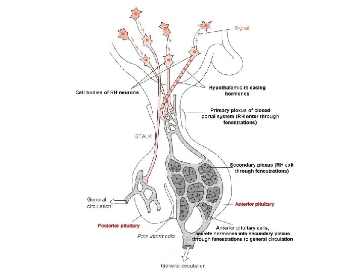
Environmental or internal signal Hormonal cascade Signal amplification CNS Electrical-chemical signal Limbic system Electrical-chemical signal Hypothalamus Releasing hormones (ng) Anterior pituitary hormone (μg) Target „gland“ The gonads, the thyroid gland, the adrenal cortex Ultimate hormone (mg) Systemic effects
 Electrical-chemical signal")
Environmental stress CRH-ACTH-Cortisol CNS A single stressor (change in temperature, noise, trauma) Electrical-chemical signal Limbic system Electrical-chemical signal Hypothalamus Portal system The corticotrophic cells Corticotropin releasing hormone (CRH) in ng, t 1/2 minutes Anterior pituitary Adrenocorticotropic hormone (ACTH) in µg, increased t 1/2 Adrenal gland Cortisol in mg, t 1/2 hours The glucocorticoid receptors in different cells Systemic effects

CNS Hormonal cascade Negative feedback system Limbic system Hypothalamus Long feedback loop Releasing hormones Anterior pituitary hormones Target „gland“ Ultimate hormone Systemic effects Short feedback loop

Clinical correlation of the hormonal cascade Testing the activity of the anterior pituitary For example infertility: which organ is at fault in the hormonal cascade? Step 1 The gonads must be considered Ø injecting LH or FSH Ø if sex hormone is elicited, the gonads function properly Step 2 The anterior pituitary must be tested Ø i. v. administration of Gn. RH (secretion of LH and FSH; by RIA) Normal response Ø The hypothalamus is nonfunctional No response Ø The anterior pituitary is nonfunctional
 Releasing hormone Number of amino acids Anterior pituitary hormone released")
Hypothalamic releasing hormones (RH) Releasing hormone Number of amino acids Anterior pituitary hormone released or inhibited Thyrotropin releasing hormone (TRH) 3 Thyrotropin (TSH) Gonadotropin releasing hormone (Gn. RH) 10 Luteinizing hormone (LH), Follicle stimulating hormone (FSH) Corticotropin releasing hormone (CRH) 41 Adrenocorticotropic hormone (ACTH), β-lipotropin, β-endorphin Growth hormone releasing hormone (GHRH) 44 Growth hormone (GH) Somatostatin 14 GH release inhibited Prolactin releasing factor (PRF) Prolactin (PRL) Prolactin release inhibiting factor (PIF), Dopamine PRL release inhibited

Hypothalamus GRH TRH CRH Dopamine PRF, PIF Gn. RH Anterior pituitary GH Liver TSH Thyroid ACTH LPH β-Endorphin MSH PRL FSH LH Ovary Mammary gland Testis Adrenal cortex Skin darkening β-Endorphin Corticosteroids Hyperglycemic effects Thyroid hormones Growth of bone, body tissues; carbohydrate and protein metabolism; production of IGFs Analgesia Ovulation, corpus luteum, progesterone Cell development, lactation Development of follicles, estradiol Interstitial cell development, testosterone Growth of seminal tubules and spermatogenesis GH-Growth hormone, TSH-Thyrotropin, ACTH-Adrenocorticotropic hormone, LPH-Lipotropin, MSH-Melanocyte stimulating hormone, PRL-Prolactin, FSH-Follicle stimulating hormone, LH-Luteinizing hormone
 Neurohypophysis Oxytocin Uterine contraction, lactation Vasopressin (ADH) Water")
Hypothalamus Axonal transport Oxytocin Vasopressin (ADH) Neurohypophysis Oxytocin Uterine contraction, lactation Vasopressin (ADH) Water balance
 Ø Axonal")
Vasopressin and oxytocin Ø Synthetized in the hypothalamus (nucleus supraopticus and paraventricularis) Ø Axonal transport with transport proteins (neurophysins) Ø Nonapeptides with disulfide bridge Cys-Tyr-Phe-Gln-Asn-Cys-Pro-Arg-Gly-NH 2 Arginine vasopressin Cys-Tyr-Phe-Gln-Asn-Cys-Pro-Lys-Gly-NH 2 Lysine vasopressin Cys-Tyr-Ile-Gln-Asn-Cys-Pro-Arg-Gly-NH 2 Oxytocin Structural similarity, overlapping functions Oxytocin: causes milk ejection in lactating female Vasopressin: increases water reabsorption from distal kidney tubule

Hypopituitarism Ø The deficiency of one or more hormones of the pituitary gland Ø The connection between the hypothalamus and anterior pituitary can be broken by 1. Trauma (automobile accidents) 2. Tumor of the pituitary gland Ø Ø Ø Decreased generation of the pituitary hormones A life-threatening situation The usual therapy involves administration of the end organ hormones (cortisol, thyroid hormone, sex hormones, progestin, growth hormone in children)

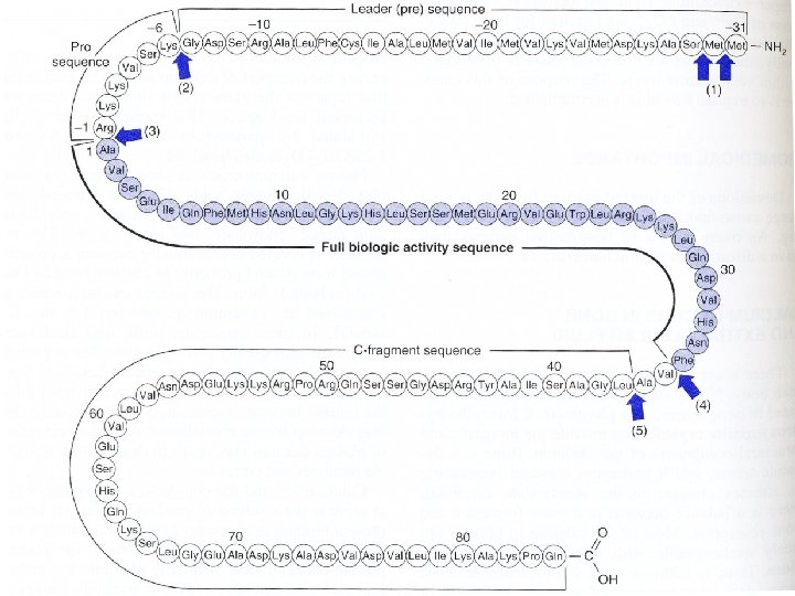
Genes and formation of polypeptide hormones Genes for polypeptide hormones contain the information for the hormone 1. More than one hormone is encoded in a gene Proopiomelanocortin peptide family Vasopressin and neurophysin II; oxytocin and neurophysin I 2. Multiple copies of a hormone are encoded in a gene e. g. Enkephalins 3. Only one hormone is encoded in a gene e. g. CRH
 is a precursor peptide for eight hormones Ø ACTH,")
Proopiomelanocortin (a single gen product) is a precursor peptide for eight hormones Ø ACTH, β-lipotropin, γ-MSH, α-MSH, CLIP, β-endorphine, enkephalins Ø Proopiomelanocortin occurs in both the corticotropic cells of the anterior pituitary and the pars intermedia cells, the products are different CLIP-corticotropin-like intermediary peptide
 and neurotransmitters Ø Precursor molecule")
Proopiomelanocortin peptide family Ø Contains hormones (ACTH, LPH, MSH) and neurotransmitters Ø Precursor molecule involves 285 amino acids Ø Gene expression in the anterior and intermediary pituitary, but also in other tissues (intestine, placenta, male reproductive system) Ø Cleavage into peptides, further modification (glycosylation, acetylation, phosphorylation) ACTH: acts on cells in the adrenal gland to increase cortisol production and secretion; excessive formation-Cushing‘s syndrome β-lipotropin: induces lypolysis, precursor of β-endorphine Endorphines: endorphines bind to the opioid receptors in CNS, analgesia MSH: acts on skin cells to cause the dispersion of melanin (skin darkening)

Multiple copies of a hormone can be encoded on a single gene ØThe gene product for enkephalins (located in the adrenal medulla) ØEnkephalins are pentapeptides with opioid activity Tyr-Gly-Phe-Met (methionine-enkephalin) Tyr-Gly-Phe-Leu (leucine-enkephalin) Model of enkephalin precursor Øencodes several met-enkephalins (M) molecules and a molecule of leu-enkephalin (L)
 Øsynthesized in the adenohypophysis, the concentration in the pituitary is 5")
Growth hormone (GH) Øsynthesized in the adenohypophysis, the concentration in the pituitary is 5 -15 mg/g Øsingle polypeptide, two disulfide bridges Øis essential for postnatal growth Biochemical actions 1. GH increases protein synthesis 2. Carbohydrate metabolism: GH antagonizes the effects of insulin (hyperglycemia); decreased peripheral utilization of glucose, increased hepatic production via gluconeogenesis 3. Lipid metabolism: GH promotes the release of free fatty acids and glycerol from adipose tissue, increases circulating free fatty acids, causes increased oxidation of free fatty acids in the liver 4. Mineral metabolism: GH promotes a positive calcium, magnesium, and phosphate balance (promotes growth of long bones) 5. Prolactin-like effects Pathophysiology: dwarfism, gigantism, acromegaly
 Ø is secreted in the adenohypophysis Biochemical actions: the initiation and maintenance")
Prolactin (PRL) Ø is secreted in the adenohypophysis Biochemical actions: the initiation and maintenance of lactation Pathophysiology: tumors of prolactin-secreting cells cause amenorrhea and galactorrhea in women, gynecomastia and impotence in men
, luteinizing hormone (LH), follicle-stimulating hormone (FSH)")
The pituitary and placental glycoproteins: Thyroid-stimulating hormone (TSH), luteinizing hormone (LH), follicle-stimulating hormone (FSH) a chorionic gonadotropin (CG) Ø structural similarities (common ancestral gene): 2 subunits-α (identical for all of these hormones) and β (determines the specific biologic activity) Ø synthesized as preprohormones and are subject to posttranslational processing (glycosylation) Ø LH and FSH are responsible for gametogenesis and steroidogenesis in the gonads h. CG is synthesized in the syncytiotrophoblast cells of the placenta; increases in blood and urine shortly after implantation; its detection is the basis of many pregnancy tests h. CG- β subunit

Biosynthesis of catecholamines in the adrenal medulla 1 2 3 4 1. Tyrosine hydroxylase: oxidoreductase, cofactor tetrahydropteridine; inhibition by the catecholamines, tyrosine derivates, and by chelating iron 2. Dopa decarboxylase: cofactor pyridoxal phosphate; inhibitors α-methyldopa 3. Dopamine β-hydroxylase: mixed function oxidase, ascorbate as an electron donor, copper at the active site 4. Phenylethanolamine-N-methyltransferase: the synthesis is induced by glucocorticoid hormones
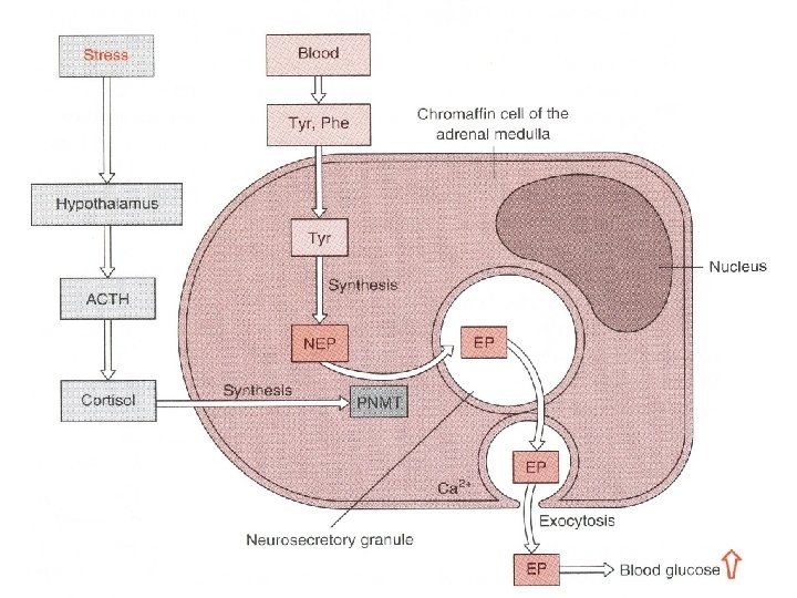
 and monoamine oxidase (MAO) Different metabolites are")
Catecholamines are rapidly metabolized by catechol-O-methyltransferase (COMT) and monoamine oxidase (MAO) Different metabolites are formed; two classes have diagnostic significance: metanephrines and 3 -methoxy-4 -hydroxymandelic acid (vanillylmandelic acid); measurable in urine; elevation in pheochromocytoma
 Pre 31 Pro PTH 6 1 84 Endoplasmic reticulum Parathyroid gland")
Parathyroid hormone (PTH) Pre 31 Pro PTH 6 1 84 Endoplasmic reticulum Parathyroid gland Golgi apparatus Blood (biological active) Liver Ø PTH affects calcium homeostasis Ø Increases the rate of dissolution of bone, reduces the renal excretion of Ca 2+, increases the efficienty of calcium absorption from the intestine by promoting the synthesis of calcitriol

Insulin Ø polypeptide consisting of 2 chains linked by 2 disulfide bridges
 is cleaved after transporting")
Synthesis and posttranslational modification of insulin ØHydrophobic pre-sequence (signal peptide) is cleaved after transporting to ER ØProinsulin is further transported to GA and cleaved by trypsin-like enzymes and carboxypeptidase E ØHeterodimer and C-peptide are formed

The human insulin gene has been isolated The synthesis of human insulin in bacterial expression systems, using recombinant DNA technology, affords an excellent source of this hormone for diabetic patients Diagrammatic structure of the human insulin gene Areas with diagonal stripes correspond to untranslated regions, open regions correspond to intervening sequences, and stippled regions correspond to coding sequences

Inactivation and degradation of peptide hormones Ø Most polypeptide hormones are degraded to amino acids by hydrolysis in the lysosome Ø Certain hormones contain modified amino acids The hypothalamic releasing hormones Pyroglutamic acid (p. Glu) C-terminal amino acid amide (Gly-NH 2, Ala-NH 2, Leu-NH 2) p. Glu C---Peptide O NH O Breakage of the p. Glu or cleavage of the C-terminal amide can lead to inactivation of these hormones (this probably accounts for the short halflife of many of these hormones)
")
Some hormones contain a ring structure joined by a disulfide bridge (oxytocin, vasopressin, somatostatin) 2. Glutathione transhydrogenase Cys-Tyr-Ile-Gln-Asn-Cys-Pro-Arg-Gly-NH 2 1. Cystine aminopeptidase Step 1: Breakage of the ring structure Step 2: Cleavage of cystine Octapeptide further degradation amino acids Oxytocin
")
Summary Ø Peptide hormones are synthetized in the transcription and translation process (DNA-m. RNA-peptid) and further modified (posttranslational modification) Ø Peptide hormones interact with specific receptors on the cell surface and trigger a cascade of secondary effects within the cytoplasm (c. AMP, second messengers) Ø Peptide hormones form gene families that originate from a common ancestral gene Ø Several important peptide hormones are secreted from the hypothalamus-pituitary cascade (signal amplification, negative feedback interaction) Ø Peptide hormones are produced by many different organs and tissues
- Slides: 35