Biochemistry of biological fluids BIOCH 472 DR MANSOUR
 DR. MANSOUR GATASHEH Biochemistry Department, Science College King")
Biochemistry of biological fluids (BIOCH 472) DR. MANSOUR GATASHEH Biochemistry Department, Science College King Saud University

Class 11: Mucus / Phlegm / Sputum

Objectives for this lecture l State the functions of

Function of Mucus l l Sputum is the mucus produced in the lungs. Properties : – – l Protective Lubricative Waterproofing Entraps microorganisms Failure may result in mechanical obstruction of the airway

Physiology of the Mucociliary System l Source of airway secretions – – – Gel layer Periciliary layer Surface epithelial cells l l l – Pseudostratified, columnar, ciliated epithelial cells Surface goblet cells Clara cells Submucosal glands l l Provide airway surface mucin Under parasympathetic control
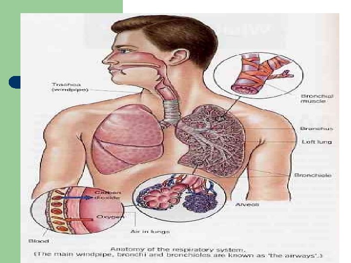

l Thin surfactant layer between the periciliary fluid and the mucous gel – – Prevents airway dehydration Permits mucus spreading on extrusion from glands Allows efficient ciliary coupling with mucus More importantly, allows ciliary release from mucus once kinetic energy is transmitted

Clinical Indication for Use l l To reduce accumulation of airway secretions, improve pulmonary function/gas exchange, reduce infection/damage Diseases: – – – – Cystic fibrosis (CF) Chronic bronchitis Pneumonia Diffuse panbronchiolitis (DPB) Primary ciliary dyskinesia Asthma Bronchiectasis

l Factors affecting mucociliary transport – – – l Airway trauma Tracheostomy Cigarette smoke Atmospheric pollutants (SO 2, NO 2, ozone) may transiently increase transport, especially at low concentration. At higher, toxic concentrations or with prolonged exposure these decrease transport rates Hyperoxia and hypoxia In infection, produce some phlegm and if that phlegm becomes darker in colour.

Pathophysiology

Nature of Mucus Secretion l Healthy person secretes 100 ml/day – – l Clear, viscoelastic, sticky All but 10 ml reabsorbed in lung Structure and composition of mucus – Two major classes of mucins (glycosylated proteins) l l Secreted mucins Membrane-tethered mucins

Lab tests for inflammation l identify bacteria, viruses or fungi that may be causing chest problems.
. Serum Ig. E")
Lab tests for inflammation l l Eosinophilia ( >250 cells/mm 3). Serum Ig. E may be increased. Arterial Blood Gases (in severely ill child): - PO 2 is decreased, - PCO 2 is decreased, at first due to hyperventilation, later it increase p. H tends to decrease (respiratory acidosis at first, later metabolic acidosis).

Specimen Collection Kit l Should be dray on, rayon, or polyester fiber swabs l Do not use calcium alginated or cotton swabs nor ones with wooden sticks; they inhibit PCR

Nasopharyngeal Swab l Store specimen collection kits in a dry, cool place

Oropharyngeal Swab 1. Swab the posterior pharynx 2. Avoid the tonsils

Nasopharyngeal Aspirate Collection 1. 2. 3. 4. 5. 6. Attach mucus trap to vacuum source Place catheter into nostril parallel to palate Apply vacuum Slowly remove catheter while slightly rotating it Repeat with other nostril using the same catheter After collection, flush catheter with 3 ml VTM and return VTM to a plastic vial

What to Collect Preferred specimens l Oropharyngeal swabs l Lower respiratory tract specimens – Bronchoalveolar lavage or tracheal aspirates Other specimens l Nasopharyngeal swabs l Nasal swabs and aspirates l Acute and convalescent sera l Sputum specimens Collect samples on several different days

Store Respiratory Specimens l Transport to laboratory as soon as possible l Store specimens at 4 °C before and during transportation within 48 hours l Store specimens at -70 °C beyond 48 hours l Do not store in standard freezer – keep on dry ice or in refrigerator l Avoid freeze-thaw cycles – Better to keep on ice for a week than to have repeat freeze and thaw

Laboratory Diagnosis l l PCR-based techniques Virus isolation Immunofluorescence Rapid antigen detection
- Slides: 20