
Biochemistry and molecular cell biology of diabetic complications

Biochemistry and molecular cell biology of diabetic complications A unifying mechanism 1

Pathophysiology of microvascular complication ± Chronic hyperglycemia ® Initiating factor of microvascular diseases ® Magnitude & duration => positively correlates to diabetic microvascular complication 2

Pathophysiology of microvascular complication ± Early DM hyperglycemia blood flow , intracapillary pressure ® NO activity , ® ET-1, angiotensin II , ® VEGF permeability ® Retinal capillary damage and albumin excretion in glomerular capillary 3
Pathophysiology of microvascular complication ± Hyperglycemia ® Decrease production of trophic factor for endothelial and neuronal cells ® Connective tissue growth factor(CTGF) Key intermediate molecule involved in the pathogenesis of fibrosing chronic disease in diabetic animal(kidney, myocardium, aorta) ® Micro, macrovascular disease caused by DM ® 4
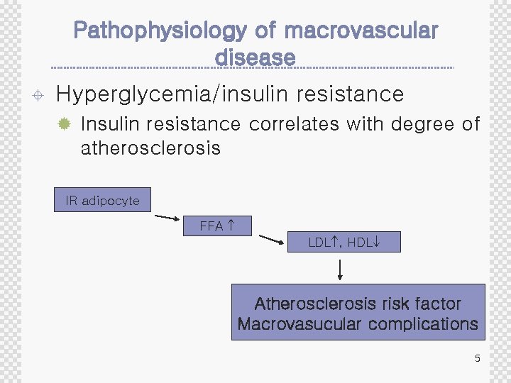
Pathophysiology of macrovascular disease ± Hyperglycemia/insulin resistance ® Insulin resistance correlates with degree of atherosclerosis IR adipocyte FFA LDL , HDL Atherosclerosis risk factor Macrovasucular complications 5
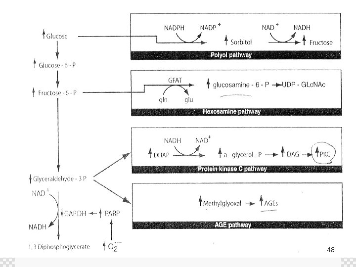
Mechanisms of hyperglycemia induced damage ± Increased polyol pathway ± Increased intracelllular Advanced Glycation End Product(AGE) formation ± Activation of PKC isoforms ± Increased hexosamine pathway 6
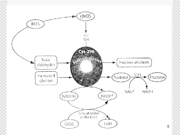
Increased polyol pathway ± Aldose reductase(AR) ® First enzyme in Polyol pathway ® Monomeric oxidoreducatese ® Catalyze reduction of carbonyl compound(e. g glucose) ® Low affinity for glucose ® Contribute to glucose utilization in small percentage ® In hyperglycemia => increased emzymatic conversion to the polyalcohol sorbitol 7
8

Increased polyol pathway ± ± Sorbitol is oxidized to fructose by sorvitol dehydrogenase(SDH) with NAD+ reduce to NADH Flux through polyol pathway during hyperglycemia varied form 33% in rabbit lens to 11% in human erythrocyte ® The contribution of this pathway to diabetic complications : site, species, tissue specific 9
Increased polyol pathway ± AR deplete reduced glutathione(GSH) ® Consume NADPH ® Intracellular oxidative stress ® Transgenic mice(AR overexpression) ® ® Decreased GSH in lens Homozygous KO mice : diabetic 10
Increased polyol pathway ± NO maintain AR in inactive ® This suppression is relieved in diabetic tissue ® NO-derived adduct formation is cys 298=> inhibition of AR ® Diabetic => decreased NO => polyol flux ± AR inhibition in dogs ® prevent diabetic nephropathy ® but failed to prevent retinopathy, capillary basement membrane thickening in the retina, kidney, muscle ± AR inhibition in human ® Zenarestat(AR inhibitor) =>positive effect on neuropathy 11

Mechanisms of hyperglycemia induced damage ± Increased polyol pathway ± Increased intracelllular Advanced Glycation End Product(AGE) formation ± Activation of PKC isoforms ± Increased hexosamine pathway 12
Increaed intracellular AGE formation ± Advanced Glycation End product(AGE) ® Increased in diabetic retinal vessle, renal glomeruli ® Hyperglycemia is primary initiating event in the formation of extra/intracellular AGEs ® AGE precursors(methylglyoxal) damage target cells 13
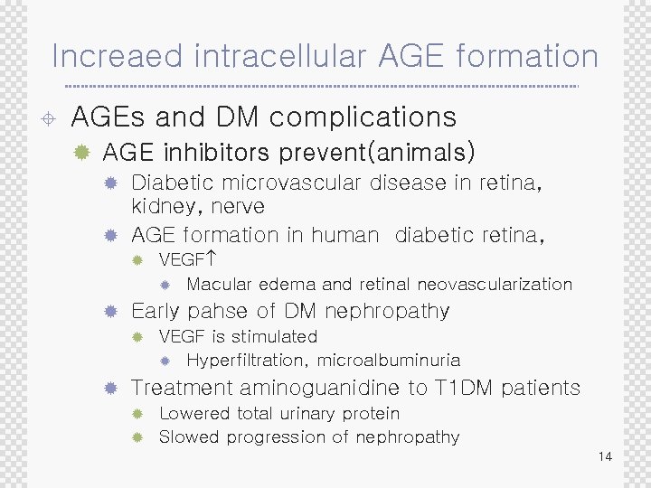
Increaed intracellular AGE formation ± AGEs and DM complications ® AGE inhibitors prevent(animals) Diabetic microvascular disease in retina, kidney, nerve ® AGE formation in human diabetic retina, ® ® ® Early pahse of DM nephropathy ® ® VEGF ® Macular edema and retinal neovascularization VEGF is stimulated ® Hyperfiltration, microalbuminuria Treatment aminoguanidine to T 1 DM patients Lowered total urinary protein ® Slowed progression of nephropathy ® 14
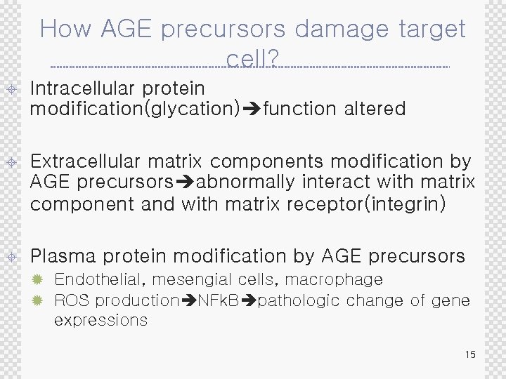
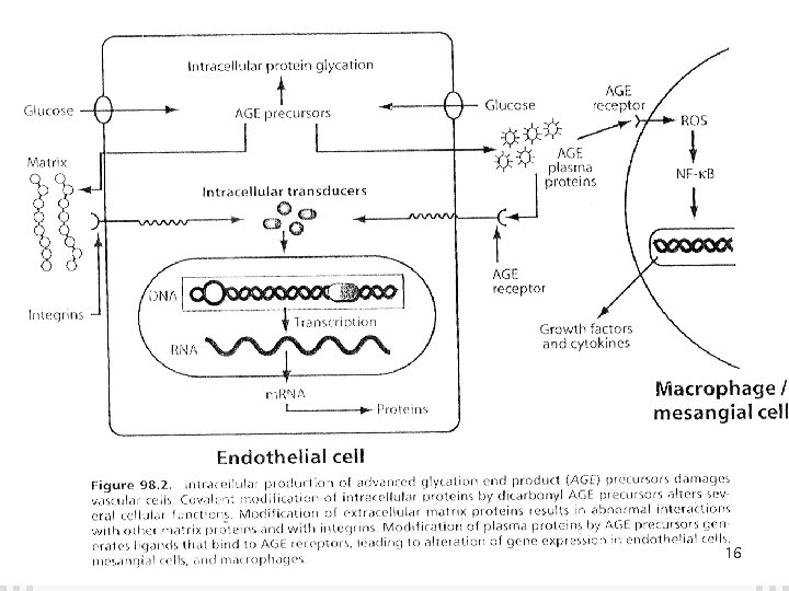
How AGE precursors damage target cell? ± Intracellular protein modification(glycation) function altered ± Extracellular matrix components modification by AGE precursors abnormally interact with matrix component and with matrix receptor(integrin) ± Plasma protein modification by AGE precursors ® Endothelial, mesengial cells, macrophage ® ROS production NFk. B pathologic change of gene expressions 15
16

Increaed intracellular AGE formation ± Methylglyoxal(AGE precursor) ® Diabetic patient( ) 3~5 times : 8 u. M ® Induction of apoptosis by DNA damage and oxidative stress ® Changes matrix molecule functional properties ® Tyep I collagen : decreased elasticity 17
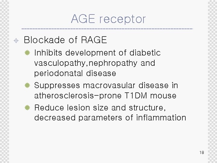
AGE receptor ± Blockade of RAGE ® Inhibits development of diabetic vasculopathy, nephropathy and periodonatal disease ® Suppresses macrovasular disease in atherosclerosis-prone T 1 DM mouse ® Reduce lesion size and structure, decreased parameters of inflammation 18
Mechanisms of hyperglycemia induced damage ± Increased polyol pathway ± Increased intracelllular Advanced Glycation End Product(AGE) formation ± Activation of PKC isoforms ± Increased hexosamine pathway 19
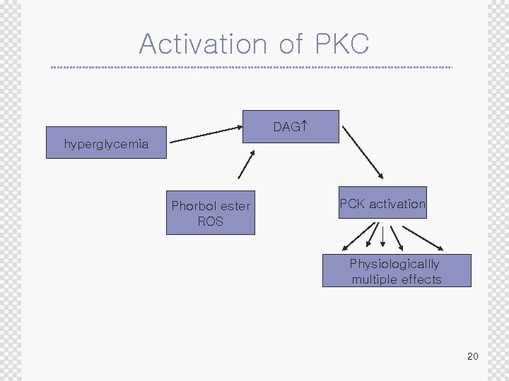
Activation of PKC DAG hyperglycemia Phorbol ester ROS PCK activation Physiologicallly multiple effects 20
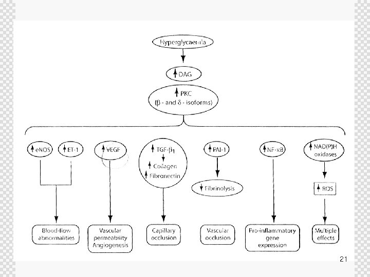
21
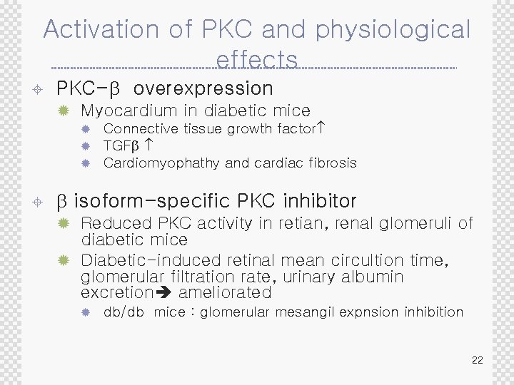
Activation of PKC and physiological effects ± PKC-b overexpression ® Myocardium in diabetic mice ® ® ® ± Connective tissue growth factor TGFb Cardiomyophathy and cardiac fibrosis b isoform-specific PKC inhibitor ® Reduced PKC activity in retian, renal glomeruli of diabetic mice ® Diabetic-induced retinal mean circultion time, glomerular filtration rate, urinary albumin excretion ameliorated ® db/db mice : glomerular mesangil expnsion inhibition 22
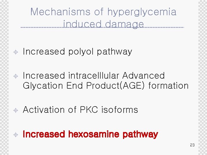
Mechanisms of hyperglycemia induced damage ± Increased polyol pathway ± Increased intracelllular Advanced Glycation End Product(AGE) formation ± Activation of PKC isoforms ± Increased hexosamine pathway 23
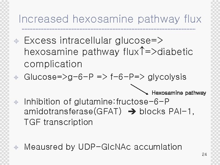
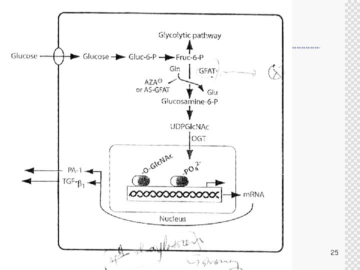
Increased hexosamine pathway flux ± Excess intracellular glucose=> hexosamine pathway flux =>diabetic complication ± Glucose=>g-6 -P => f-6 -P=> glycolysis Hexosamine pathway ± Inhibition of glutamine: fructose-6 -P amidotransferase(GFAT) blocks PAI-1, TGF transcription ± Meausred by UDP-Glc. NAc accumlation 24
25
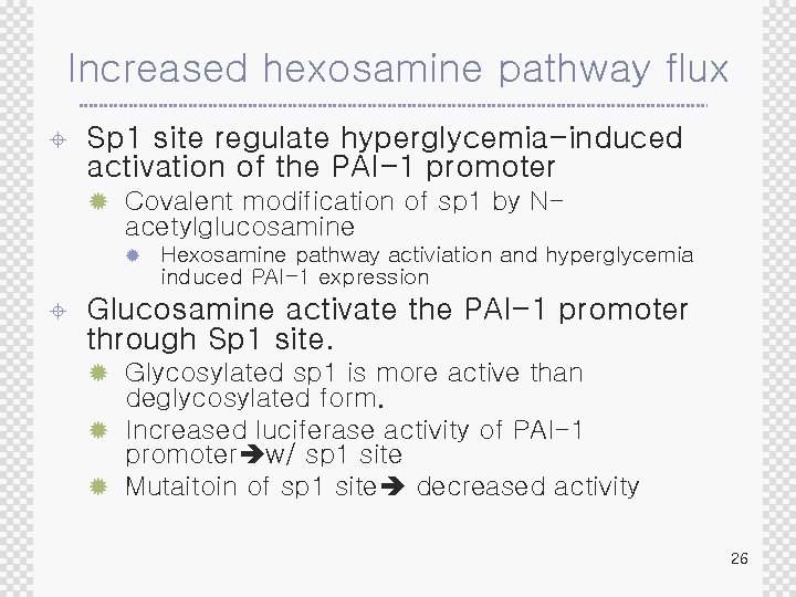
Increased hexosamine pathway flux ± Sp 1 site regulate hyperglycemia-induced activation of the PAI-1 promoter ® Covalent modification of sp 1 by N- acetylglucosamine ® ± Hexosamine pathway activiation and hyperglycemia induced PAI-1 expression Glucosamine activate the PAI-1 promoter through Sp 1 site. ® Glycosylated sp 1 is more active than deglycosylated form. ® Increased luciferase activity of PAI-1 promoter w/ sp 1 site ® Mutaitoin of sp 1 site decreased activity 26
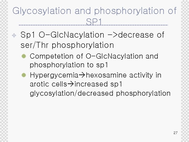
Glycosylation and phosphorylation of SP 1 ± Sp 1 O-Glc. Nacylation ->decrease of ser/Thr phosphorylation ® Competetion of O-Glc. Nacylation and phosphorylation to sp 1 ® Hypergycemia hexosamine activity in arotic cells increased sp 1 glycosylation/decreased phosphorylation 27
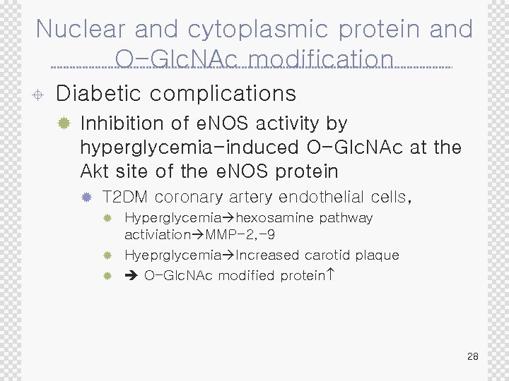
Nuclear and cytoplasmic protein and O-Glc. NAc modification ± Diabetic complications ® Inhibition of e. NOS activity by hyperglycemia-induced O-Glc. NAc at the Akt site of the e. NOS protein ® T 2 DM coronary artery endothelial cells, Hyperglycemia hexosamine pathway activiation MMP-2, -9 ® Hyeprglycemia Increased carotid plaque ® O-Glc. NAc modified protein ® 28
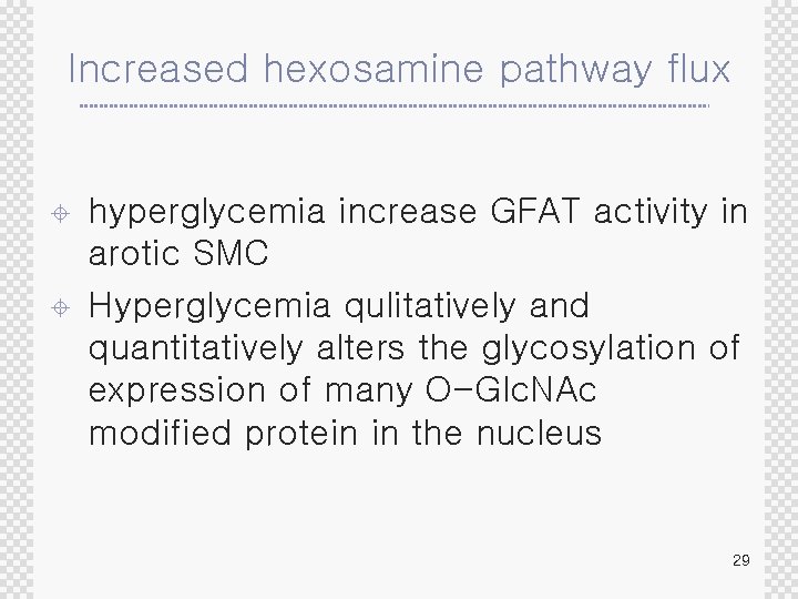
Increased hexosamine pathway flux ± ± hyperglycemia increase GFAT activity in arotic SMC Hyperglycemia qulitatively and quantitatively alters the glycosylation of expression of many O-Glc. NAc modified protein in the nucleus 29
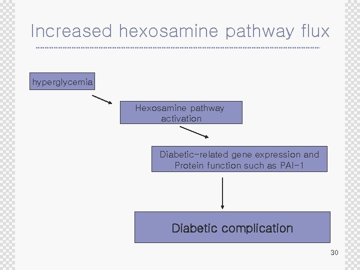
Increased hexosamine pathway flux hyperglycemia Hexosamine pathway activation Diabetic-related gene expression and Protein function such as PAI-1 Diabetic complication 30
Other possible mechanisms of hyperglycemia-induced damage ± Inactivation of glucose-6 -phosphate dehyrogenase ± Decreased c. AMP-response element-binding protein(CREB) activity and content ± Mechanism of macrovascular damage induced by FFA 31
Inactivation of glucose-6 -phosphate dehyrogenase ± G 6 P-Dehydrogenase ® First rate-limiting enzyme in glycolysis ® Produce NADPH ® NADPH : critical intracellular reducint equivalent reduction of oxidized glutathione(against oxidative stress) ® Act as cofactor for e. NOS activity 32
Inactivation of glucose-6 -phosphate dehyrogenase ± Hyperglycemia inhibits G 6 PDH in bovine aortic endothelial cell by PKA inhibit by phosphorylation of G 6 PDH ® These inhibition increase oxidative stress ± Decreased G 6 PDH activity decrease endothelium derived bioavailable NO 33
Decreased c. AMP-response elementbinding protein(CREB) activity and content ± CREB ® Located in c. AMP signal downstream ® Important roles in VSMC Inhibition of proliferation and migration ® Decrease expression of GF-receptor for PDGF, endothelin-1, IL-6 ® 34
Decreased c. AMP-response elementbinding protein(CREB) activity and content ± Hyperglycemia in VSCM ® CREB content , function increase of migration and proliferation ± CREB overexpression ® Completely restore hyperglycemia-induced proliferation and migration ± DM ® CREB macrovascular complication 35
Decreased c. AMP-response elementbinding protein(CREB) activity and content ± Decreased level of CREB ® Insulin resistant/deficient mice ® Nervous system in DM ® STZ animal’s hippocampus and nerve ± Thus, ® Change and function of CREB represent a pivotal consequence of glycemiamediated dysfunction in complications target tissue of diabetic complication 36
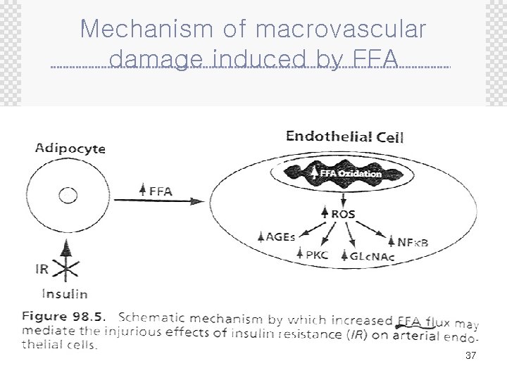
Mechanism of macrovascular damage induced by FFA 37
Mechanism of macrovascular damage induced by FFA ± In vitro ® Low glucose cultured arotic endothelial cell and elevated FFA ® AGE , PKC activation, hexosamine pw , NFk. B ® ± The same extent as hyperglycemia In vivo ® Fatty Zuker rat(insulin resistant but no DM) ® ± Above pathway blocked by inhibition of lipolysis with nicotinic acid Thus, ® Increased of FFA from visceral adipocyte to arterial endothelia cells metabolic linkage between IR and macrovascular disease 38
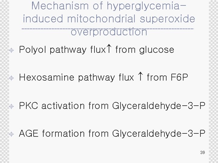
Mechanism of hyperglycemiainduced mitochondrial superoxide overproduction ± Polyol pathway flux from glucose ± Hexosamine pathway flux from F 6 P ± PKC activation from Glyceraldehyde-3 -P ± AGE formation from Glyceraldehyde-3 -P 39
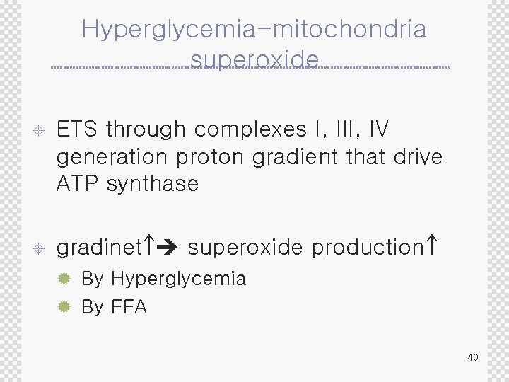
Hyperglycemia-mitochondria superoxide ± ETS through complexes I, III, IV generation proton gradient that drive ATP synthase ± gradinet superoxide production ® By Hyperglycemia ® By FFA 40
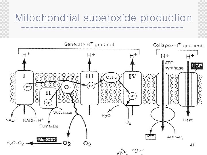
Mitochondrial superoxide production 41
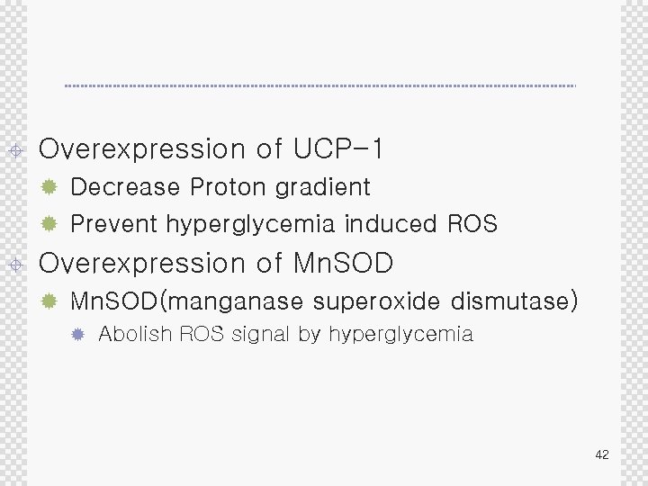
± Overexpression of UCP-1 ® Decrease Proton gradient ® Prevent hyperglycemia induced ROS ± Overexpression of Mn. SOD ® Mn. SOD(manganase superoxide dismutase) ® Abolish ROS signal by hyperglycemia 42
UCP-1 / Mn. SOD and polyol pathway ± Inhibition of hyperglycemia induced superoxide production by UCP 1 and Mn. SOD ® Prevent incresed polyol pathway flux in endothelia cells ® Sorbitol accumulation increased Cultured cell, 5 30 m. M glucose media ® Mt superoxide production inhibition no change of sorbitol in 30 m. M glucose media ® 43
UCP-1 / Mn. SOD and GAPDH activity ± Hyperglycemia-induced superoxide by inhibition of UCP 1 and Mn. SOD ® 66% decrease of GAPDH activity ® GAPDH inhibition ROS induced DNA strand break ® Polyol flux increased 44
UCP-1 / Mn. SOD and AGE formation ± Hyperglycemia-induced superoxide by inhibition of UCP 1 and Mn. SOD ® Decrease AGE formation in endothelial cell ® Hyperglycemia Methylglyoxal-derived AGE 5 m. M 30 m. M glucose medium : AGE ® Mt superoxide prevented 30 m. M: AGE was not increased ® GAPDH inhibition by hyperglycemia triose increased methylglyoxal formation AGE ® 45
UCP-1 / Mn. SOD and PKC activation ± Hyperglycemia-induced superoxide by inhibition of UCP 1 and Mn. SOD ® Decrease PKC activation in endothelial cells ® Hyperglycemia PKC activation ® ® 5 m. M 30 m. M glucose medium : PKC Mt superoxide prevented 30 m. M: PKC was not increased ® Hyperglycemia GAPDH inhibition de novo synthesis of DAG PKC activation ® GAPDH antisense : activation of PKC in physiologic glucose conc. ® PKC NADPH oxidase activation superoxide production 46
UCP-1 / Mn. SOD and hexosamine pathway acitivity ± Hyperglycemia-induced superoxide by inhibition of UCP 1 and Mn. SOD ® Prevent hexosamine pathway acitivity in endothelial cells ® ® ± 5 m. M 30 m. M glucose medium : UDP-Glc. NAc Mt superoxide prevented 30 m. M: UDP-Glc. NAc was not increased Hyperlgycemia ® more F 6 P ® ROS inhibition of GAPDH F 6 P GFAT hexosamine pathway ± GAPDH antisense : increase hexosamine pathway flux in the absence of hyperglycemia 47
48
hyperglycemia and NFk. B ± Hyperglycemia-induced activation of redoxsensitive transcription factor NFk. B was prevented by inhibition of Mt superoxide overproduction 49
Overexpression of UCP-1 and Mn. SOD ± ± Prevent hyperglycemia-induced inactivation of GAPDH SOD mimetic ® Loss of CREB, PDGF recector-a reversed in NOD mice ® CREB and Bcl-2 expression restored 50
Overexpression of UCP-1, Mn. SOD and diabetic complications ® Mn. SOD : suppress the increase cllagen synthesis caused by hyperglycemia in glomerular cell ® Mn. SOD overexpressed mice: decrease programmed cell death caused by hyperglycemia in DRG neuron ® UCP-1 overexpression in embryonic DRG ® Caspase inhibition ® In aortic cells ® UCP-1/Mn. SOD blocking of hyperglycemid-induced monocyte adhesion to endothelial cells ® Anti-atherogenic enzyme ® Hyperglycemia inhibits prostacyclin synthetase prevented by overexpression of UCP 1/Mn. SOD 51
Overexpression of UCP-1 and Mn. SOD ± ± Prevent Hyperglycemia-induced e. NOS inhibition STZ animal ® STZ-wild ® STZ-human Cu++/Zn++ superoxide dismutase overexpressed transgenic mice ® ± Albumiuria, glomerular hypertrophy, TGF in glomerular was attenuated db/db mice ® SOD transgene mice ® Attenuation Glomerular mesngial matrix expansion 52
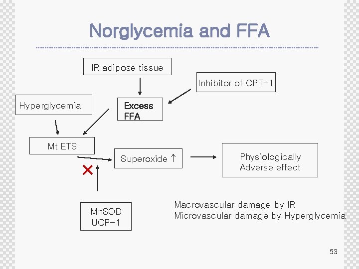
Norglycemia and FFA IR adipose tissue Inhibitor of CPT-1 Hyperglycemia Excess FFA Mt ETS Superoxide Mn. SOD UCP-1 Physiologically Adverse effect Macrovascular damage by IR Microvascular damage by Hyperglycemia 53
- Slides: 53