BIOCHEMICAL STRUCTURE OF BONE TISSUE THE COLLAGEN MATRIX

BIOCHEMICAL STRUCTURE OF BONE TISSUE, THE COLLAGEN MATRIX AND THE HYDROXYAPATITE CEMENT Dr. Mazhar Al Zoubi Dec 2020

50 © The Mc. Graw-Hill Companies, Inc, 2011

1. Describe the biochemical structure of bone tissue, the collagen matrix and the hydroxyapatite cement. 2. List bone matrix proteins and describe their function. 3. Describe the Composition of calcified tissues, calcification in bones and teeth and formation of hydroxyapatite. 4. Understand the role of alkaline phosphatase, calcium and hosphate and vitamin D: 1, 25 -Dihydroxy-vit-D in bone formation and remodeling. 5. Review calcium and phosphate homeostasis.

BONE IS A MINERALIZED CONNECTIVE TISSUE
: • Hyaluronic acid, •")
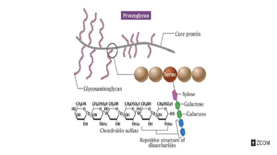
The organic matter is mainly protein. Glycos. Amino. Glycans (GAGs): • Hyaluronic acid, • Chondroitin sulfate, • Keratan sulfates I and II, • Heparin, • Heparan sulfate, • Dermatan sulfate. * CS: chondroitin sulfate
6(OH) q. With")
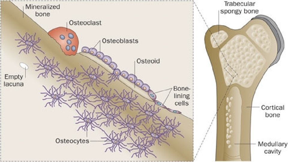
The inorganic or mineral component is mainly crystalline hydroxyapatite Ca 10(PO 4)6(OH) q. With sodium, magnesium, carbonate, and fluoride q 99% of the body's calcium is contained in bone. q. Bone is a dynamic structure: q. Resorption followed by Deposition of new bone tissue q. This remodeling system is adapted to physiological and hormonal signaling by: q Osteoclasts (Resorption) q Osteoblasts (Deposition)

Synthesizing type I collagen

Osteoclasts v Multinucleated cells derived from pluripotent hematopoietic stem cells. v Possess an apical membrane domain, exhibiting a ruffled border that plays a key role in bone resorption. v Low p. H (4. 0) or less, increases the solubility of hydroxyapatite and allowing demineralization to occur.

H 2 O ATPase

Osteoblasts v. Mononuclear cells derived from pluripotent mesenchymal precursors. v. Synthesize most of the proteins found in bone. v Produce growth factors and cytokines v Deposits new bone matrix (osteoid) and its subsequent mineralization v Mineralization is controled by regulating the passage of calcium and phosphate ions across their surface membranes. v Alkaline phosphatase (ALP) is used to generate phosphate ions from organic phosphates. Type I collagen appears to be necessary, with mineralization being first evident in the gaps between successive molecules

q Acidic phosphoproteins, such as bone sialoprotein, acting as sites of nucleation. q These proteins contain motifs (eg, poly-Asp and poly-Glu stretches) that bind calcium and may provide an initial scaffold for mineralization. q. Certain proteoglycans and glycoproteins, can also act as inhibitors of nucleation. approximately 4% of compact bone is renewed annually whereas approximately 20% of trabecular bone is replaced.

q Osteoblasts stimulated by: üparathyroid hormone ü 1, 25 dihydroxycholecalciferol v. Inhibited by: üCorticosteroids q Osteoclasts stimulated: ü Parathyroid hormone ü 1, 25 dihydroxycholecalciferol v. Inhibited by: üCalcitonin üEstrogens

BONE IS AFFECTED BY MANY METABOLIC & GENETIC DISORDERS
: Fragility of bones. üThin and translucent sclera and may appear")
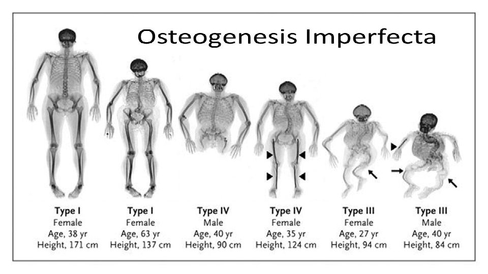
Osteogenesis imperfecta (brittle bones): Fragility of bones. üThin and translucent sclera and may appear blue owing to a deficiency of connective tissue. üFour types: (mild, extensive, severe, and variable) § Affected infants may be born with multiple fractures and not survive. § Over 90% of patients with osteogenesis imperfecta have mutations in the COL 1 A 1 and COL 1 A 2 genes, encoding pro 1(I) and pro 2(I) chains. § Over 100 mutations in these two genes have been documented. § Most frequent type: Replacement of glycine by another bulkier amino acid, affecting formation of the triple helix. "procollagen suicide" is an example of a dominant negative mutation, when a protein consists of multiple different subunits. resulting in enzymatic degradation

 Increased bone density due to inability to resorb bone. ~")
Osteopetrosis (marble bone disease) Increased bone density due to inability to resorb bone. ~ with renal tubular acidosis and cerebral calcification: due to mutations in the gene (located on chromosome 8 q 22) encoding carbonic anhydrase II (CA II)

Osteoporosis Ø Is a generalized progressive reduction in bone tissue mass per unit volume causing skeletal weakness. Ø Estrogens and the cytokines interleukins-1 and -6 appear to be intimately involved in the causation of osteoporosis.

THE MAJOR COMPONENTS OF CARTILAGE ARE TYPE II COLLAGEN & CERTAIN PROTEOGLYCANS Hyaline cartilage (the major type of cartilage) Elastic cartilage contains elastin and fibroelastic cartilage contains type I collagen. v. Cartilage is an avascular tissue and obtains most of its nutrients from synovial fluid. Slow continuous turnover. v. Various proteases (eg, collagenases and stromalysin) synthesized by chondrocytes can degrade collagen and the other proteins found in cartilage. v(IL-1) and (TNF-α) stimulate the production of such proteases. v. Transforming growth factor-β (TGF-β ) and insulinlike growth factor 1 (IGF-I) generally exert an anabolic influence on cartilage.


Schematic diagram of the aggrecan from bovine nasal cartilage

THE MOLECULAR BASES OF THE CHONDRODYSPLASIAS INCLUDE MUTATIONS IN GENES ENCODING TYPE II COLLAGEN & FIBROBLAST GROWTH FACTOR RECEPTORS FGFR Chondrodysplasias: are a mixed group of hereditary disorders affecting cartilage. A number of them are due to a variety of mutations in the COL 2 A 1 gene, leading to abnormal forms of type II collagen. One example is Stickler syndrome, manifested by degeneration of joint cartilage and of the vitreous body of the eye. The best-known of the chondrodysplasias is achondroplasia, the most common cause of short-limbed dwarfism.

Rejection letters from more than 20 medical schools A pediatric orthopedic surgeon at Johns Hopkins Hospital Michael Ain


q. Bone and cartilage are specialized forms of the ECM. Collagen I and hydroxyapatite are the major constituents of bone. q Collagen II and certain proteoglycans are major constituents of cartilage. q. The molecular causes of a number of heritable diseases of bone (eg, osteogenesis imperfecta) and of cartilage (eg, the chondrodystrophies) are being revealed by the application of recombinant DNA technology.
- Slides: 27