BIOCHEMICAL MONITORING IN CRITICALLY ILL Brodsk H Critically

BIOCHEMICAL MONITORING IN CRITICALLY ILL Brodská H.

Critically ill patient - definition u Decompensation of the status of the patient leading without therapeutic intervention to the multiorganic failure and to death
")
Basal vital function u u Central nervous system Respiration Blood circulation (optimal aerobic metabolism) Renal, hepatic function, immune systém, hemocoagulation and fibrinolysis, thermoregulation, barrier function of the gut

How to treat critically ill patient? u u u To save the basic vital function Stabilization of patient Support of insufficiency organs , prevention impending organ insufficiency Right diagnosis adequate therapy

Main clinical diagnosis of intensive medicine u u u Traumatological: polytrauma, crush sy, craniocerebral , contusion of the chest, burns Shock of various clinical causes, cardiac impairment Sepsis, acute hemorrhagia, pulmonal embolia

Diagnostic of critically ill patients u u u Clinical data, examination /vomitus, heavy smoker, DM, icterus, arytmie, hepatomegalia. . / Monitoring of basic function (blood pressure, heart rate…) Imaging methods (X-ray, Computer tomography) Bacteriology Hematology, Imunology Biochemistry

The possibilities of biochemical monitoring u u u On-line monitoring (cardiosurgery – p. H, minerals (K), the electrodes are localized on central cateter, possibility to check parameters on-line. bed side monitoring (point of care testing – glycaemia, urine /protein, p. H, blood. . /, oximeter O 2 saturation, acidobasis, drugs /dg. strips) Biochemical analysis (vital indication, statim , rutine analysis)

Biochemical parameters – statim set u u u u u Na, K, Cl, Ca, P, Mg, osmolality - blood, urine Acidobasis, lactate urea, creatinin clearence, Nitrogen balance bilirubine, ALT, AST, GMT, LD, amylase, lipase cholesterol, triglycerides, glucose – blood, urine CK, CK-MB, Troponin T, I, myoglobine, CK-MBmass Total protein, albumine, prealbumine, CHE CRP, procalcitonine TSH, b-HCG. Basic analysis are made at the first, must be done within 90 minutes

Other biochemical parameters u u u u Trace elements /Zn, Se. . / Vitamins Drugs /methotrexate, antiepileptics, antibiotics. . . / Drugs /amfetamine, methadon, opioids. . / Aminogram /glutamin. . / Interleucins, TNF… Small bowel permeability /Lactulose/mannitol/ Hormones /cortisol, glucagone, adrenaline. . /.

Range and frequency of biochem. monitoring- patient status u Diagnostic examination-a new patient u Standart examination relat. stabilized patient u Storm, shock, syncopa, coma. . u Perioperation monitoring

The Indication for examination What do I expect ? u u Confirmation of clinical diagnosis – ( ac. pancreatitis, AIM, hyperkalemie by ARI). Monitoring of therapy /Na, K, Mg, Ca deficit, glykemie, CRP, PCT, bilance. Answer for unclear status of patient- (may be I find something/ Automatizm

Na, K, Cl, Ca, P, Mg – serum levels u u u Hypernatremia – over 150 mmol/l Sec. hyperaldosteronism (capillary leak, albumin(water) moves to intersticium, intravascular hypovolemia)renin-angiot-aldost. UNa<20 mmol/l Hypothalamic damage Hypertonic hyperhydration Diabetes insipidus Brain death

Na, K, Cl, Ca, P, Mg – serum levels u u u Hyponatremia – under 130 mmol/l Na in the third space - ascites, hydrothorax Cardiac failure – increase of extracellular volume Application of solutions without electrolytes Hypersecretion of ADH – water retention

Na, K, Cl, Ca, P, Mg – serum levels u u u Hyperkalemia – over 5, 0 - 5, 5 mmol/l – p. H dependent /acidosis increases K level p. H decreased 0, 1…K increased 0, 6 mmol/l / Bigger intake, low output or both Acute renal failure Cytostatics, tumors Acute metabolic acidosis Infusion with K

Na, K, Cl, Ca, P, Mg – serum levels u u u u Hypokalemia – under 3, 5 mmol/l Low intake, bigger uptake, or both Emesis, diarrhoe / intestinal loss/ Diuretics Chemotherapy, antimycotics /renal tubules failure/ Anabolic phasis Hyperaldosteronism Acute metabolic alcalosis

Na, K, Cl, Ca, P, Mg – serum levels u Ca-total-binding protein 40%, 12% binding phosphates, hydrogencarbonate, ionizate 48%. u Ioniz. Ca the only one biological active form /influeces contractility of muscle, hemocoag. . /. u Hypoalb. -10 g /-0, 2 mmol/l, p. H +0, 1/ +0, 03 mmol/l binding protein-decrease ionizate form i. Ca-tetanie u Cl corig. Cl diagnosis of hypo chloremic alcalosis, hyperchloremic acidosis u

Na, K, Cl, Ca, P, Mg – serum levels u u u Hypophosphataemia – under 0, 6 mmol/l Acute wastage of energy after succesfully resuscitation, overfeeding sy, anabolism (energetic substrates without K, Mg, P) Hyperphosphataemia – over 1, 9 mmol/l Renal failure Cell damage

Na, K, Cl, Ca, P, Mg – serum levels u u u Mg – together with potassium Hypomagnesaemia – under 0, 6 mmol/l / in renal failure cause impaire of renal tubular cells, low intake. . / Preventive application during cardiac surgery (metabolic resuscitation of myocardial cells – K, P, Mg, glucose

Na, K, Cl, Ca, P, Mg – urine levels u u u Hypernatriuria – over 200 mmol/l High input of Na (food, drugs) Na mobilisation from the 3 rd space during anabolic phase Hyponatriuria – under 20 mmol/l Hyperaldosteronism, Hypovolemia Diferential diagnosis between prerenal and renal failure

Na, K, Cl, Ca, P, Mg – urine levels u u u Hyperkaliuria – hyperaldosteronism Na – K change Hypokaliuria Hypocorticoidism The risk of hypokalemia Metabolic balance of electrolytes/24 hours – monitoring of parenteral nutrition Clearence Na, K, fraction
 u u u All osmotic active particles (measured x counted) 2")
Osmolality (blood, urine) u u u All osmotic active particles (measured x counted) 2 x Na + glykemia + urea 380 mosmol/l x 265 mosmol/l Quick changes- danger for neurons Urine osmo- tubular function, hydratation

Acidobasis, lactate u u u We evaluate several aspects, Clinical aspects anamnesis-emesis, diarhoea, smoker , renal failure… Electrolytes, total protein, albumine, Hemoglobine, respiration functions, hepatic and renal function, hydration, Metabolic acidosis – cardiopulmonal resuscitation, Diabetes mellitus, renal insuficiency, hypoxia, hypoperfusis Metabolic alcalosis – blood transfusions, hyperaldosteronism, stomach output

Acidobasis, lactate u u Respiratory acidosis – Chronic obstructive bronchopulmonary disease, hydrothorax, malnutrition, high intake of glucose in parenteral nutrition Respiratory alcalosis – hypoxia, microembolisation into pulmonary arteria, shock, craniocerebral trauma

New aspects i evalution ABB Steward-Fencl Decomposition of finding on examination ABB into separate failures- possibility causally therapy /90% mixed failures ABB/ u p. H, p. CO 2, HCO 3, BE…. u SID, Cl corig. , UA, P, Alb. , . AG corig. u Alb 44, P 0. 9 -1. 1, Na 140 u
-Cl+UA/ 38 -40 mmol/l…HCO 3+Alb. +P Cl")
New markers u u u SID = Na(sum)-Cl+UA/ 38 -40 mmol/l…HCO 3+Alb. +P Cl corig. = Cl akt. x Na akt. /Na norm. 104 -108 mmol/l AG= Na + K - /Cl +HCO 3/. . RA AGcorig for act. albumin UA=/Na+ K+ Ca+Mg/ -/Cl+ SID/ UAcorig for act. Na Cl Cl HCO 3 Na K, Ca, Mg. . ALB UA SID ALB UA AG

Interpretation of new markers u u Changes of SID –decrease MAC /increase Cl, UA, water dilution. . / Increase SID- MAL /decrease Cl, water loss. . / UA inrease- MAC /lactate, hydroxybutyrate, sulfate. . / AG increase MAC- UA, hyperfosfatémia, decrease- MAL hypoproteinémia Cl Cl HCO 3 Na K, Ca, Mg. . ALB UA SID ALB UA AG

Case No. 1 u u u p. H p. CO 2 akt. HCO 3 BE p. O 2 SO 2 7, 392 2, 93 13, 1 -9, 6 5, 38 76, 8 u u u u u MAC with increase UA / ren. insuf. , ketones/ RAL MAL hypoproteinemic Na 135 K 5, 3 Cl 102 Alb 21 P 1, 2 SID 22, 3 Clcor 106 UA 35 AG 25 38 -40 104 -108 6 -10 14 -18 Urea 32, 1 kreat 475 lactate 2, 3 Glykémia 12, 6

Case No. 2 u u u p. H p. CO 2 akt. HCO 3 BE p. O 2 SO 2 7, 400 5, 7 25 0 5, 38 76, 8 u Na 143 K 4, 5 Cl 111 Alb 18 P 1, 2 u SID 32 Clcor. 110 UA 9 AG 11 38 -40 104 -108 6 -10 14 -18 MAC hyperchloremic / decrease SID/ MAL hypalbuminemic /decrease AG/ norm. value BE, HCO 3

Parameters need for calculation u Na, K, Cl, P, Ca, /Mg/ u Albumin u p. H, p. CO 2, akt. HCO 3, BE, p. O 2

Causal treatment Improve perfusion, hydratation, saturation of O 2/ u Correction ionts dysbalance Na, Cl, K u Compensation of renal failure, DM, hepatic functions u Correction hypoproteinémia u Correction lactic acid MAC u Correction respiratory component u

ABR, lactate u Hyperlactataemia – over 2, 2 mmol/l /1, 0/ u Bigger offer- move the balance of lactate-pyruvate to the part of lactate Hypoxia – cardiopulmonal resuscitation low level of B 1 /koenzyme pyruvatedehodrogenaze complex. . Krebs cycle/ Metabolic failure – diabetic coma, intoxication, Low output hepatopathy, leucaemia, low level of B 1… u u u

Urea, creatinine clearence, N-balance, uratic acid u u The level of urea – together with nutritional status, hydration High level of urea – high intake of N, increase catabolism /polytrauma- 500 gr of musceles per day loss/ GIT bleeding, dehydration low output- renal failure, Low level – malnutrition, serious hepatic failureureosyntetic cycle and gluconeogenesis dysfunction, pregnancy- increase ECF

Urea, creatinine clearence, N-balance, uratic acid u u u Urea in urine Increase – catabolism, prerenal failure Decrease – chronic malnutrition, acute renal failure

Urea, kreatinin, clearence kreatininu, N-balance, uratic acid u u u Serum levels of creatinine evaluation together with muscle mass, age, gender Increase – bigger offer- destruction of muscle mass, low output-renal failure Decrease- low offer-low muscle mass-seniores, malnutrition Creatinine clearence, excretion fraction -renal function N-balance – catabolism – the need of nitrogen Uratic acid – cell damage, arthritis uratica

NGAL neutrophil gelatinaseassociated lipocalin u u in renal tubules- increase during ischemia and nefrotox. demage. /elevation after 2 houres/ Blood and urine Early, sensitive and non invasive parameter of acute renal demage Elevace above 350 -400 ng/ml positive prediction ARI /90%/

bilirubin, ALT, AST, GMT, ALP, LD, amonium, amylase, lipase u Bilirubine direct, indirect u High level – neonatal hyperbilirubinaemia, massive blood transfusion, hepatocellular damage /drugs, hepatitis, hypoperfusion, necrosis, obstruction – extra-, intrahepatic

Metabolismus of bilirubin

bilirubine, ALT, AST, GMT, ALP, LD, amoniak, amylase, lipase u u u ALT – high level – hepatopathia, steatosis, hepatitis, cell damage, AST – high level – hypoperfusion, hepatitis, cell necrosis, muscles damage both aminotransferases increase during damage of hepatic cells during inf. hepatitis.

bilirubine, ALT, AST, GMT, ALP, LD, amoniak, amylase, lipase u ALP – increase – extrahepatal obstruction, isoenzymes – intestinal, bone, placental u GGT – increase – obstruction intra, extrahepatal, intoxication – ethanol, amanita. . u Both enzymes increase during parenteral nutrition. Impaire enterohepatic cycle of bile acids

bilirubín, ALT, AST, GMT, LD, amoniak, amylase, lipase u amoniac-increase during hepatic insufficiency /decompenzation of cirrhosis, premature / u amyláza-serum, or plasma and urine, drain pancreatic izoenzyme Increase- irritation of pancreas, hypoperfusion, treatment with opiates, inflamation-pancreatitis, tumor u u u lipasa- specific for pancreas

cholesterol, TG, glukóza u TG-increase- during sepsis, mainly on the begining, monitorate during parenterál nutrition with lipids more than 5 mmol/l contraindication to take lipid emulsion u Glycemia, serum, urine, Hypoglycemia below 2, 5 mmol/l-vital danger hyperglycemia- insulin. rezistence, recomendation level of glycemia 4, 5 -8, 2 /2006/ better survive in ICU patient u u

Biochemical markers of nutrition status: u Plasmatic proteins with short biologic half-life u Albumin -syntetizate in liver, half-life time is 21 days u Fyziolog. value 35 -45 g/l. Decrease of alb. level is typical for malnutrition kwashiorkor. /chronical maln. /Trends of changes alb. levels during realimentation are criterium of succesfull terapy. Acute decrease during inflammatory- increase of capilary leak, escape fluid from IVspace to intersticium. Hypalbuminemiae-marker of systemic responce. u

Biochemical markers of nutrition status: u Transferin-syntetizate in liver, biolog. half-life is 8 days. Fysiolog. value 2 -4 g/l, u u Prealbumin-syntetizate in liver, biolog. half-life is 1, 5 days. Fysiolog. Value 0, 15 -0, 4 g/l. Decrease in failure of proteosyntesis-indicator of acute protein malnutrition. RBP - biolog. half-life time is 12 hod. , fysiolog. value 0, 03 -0, 006 g/l.

Biochemical markers of nutrition status: u Prognostic index: u Concentration of plasmat. proteins depends on nutrition parameters and reaction of acute phase /acute inflammation- IL 1, IL 6, TNF, PAF. . / u PINI= CRPx Oroso / Prealb. x albumin. u 1 and less very good nutrition status, infl. 0 1 -10 probably subacute infl. with low degree of malnutrition 11 -20 middle degree of malnutrition, acute infl. 21 -30 acute infl. high degree of malnutrition, high risk of complication, nutrition support is necessery. Above 30 -high risk, vital danger. u u

Biochemical markers of nutrition status: u PINI / prognostic inflanatory nutrition index / u Acute infect /tonsilitis/CRP-83, AGP-2. 4, prealb. 0, 35, alb. 42 PINI=10 Chron. malnutrition patient CRP 40, AGP 2, 0, prealb. 0. 1, alb. 27, PINI=29 Polytrauma, sepse CRP 240, AGP 2, 5, prealb 0, 07, alb 25, PINI=600. u u

CK, , Troponin T, I, Myoglobin, CKMB mass, BNP, pro BNP u u u Myoglobin-2 -12 hours, nonspecific for myocard, increase injury muscels mass, renal insufficiency CK-MB mass-3 -36 hours-better specific than myoglobin, less than troponin T, I-4 h-7 days, absolut specifity for myocard dg. AIM combination 3 markers CK and CRP for monitoring dynamic and range of damage BNP, pro. BNP diagnosis heart failure, exclude noncardiac dyspnoe.

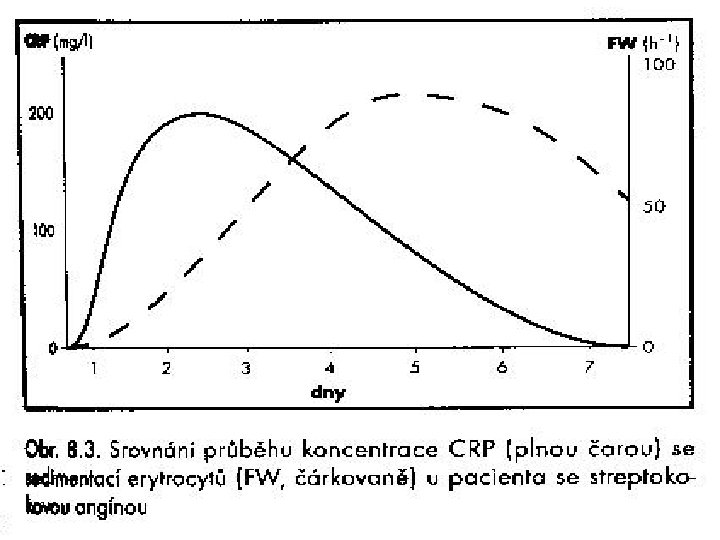
CRP, procalcitonin CRP /C-reactive protein/ 6 -12 hours to increase, nespec. increase-viral inf. , tumors, monitoring of ATB therapy. u Normal value is less 7 mg/l / bact. inf. more than 100 mg/l u u PCT /procalcitonin/-more sensitive and earlier marker for systemic bacterial inflammation depends on degree of MODS and stages of sepsis u Combination both PCT and CRP

Obr. 3. Srovnání diagnostické efektivity PCT a CRP ve vztahu k MODS ROC křivka pro CRP: Plocha pod ROC křivkou = 0, 511 ROC křivka pro PCT: Plocha pod ROC křivkou = 0, 698 Hladina významnosti: P = 0, 000

Koncentrace PCT během různých stadií sepse, klasifikovaných dle kritérií ACC/SCCM (Zeni et al. Clin. Intensive Care 1994, Suppl. 2, s. 89 -98, cit. Meisner 2002)

Proteiny akutní fáze – dynamika tvorby Krejsek et al. , 2004

Wegwnerova granulomatoza, lob. pneumonie, sepse Na urea ALT Alb p. A CRP PCT chol 3. 10 150 17 1, 4 25 0, 07 458 23 2, 6 4. 10 155 26, 7 1, 5 20 0, 07 718 71 2, 6

bili ALT AST GGT ALP LD p. A CRP PCT CMV 14. 5. 23 6, 0 3, 7 1, 3 2, 5 15 0, 20 40 0, 05 30. 5 0, 6 0, 8 2, 2 9, 0 0, 25 20 0, 05 19 2, 5
- Slides: 54