BIO 122 Week 3 Acellular and Prokaryotic Microbes

BIO 122 - Week 3 Acellular and Prokaryotic Microbes New England Institute of Technology Shellee Morehead Ch 4

Episode 1 ACELLULAR MICROORGANISMS

Introduction l Acellular microorganisms – – – l Viruses Viroids Prions Cellular microorganisms – – – Bacteria (procaryotic) Archaeans (procaryotic) Algae (eucaryotic) Protozoa (eucaryotic) Fungi (eucaryotic)

Viruses l l l AKA virions, when intact Size, viewing Organisms affected – – – Animals Plants bacteria

Virus Sizes vs RBC

Specific Viral Characteristics l Viruses differ from living cells: – – – Either RNA or DNA Cannot reproduce independently Do not divide by binary fission, mitosis or meiosis Can’t produce their own energy Rely on host cell’s organelles for metabolism
 Capsid- determines organism the virus will")
Virus Structure l l Genome (RNA or DNA) Capsid- determines organism the virus will infect Some are “enveloped”- having outer coat of sugars & fats Some have tails & fibers for attachment

Virus Classification l l l l RNA or DNA Capsid shape & size Enveloped or non-enveloped Host it affects Disease produced Target cell(s) Immunologic responses to virus

Categories of Viruses l Based on Genome – – Double stranded DNA Single stranded RNA Single stranded DNA Double stranded RNA

Virus Classification System
 Helical (coiled) Round combinations")
Capsid Shapes l l Polyhedral (20 sided) Helical (coiled) Round combinations

Influenza Virus

Ebola Virus

Bacteriophage

Bacteriophages l l l Infect only bacteria Obligate intracellular pathogens 3 types; based on capsid shapes Also categorized as RNA or DNA viruses Categorized as being “virulent” bacteriophages or “temperate” bacteriophages

Virulent vs Temperate Bacteriophages l l Both inject their genetic material into a cell to invade it Process that follows is different

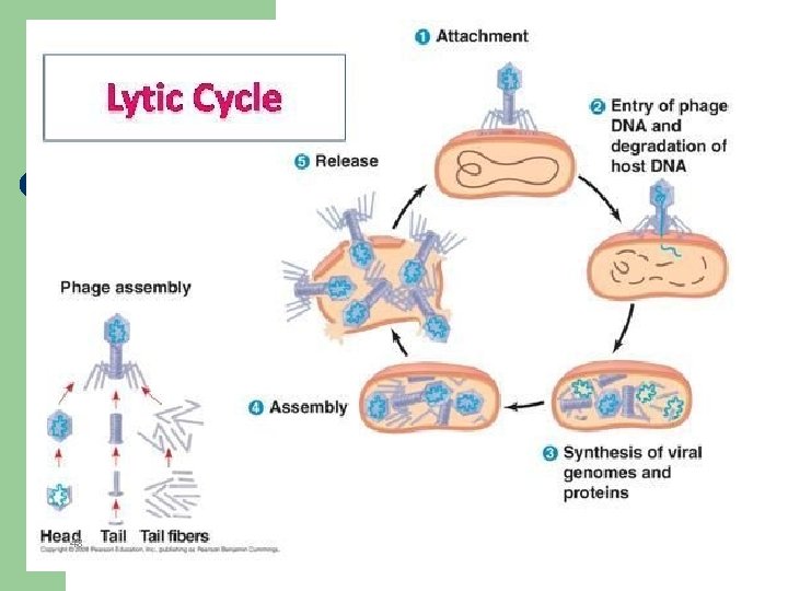
Virulent Bacteriophages l l Always go through “lytic cycle”, destroying host bacterium; usually within an hour Steps in the lytic cycle: (viral replication) – – – Attachment (adsorption) Penetration Biosynthesis (replication) Assembly (maturation) Release (lysis) of virions

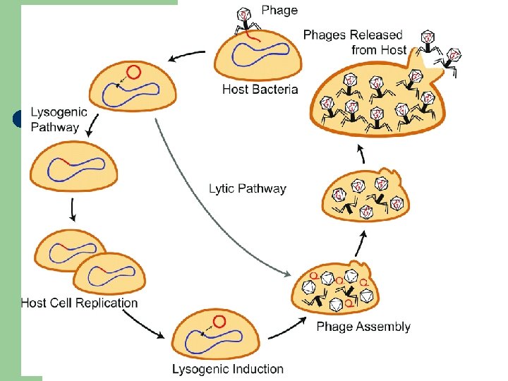
Temperate Bacteriophages l l l AKA “lysogenic phages” Do not proceed immediately through lytic cycle Remain genetically integrated into host bacterium’s genome for many generations

Animal Viruses l l l RNA or DNA Some are only a capsid and genetic material; others more complex Viral replication in animal cells: – – – Attachment (adsorption) to specific cells Penetration (of entire virion) Uncoating (shedding of capsid within the infected cell) Biosynthesis (replication) Assembly release

Inclusion Bodies l l Remnants of viruses within infected cells; used to ID viruses, diagnose disease Negri bodies are the inclusion bodies found in rabies infection AIDS infection & smallpox infections also yield inclusion bodies within cell’s cytoplasm Some diseases (herpes, polio) produce intranuclear inclusion bodies

Negri Bodies

B-cell inclusion bodies – smallpox

Examples of Human Viral Infections l l l l l AIDS Chickenpox Smallpox Cold sores Measles Mumps Influenza Hantavirus Ebola l l l l Genital herpes Warts Mononucleosis Polio Rabies SARS Encephalitis Meningitis (viral)

Latent Viral Infections Herpes, cold sores Shingles

Antiviral Medications l l **Antibiotics are NOT effective against viral infections** There are some medications that are somewhat effective at reducing the replication rate of viruses; antiviral agents
,")
Oncogenic Viruses l l Some human cancers are caused by viruses Epstein-Barr virus (mono), causes nasopharyngeal carcinoma, Burkitt lymphoma, B-cell lymphoma Kaposi’s sarcoma is viral in origin Hepatitis B & C related to liver cancers

HIV l l l Human Immunodeficiency virus AIDS Enveloped, RNA virus Attaches to “CD 4” cells, one of which is known as the “helper T cell” These usually fight off viral infections

Mimiviruses l l LARGE Double stranded DNA virus Capable of some functions of cellular organisms Possible cause of pneumonia

Viroids l l Smaller and simpler than viruses Short, bare RNA pieces Interfere with plant metabolism Thus far, no known animal infections

Prions l l Tiny infectious agent; resistant to disinfectants Fatal neurological involvement in many species – – – Scrapie BSE Kuru CJD Fatal familial insomnia

Episode 2 COVID-19

COVID-19 l l l COVID-19 is caused by a virus in the coronavirus group. This large group of viruses usually cause mild to moderate upper respiratory tract illnesses, and include the common cold Co. Vs mainly affect birds and mammals

Structure of coronaviruses

Previously 6 Co. Vs that infect humans l l l HCo. V-229 E HCo. V-OC 43 HCo. V-NL 63 HKU 1 SARS-Co. V MERS-Co. V l SARS-Co. V and MERS-Co. V cause server respiratory syndrome in humans

COVID-19 l l l COVID-19 is a novel Coronavirus likely zoonotic in origin Originated in Wuhan City, in the Hubei Province of China World Health Organization named it coronavirus disease 2019 on 11 Feb 2020 (Situation Report, 11 Feb 2020, WHO, Geneva. )

Signs and Symptoms l l l Occur between 2 - 14 days following exposure Fever Tiredness Dry cough Less common symptoms include – – – Loss of smell Loss of taste Rash www. CDC. gov and A Case of COVID-19 Pneumonia in a Young Male with Full Body Rash as a Presenting Symptom Hunt, Madison; Koziatek, Christian. Clin Pract Cases Emerg Med ; 2020. Article | COVIDWHO | ID: covidwho-55852

Treatment No known pharmaceutical treatment l l Respiratory Distress Reduce respiratory symptoms with fluids and bed rest Use paracetamol (acetaminophen) for fever reduction l l CPAP machines Ventilation www. cdc. org

Prevention • • • Handwashing Isolation from infectious people Proper use of Personal Protective Equipment (PPE)

Episode 3 PROKARYOTIC MICROORGANISMS

Prokaryotes l Bacteria – – – l Gram negative Gram positive Cell wall Archaea – – Varying shapes Ancient forms

Bacterial Characteristics Used for Classification l l l l Morphology Staining characterisitcs Motility Colony morphology Atmospheric requirements Nutritional requirements Metabolic activities pathogenicity

Bacterial Cell Morphology l l Reproduce by binary fission; may remain connected in specific arrangements Cocci – – Single, diplococci, streptococci, staphylococci, tetrads, octads Examples: Enterococci species, Neisseria species, Staphylococcus species, Streptococcus species

Nesseria gonnorhoeae

Bacterial Cell Morphology l Bacilli – – – “rods” Single, diplobacilli, streptobacilli Examples: Enterobacter species, Escherichia species, Salmonella species, Bacillus species, Clostridium species

Enterobacter spp – bacteriemia, UTIs,

Bacterial Cell Morphology l Spirilla – May be commashaped, such as Vibrio cholera – Some are tightly coiled, such as the syphilis organism – May be less tightly coiled, such as Borrelia burgdorferi

Vibrio cholera- causes cholera l SEM

Treponema pallidum - Syphylis l SEM

Borrelia burgdorferi – Lyme

Bacterial Staining l l Bacteria are smeared onto a slide, fixed, stained and dried before viewing Heat fixation or methanol fixation – – – Kills bacteria Maintains morphology Attaches smear to slide

Simple Staining l l l Methylene blue Only 1 stain used Demonstrates morphology and arrangement

Gram Staining l l l Differential stain Main stains are crystal violet and safranin Structure of the bacterial cell wall determines which stain will remain Gram positive bacteria (purple) have a thick proteinsugar layer that causes the 1 st stain to “stick” Gram negative bacteria have thinner walls, so violet rinses out, and safranin remains

Gram-Variable Bacteria l l l Mycobacterium tuberculosis – causes TB Mycobacterium leprae – leprosy or Hanson’s disease Acid-fast stain used to identify these bacteria

Bacterial Motility l l l Motile vs nonmotile Most spirilla, 50% of bacilli are motile Cocci nonmotile

Colony Morphology l l l Colonies are millions of bacteria attached as they grow Visible to naked eye Colony morphology varies between species – – Size Color Shape Elevation

Colony Morphology

Atmospheric Requirements of Bacteria l l l Obligate aerobes; room air Microaerophilic bacteria; 5% oxygen: gonorrhea Facultative anaerobes; common in clinical specimens Aerotolerant anaerobes- prefers no oxygen, but can tolerate some Obligate anaerobes-only grows without oxygen; ex- botulism

Nutritional Requirements of Bacteria l l l All bacteria need C, H, O, S, P, N to grow Fastidious bacteria require other nutrients also Enriched media are used in the lab to grow them

Metabolism of Bacteria l Many secrete waste products or toxins or enzymes that are the cause of disease symptoms in the host

Pathogenicity of Bacteria l Pathogenic characteristics include – – – Pili Capsules toxins

Atypical Bacteria l l Formerly thought to be viruses, as they are smaller than usual bacteria Lack many characteristics of usual bacteria

Rickettsias and Chlamydias l l l Gram negative cell wall Obligate intracellular parasites Rickettsial diseases are transmitted by arthropod vectors – l Rocky Mt Spotted Fever Chlamydias are called energy parasites also; use host cells’ energy; transferred by aerosol or direct contact – Eye infections, urethral infections, pneumonias

Mycoplasmas l l Smallest known cellular microbe Pleomorphic Pneumonia, GU infections Don’t have a cell wall, so can’t be treated with penicillin – Said to be CWD (cell wall deficient)

Archaea l l l Discovered in 1977 Ancient bacterial forms, actually similar in some ways to eucaryotes Morphology varies – – – l Cocci Bacilli filamentous Often “extremophiles” (hot, saline environments)

Quick Review l l What is the difference between a lytic and a lysogenic virus? What are the 3 shapes of bacteria? Do all the shapes have the same arrangements possible? How do chlamydias and ricketsias differ from prokaryotes, and how are they similar to viruses?
- Slides: 67