
Beyond TME why do an exenteration Gina Brown
Beyond TME : why do an exenteration? Gina Brown Department of Radiology Royal Marsden Hospital Imperial College, London gina. brown@rmh. nhs. uk

The Royal Marsden Beyond TME • In a significant proportion of patients (15 -20%), tumour extends beyond the achievable TME planes and requires more extensive surgery to achieve clear margins.

The Royal Marsden Exenterative surgery • Radical resection can achieve complete tumor clearance. Reported R 0 rates range from 22%-67%. • Can significantly increase survival, enhancing the prospects for long term cure. • High rate of post-operative adverse effects/morbidity. A. G. Heriot. Colorectal Dis, 2006; 8(9): 733 -747

The Royal Marsden

The Royal Marsden Beyond TME Collaborative Trial : Multicentre open observational study • To demonstrate that a validated staging system can be employed, • Establish scale and scope of pelvic exenterative surgery in advanced rectal cancer beyond TME • Assess quality of life and outcomes • Develop prognostic classification to assist in counselling patients

The Royal Marsden Beyond TME trial Eligibility: Any rectal tumour extending beyond TME plane (primary or recurrent) Participating sites: Royal Marsden St Marks Oxford Southampton Swansea Edinburgh (tbc) Pls contact: Gina. brown@rmh. nhs. uk if you wish to recruit your patients to this study
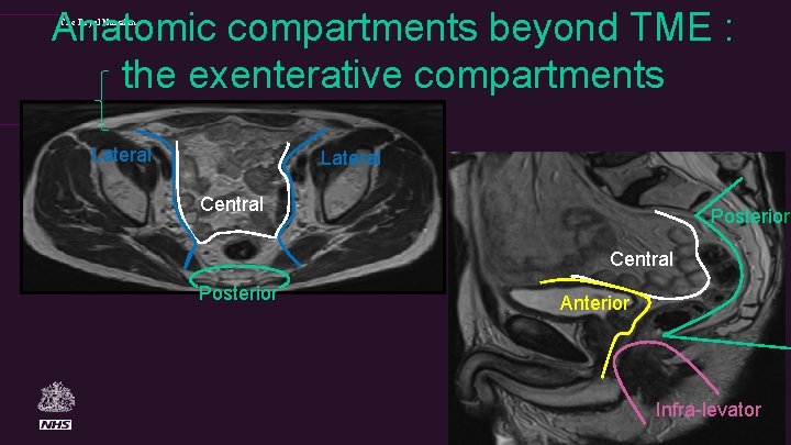
Anatomic compartments beyond TME : the exenterative compartments The Royal Marsden Lateral Central Posterior Anterior Infra-levator

The Royal Marsden Reporting Proforma Advanced Tumour Requiring Surgery Beyond TME Primary tumour The primary tumour is demonstrated as an [ Annular | Semi-annular | Ulcerating | | Polypoidal | Mucinous] mass with a [nodular / smooth] infiltrating border. The distal edge of the luminal tumour arises at a height of [ ] mm from anal verge: The distal edge of the tumour lies [ ]mm [Above, at, below] the top of the puborectalis sling The tumour extends craniocaudally over a distance of [ ] mm The proximal edge of tumour lies [above at below] the peritoneal reflection Invading edge of tumour extends from [ to ] O’clock Tumour is [confined to] [extends through] the muscularis propria: Extramural spread is [ ] mm mr. T stage: [T 1 ] [ T 2 ] [ T 3 a] [ T 3 b ] [ T 3 c] [ T 3 d ] [T 4 visceral ] [T 4 peritoneal] Lymph node assessment Only benign reactive and no suspicious nodes shown [N 0] [ ] mixed signal/irregular border nodes [N 1/N 2] Extramural venous invasion: [ No evidence ] [ Evidence] [ ] Small [ ]Medium [ ]Large vein invasion is present Peritoneal deposits: [ No evidence] [ Evidence] Pelvic side wall lymph nodes: [ None] [ Benign] [ Malignant mixed signal/irreg border] Location: [Obturator fossa • R • L ]. [External Iliac Nodes • R • L]. [internal iliac • R • L ]
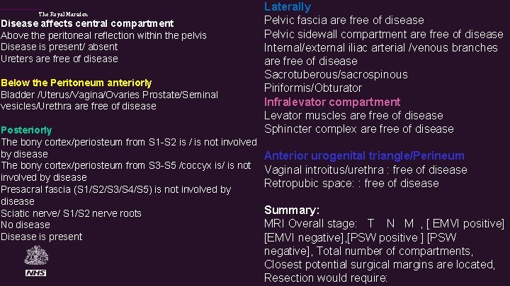
The Royal Marsden Disease affects central compartment Above the peritoneal reflection within the pelvis Disease is present/ absent Ureters are free of disease Below the Peritoneum anteriorly Bladder /Uterus/Vagina/Ovaries Prostate/Seminal vesicles/Urethra are free of disease Posteriorly The bony cortex/periosteum from S 1 -S 2 is / is not involved by disease The bony cortex/periosteum from S 3 -S 5 /coccyx is/ is not involved by disease Presacral fascia (S 1/S 2/S 3/S 4/S 5) is not involved by disease Sciatic nerve/ S 1/S 2 nerve roots No disease Disease is present Laterally Pelvic fascia are free of disease Pelvic sidewall compartment are free of disease Internal/external iliac arterial /venous branches are free of disease Sacrotuberous/sacrospinous Piriformis/Obturator Infralevator compartment Levator muscles are free of disease Sphincter complex are free of disease Anterior urogenital triangle/Perineum Vaginal introitus/urethra : free of disease Retropubic space: : free of disease Summary: MRI Overall stage: T N M , [ EMVI positive] [EMVI negative], [PSW positive ] [PSW negative], Total number of compartments, Closest potential surgical margins are located, Resection would require:
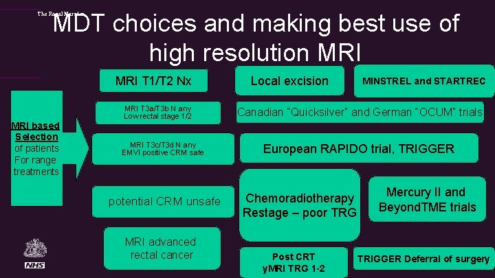
MDT choices and making best use of high resolution MRI The Royal Marsden MRI T 1/T 2 Nx MRI based Selection of patients For range treatments MRI T 3 a/T 3 b N any Low rectal stage 1/2 MRI T 3 c/T 3 d N any EMVI positive CRM safe potential CRM unsafe MRI advanced rectal cancer Local excision MINSTREL and STARTREC Canadian “Quicksilver” and German “OCUM” trials European RAPIDO trial, TRIGGER Chemoradiotherapy Restage – poor TRG Post CRT y. MRI TRG 1 -2 Mercury II and Beyond. TME trials TRIGGER Deferral of surgery
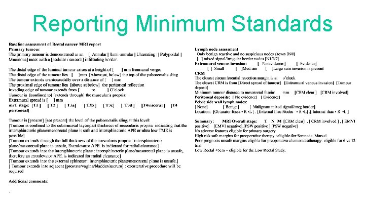
Reporting Minimum Standards
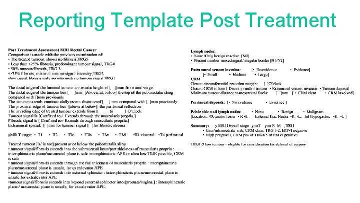
Reporting Template Post Treatment
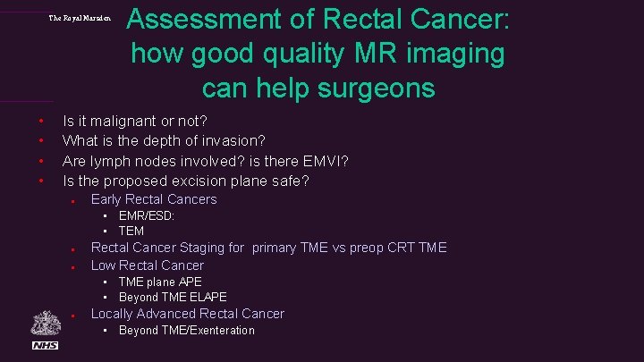
The Royal Marsden • • Assessment of Rectal Cancer: how good quality MR imaging can help surgeons Is it malignant or not? What is the depth of invasion? Are lymph nodes involved? is there EMVI? Is the proposed excision plane safe? l Early Rectal Cancers • EMR/ESD: • TEM l l Rectal Cancer Staging for primary TME vs preop CRT TME Low Rectal Cancer • TME plane APE • Beyond TME ELAPE l Locally Advanced Rectal Cancer • Beyond TME/Exenteration
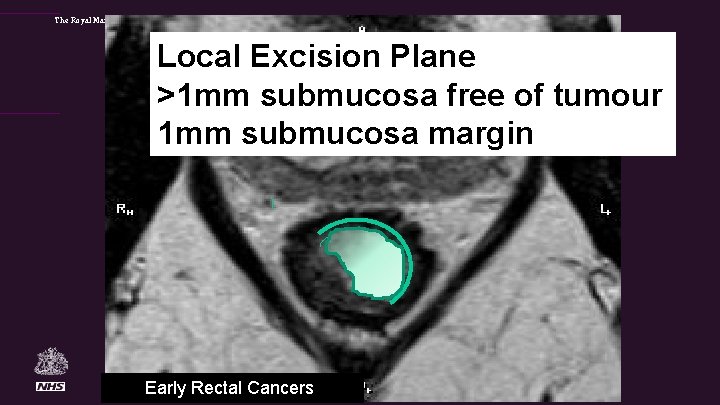
The Royal Marsden Local Excision Plane >1 mm submucosa free of tumour 1 mm submucosa margin Early Rectal Cancers
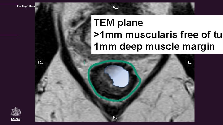
The Royal Marsden TEM plane >1 mm muscularis free of tum 1 mm deep muscle margin
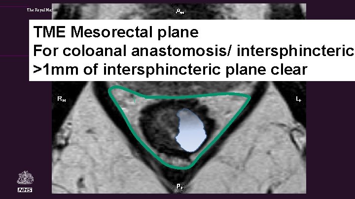
The Royal Marsden TME Mesorectal plane For coloanal anastomosis/ intersphincteric >1 mm of intersphincteric plane clear
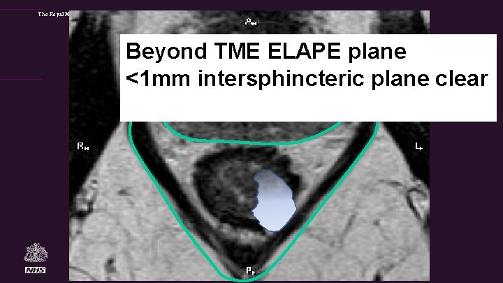
The Royal Marsden Beyond TME ELAPE plane <1 mm intersphincteric plane clear
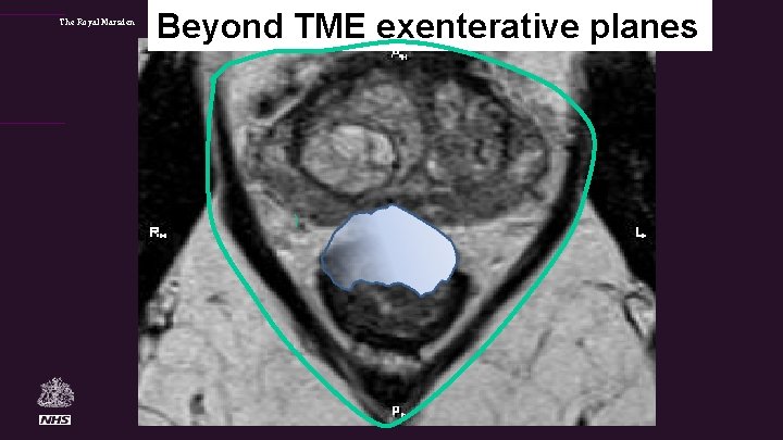
The Royal Marsden Beyond TME exenterative planes
The Royal Marsden Evidence base for MRI as a gold standard • CRM involvement on MRI prognostic predictor for recurrence • Depth of extramural spread >5 mm risk factor for poor DFS • Presence of MRI detected venous invasion – risk factor for local and distant recurrence and seen more frequently than path EMVI • MRI detected mucinous tumours • Tumour spread into or beyond the intersphincteric plane: risk of local recurrence • MRI TRG status: independent prognostic predictor for overall survival and disease free survival and seen more frequently than the pathologic gold standard of p. CR
Acknowledgements: • Pelican Cancer Foundation • European Mercury Study Group: Prof Bill Heald, Brendan Moran, Phil Quirke, I Swift, P Tekkis, S Stelzner, G Branagan, M Gudgeon, J Strassburg, S Laurberg, T Holm • Radiologists in MERCURY I and II: Nicola Bees, Helena Blake, Rob Bleehan, Lennart Blomqvist, Alan Chalmers, Mike Creagh, Hanne. Linne Emblemsvaag, Sarah Evans, Ashley Guthrie, Chris George, Knut Håkon Hole, Nick Hughes, Shaun Mc. Gee, Petra Knuth, Delia Peppercorn, Clemens Schubert, Andrew Thrower, Turid Vertrus • Research fellows: Sarah Burton, Neil Smith, Gisella Salerno, Fiona Taylor, Shwetal Dighe, Oliver Shihab, Peter How, Uday Patel, Jessica Evans, Chris Hunter, Panagiotis Georgiou, Vera Tudyka, Rafay Siddiqui, Jemma Bhoday, James Read, Manish Chand, Anita Wale, Alistair Slesser, Nick Battersby, Svetlana Balyasnikova , Anisha Patel and Mit Dattani
Email: Gina. Brown@rmh. nhs. uk To receive further details
- Slides: 21