Benign tumors of the liver Tips and Tricks

Benign tumors of the liver: Tips and Tricks Laurence BARANES, Pierre ZERBIB, Frédéric PIGNEUR, Alain LUCIANI, Alain RAHMOUNI CHU HENRI MONDOR, CRETEIL, FRANCE MR of Liver imaging : How I do it? AFIIM -ISRA 2016

Aims To know the role of imaging techniques for the diagnosis of benign liver nodules To know when to biopsy a benign liver nodule

Principles Common situation Need a definitive diagnosis High specificity Sensitivity ? New developments in MRI Hepatospecific contrast agents

yes HCC ? Hemangioma Focal nodular hyperplasia Chronic liver disease ? Single nodule Adenoma No Cysts Metastases ? Multiple nodules Multiple benign nodules?

yes HCC ? Hemangioma Focal nodular hyperplasia Chronic liver disease ? Single nodule Adenoma No Cysts Metastases ? Multiple nodules Multiple benign nodules?

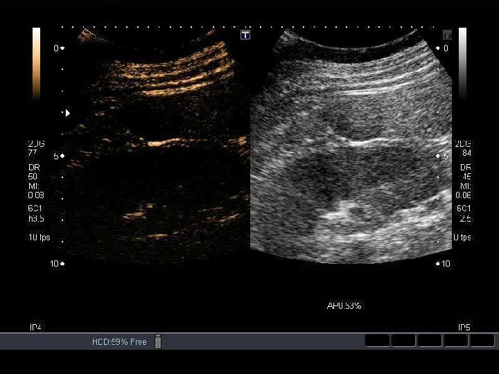
Liver hemangioma Diagnosis based on US if No history of cancer Normal liver tests Less than 3 nodules Typical features Hyper-echoic Homogeneous Posterior acoustic enhancement No doppler signal

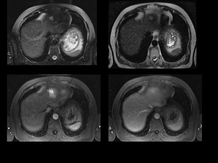
Liver hemangioma What to do if there atypical features? Constrast enhanced US MRI Typical enhancement: arterial, peripherical, discontinuous, centripetal enhancement, uniform filling on the venous phase
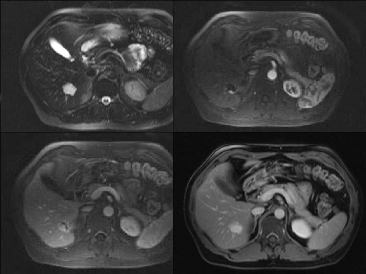

Liver hemangiomas Capillary or fast flow hemangioma 16% of all hemangiomas Often small lesions Enhancement: arterial, intense, homogeneous similar to the aortic enhancement during all phases Common association with an arterioportal shunt: transient arterial perilesional enhancement

Focal nodular hyperplasia: MRI typical features Signal: Homogeneous Slighty hyperintense on T 2 Slightly hypointense on T 1 Hyperintense central scar on T 2 Se = 74% Enhancement : = 100% Homogeneous (central. Sp scar excepted) Arterial Late enhancement of the central scar T 2 IV- No capsule Mathieu et al. Lancet. 1998 Nov 21; 352(9141): 1679 -80

Focal nodular hyperplasia: atypical features and traps Nodules <3 cm often display atypical features Which explain the low sensitivity (75%) What to do? Technical optimization b=800 s/mm 2 CEUS MRI with injection of hepatospecific contrast agent b=100 s/mm 2 Nguyen BN, Flejou JF, Terris B, Belghiti J, Degott C. Focal nodular hyperplasia of the liver: a comprehensive pathologic study of 305 lesions and recognition of new histologic forms. Am J Surg Pathol. 1999 Dec; 23(12): 1441 -54.

Focal nodular hyperplasia: atypical features and traps CEUS Characteristics Centrifugal enhancement Centripetal enhancement Mixed enhancement b=100 s/mm 2 FNH 74 -91% 2 -7% 7 -19% Adenoma 16% 47 -53% b=800 s/mm 2 32 -37%

Focal nodular hyperplasia: atypical features and traps MRI with injection of hepatospecific contrast agent Blood Vessel Bile duct OATP 1 Hepatocyte Bilirubin MRP 2 OATP 1 = Organic Anion Transporting Polypeptide 1 MRP 2 = Multridrug Resistance associated Protein 2 - extracellular distribution - hepatocellular captation via OATP 1 receptor (same as bilirubin) - biliary excretion via MRP 2 transporter. Planchamps et al. Mol Pharmacol 2007 Pastor et al. Radiology 2010

Focal nodular hyperplasia: atypical features and traps MRI with injection of hepatospecific contrast agent Gd-BOPTA - Gadobenate Dimeglumine Focal nodular hyperplasia enhancement on biliary phase No enhancement of adenoma Se 97% / Sp 100% Grazioli et al. Radiology 2005; 236: 166 -177

Focal nodular hyperplasia: atypical features and traps MRI with injection of hepatospecific contrast agent T 1 IP T 1 VIBE IV- T 1 OP T 1 VIBE arterial T 2 FS T 1 VIBE portal

Focal nodular hyperplasia: atypical features and traps MRI with injection of hepatospecific contrast agent T 1 IP T 1 VIBE T 1 OP T 1 FS

Focal nodular hyperplasia: atypical features and traps How to choose the diagnostic modality? Importance of the size of the nodule on CEUS Local experience (Henri Mondor) n=40 patients FNH or HCA with CEUS and MRI-HBP CEUS Sensitivity % For all lesions(n=43) 67, 7 For lesions > 35 mm(n=13) For lesions ≤ 35 mm(n=30) 7, 7 93, 3 Specificity % For all lesions 100 For lesions > 35 mm For lesions ≤ 35 mm 100

Adenoma Genotype/phenotyp e classification HNF 1α mutated β catenin mutated Inflammatory % Characteristic Histology Immunohistoche mistry HCC transformation diabetes MODY 3 Steatosis LFABP - no man Cytologic abnormalities glutamin synthetase : overexpression β catenin : nuclear localisation 35 -45 15 -20 b=800 s/mm 2 35 -40 dysmetabolic syndrom gp 130 mutated 66% Non mutated 33% bleeding risk Non mutated and non inflammatory ? frequent (3040%) 10 -20 b=100 s/mm 2 Inflammatory infiltration Dystrophic vessels ? non specific SAA, CRP : overexpression Only if β catenin mutation association with gp 130 (10%) non specific unknown
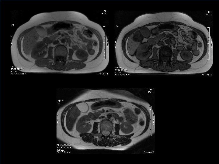

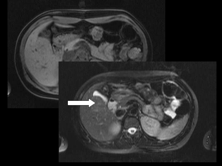
HNF 1α mutated adenoma

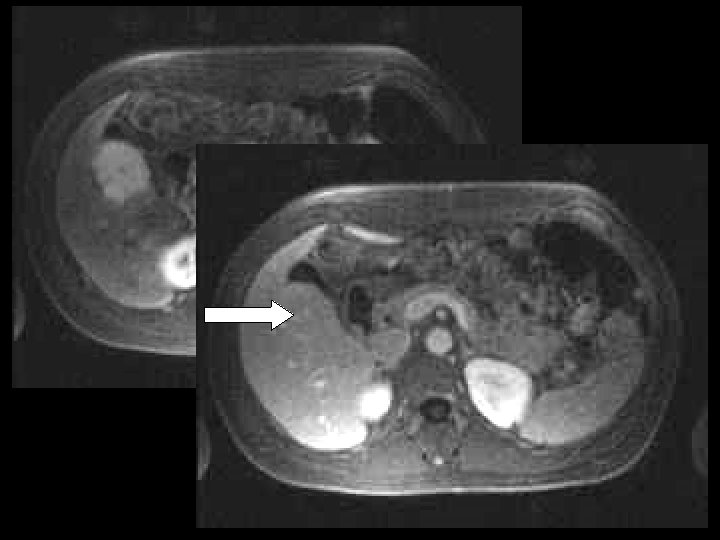
Inflammatory adenoma

Liver MRI Typical FNH Stop « Atypical » FNH Adenoma Biopsy Stop MRI with hepatospecific contrast agent CEUS Steatotic adenoma (HNF 1αmutated) Male sex or βcatenin Inflammatory adenoma Stop oral contraception Multidisciplinary meeting Follow up > 5 cm Resection resection Non specific adenoma Hemangiom a
- Slides: 26