Benign or low grade malignancy Salivary gland lesions


Benign or low grade malignancy

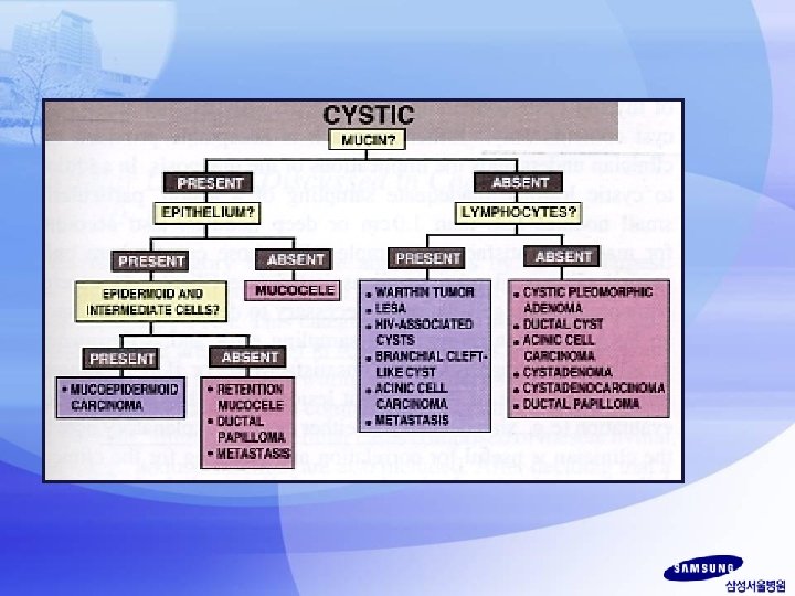
Salivary gland lesions that may have Mucin Salivary gland neoplasm that may have a cystic component • Mucocele or retention cyst • Warthin’s tumor • Pleomorphic adenoma • Mucoepidermoid tumor • Adenoid cystic carcinoma • Pleomorphic adenoma • Warthin’s tumor • Low grade mucoepidermoid carcinoma • Papillary cystic acinic cell carcinoma • Metastatic keratinizing squamous cell carcinoma

Differential diagnosis • Low grade mucoepidermoid carcinoma • Acinic cell carcinoma • Sclerosing polycystic adenosis • Warthin’s tumor • Metastatic renal cell carcinoma

ACC LG-MEC mucoid background - + finely vacuolated cytoplasm + -

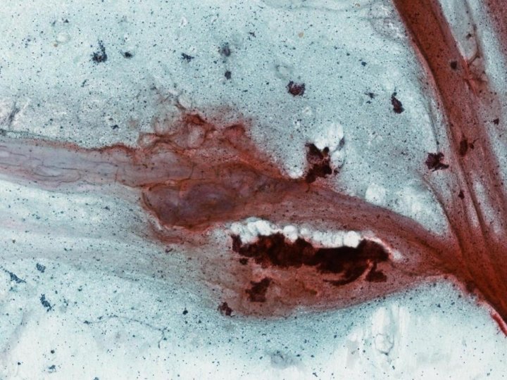
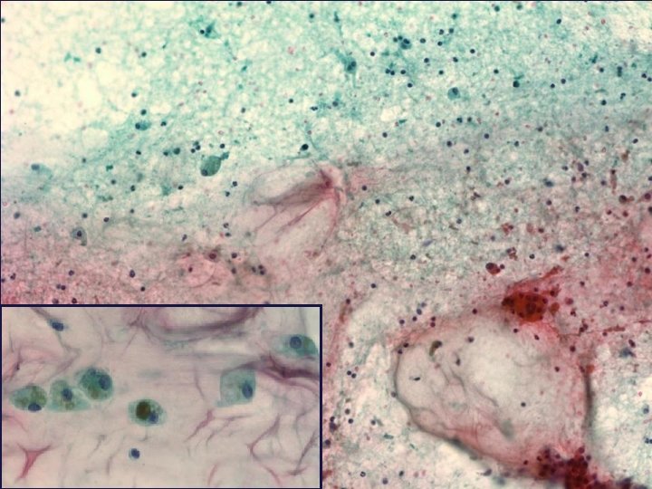
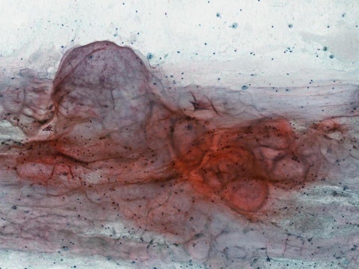
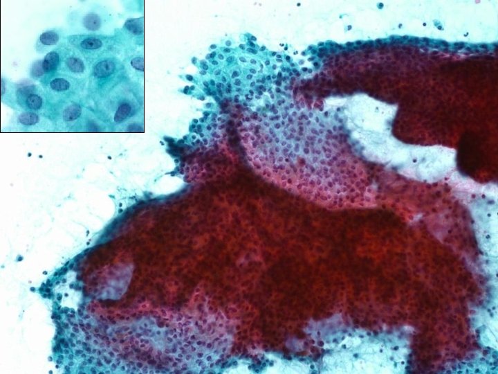
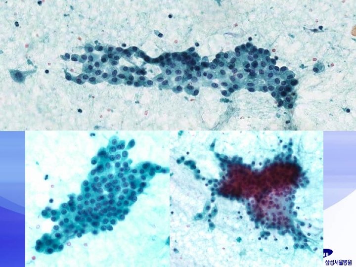
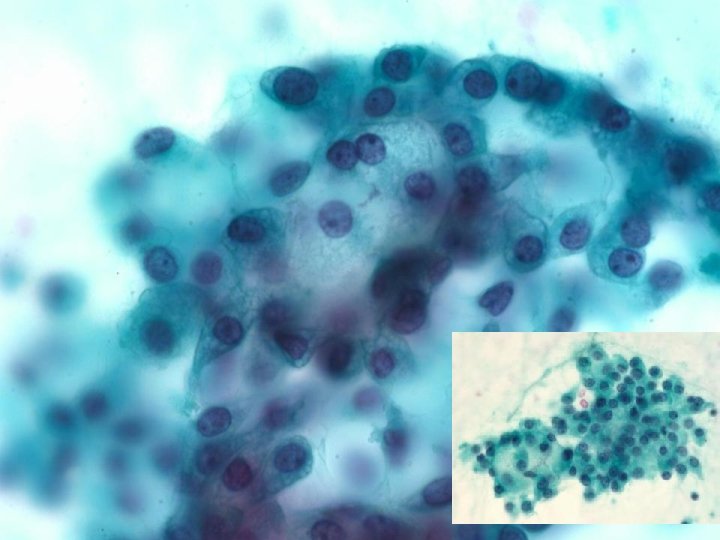
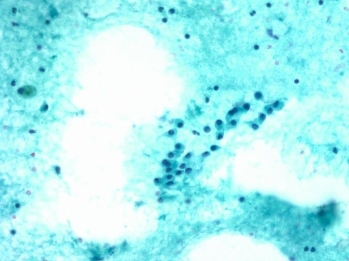
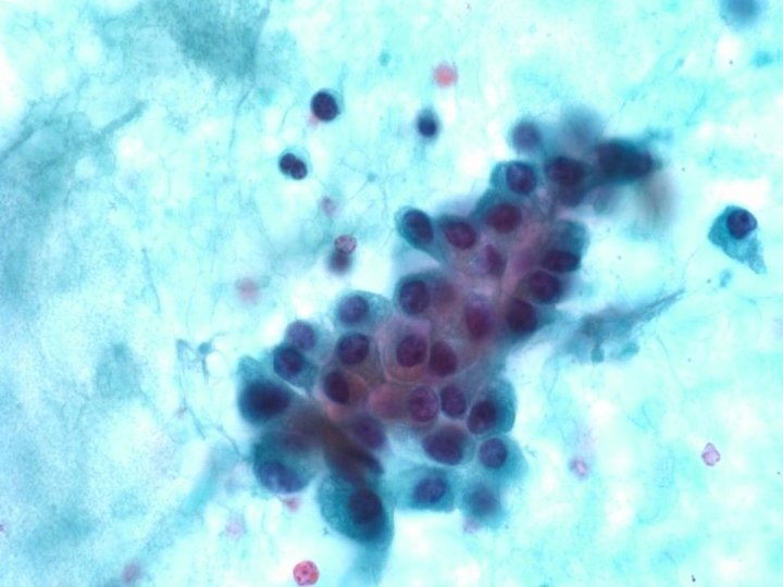
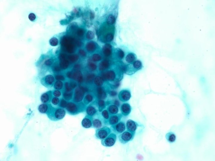
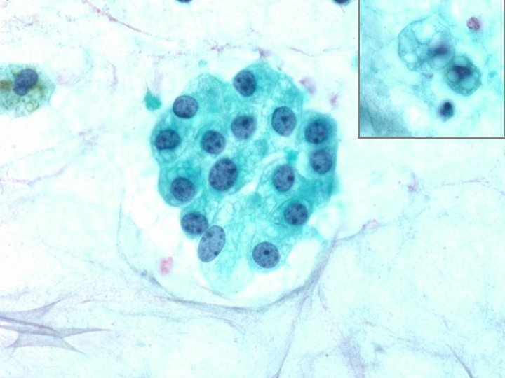
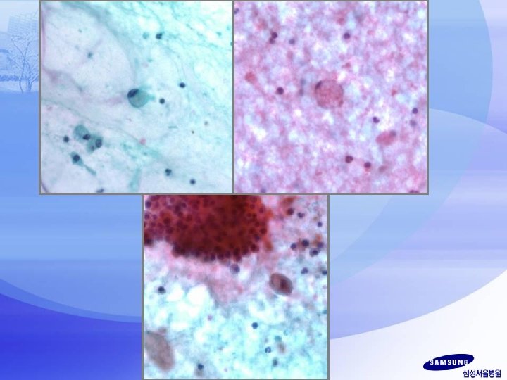
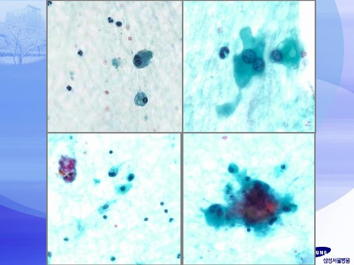
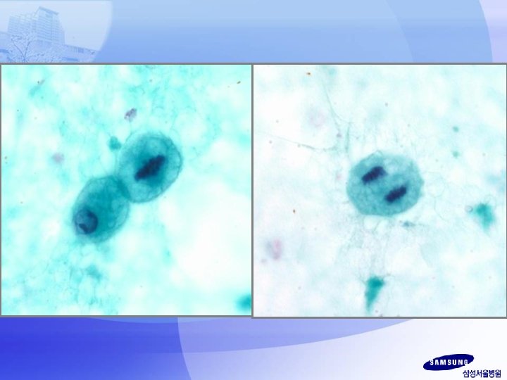
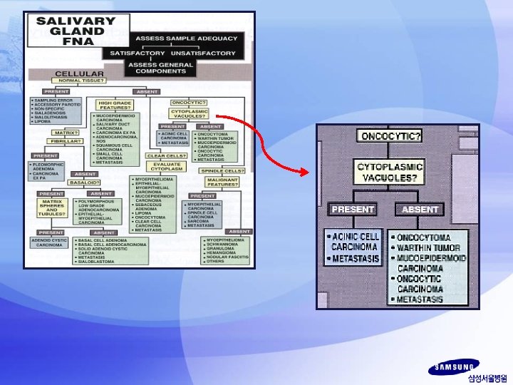
Acinic cell carcinoma Ø Delicate vacuolated cytoplasm - the key to the cytologic diagnosis of acinic cell carcinoma Ø At least focal serous acinar differentiation with its characteristic cytoplasmic zymogen granules; while they are sometimes abundant, they are more often sparse and difficult to find Ø Cell types seen in acinic cell carcinomas – – – Serous acinar Intercalated duct Vacuolated Clear Non-specific glandular
 1. ACC-PCV : small, distinct, and evenly distributed")
Acinic cell carcinoma, papillary cystic variant(ACC-PCV) 1. ACC-PCV : small, distinct, and evenly distributed vacuoles MEC : larger and irrrecular vacuoles 2. predominantly cystic Cystic material in ACC-PCV is rarely of mucinous type, though it may still lead to an erroneous diagnosis of mucoepidermoid carcinoma 3. Cytomolphologic findings a/w conventional type ACC but notably absent in ACC-PCV -predominant single discohesive cell population -delicate branching capillaries intimately a/w nests of proliferating tumor -Acinar or rosette-like formations -Bare malignant nuclei -cells with densely granular cytoplasm -Background lymphocytes Diagn Cytopathol 2002; 27: 244 -50 Cytopathology 2009, 20, 96– 102

Mucoepidermoid carcinoma Ø Combination of 3 epithelial cell types epidermoid, mucus, intermediate Ø Epidermoid – bland cohesive but crowded sheets with squamoid features including well-defined intercellular borders and abundunt dense waxy cytoplasm Ø Mucus- abundant delicate mucinous cytoplasm with a peripherally placed and indented nucleus (easily be mistaken for foamy histiocytes and muciphages) Ø Intermediate cells resemble the suprabasal cells of the epidermis or metaplastic squamous cells of the cervix

Oncocytic variant of mucoepidermoid carcinoma –rare -potential pitfall in the evaluation of oncocytic salivary gland lesions -Sheets of bland cells with densely granular oncocytic cytoplasm -Rare admixed mucus cells -partially cystic and mucoid background
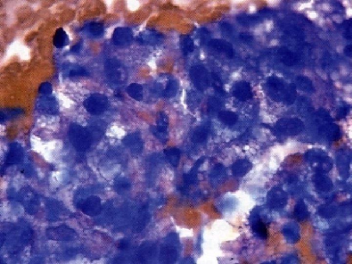

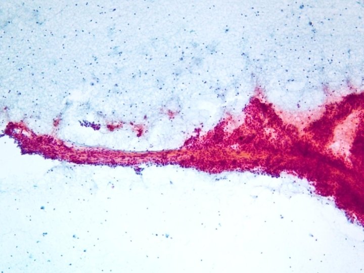
Parotid gland, Lt, FNA : Acinic cell carcinoma
- Slides: 28