BENIGN AND MALIGNANT TUMORS OF ORAL CAVITY DR

BENIGN AND MALIGNANT TUMORS OF ORAL CAVITY - DR GITANJALI KHULBE MDS (ORAL MEDICINE AND RADIOLOGY)

ODONTOGENIC TUMORS: § Epithelial tumors: - Ameloblastoma - Adenomatoid odontogenic tumor - Squamous odontogenic tumor - Calcifying cystic odontogenic tumor - Calcifying epithelial odontogenic tumor - Keratocystic odontogenic tumor

§ Mesenchymal tumors: - Cementoma - Benign cementoblastoma - Cemento-ossifying fibroma - Odontogenic myxoma - Odontogenic carcinoma - Fibro-osseous lesions

§ Mixed tumors: - Ameloblastic fibroma - Ameloblastic fibro-odontoma - Ameloblastic odontoma - Giant cell granuloma - Metastatic tumors to the jaws

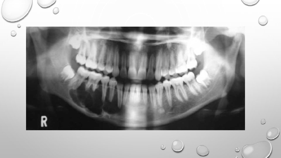
AMELOBLASTOMA: • It is an odontogenic tumor representing approx. 11% to 13% of the odontogenic tumors. • It is bony-hard and non-tender on palpation. • Unicystic or mural ameloblastoma forms in the wall of a dentigerous cyst. • Radiographically, a unilocular or multilocular radiolucency is seen. Soap-bubble or honey-comb appearance is characteristic.

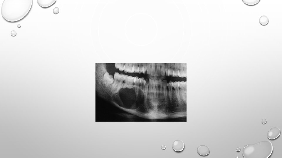
ADENOMATOID ODONTOGENIC TUMOR: • It is an uncommon, benign tumor which makes up approximately 3% of all odontogenic tumors. • It is a slow-growing tumor which is locally invasive, tends to displace teeth and cause mobility. • Usually occurring in the second decade of life, is more frequent in the maxilla than in mandible, mostly in the anterior region. • Enucleation is the treatment of choice.
: • A rare, well-circumscribed, solid or cystic tumor")
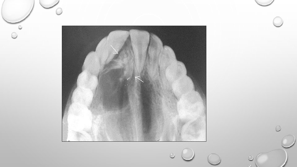
CALCIFYING CYSTIC ODONTOGENIC TUMOR (GORLIN CYST): • A rare, well-circumscribed, solid or cystic tumor which contains ‘ghost cells’ and spherical calcifications. • It is a slow-growing tumor with equal frequency in maxilla and mandible. • Majority of the lesions are located in incisor-canine region. • Cyst-like radiolucency containing radiopaque foci can be seen. • Surgical enucleation is the treatment.

CALCIFYING EPITHELIAL ODONTOGENIC TUMOR: • It is a rare tumor, representing approx. 1% of all odontogenic tumors. • Occurs as a painless, slowly increasing expansion of the jaws, with a marked predilection for molar region of mandible. • Characteristic radiographic presentation is “driven snow” appearance.

KERATOCYSTIC ODONTOGENIC TUMOR: • It is a cystic neoplasm, characterized by its unique histologic appearance. • May occur in any location, posterior mandible is the most common site of occurrence. • It can occur as a component of ‘Nevoid Basal Cell Carcinoma’ syndrome. • Radiographically, presents as a small, asymptomatic, unilocular radiolucency.

ODONTOGENIC MYXOMA: • These are slow-growing, invasive tumors composed of very loose cellular connective tissue (acid mucopolysaccharide). • It consists of small, rounded cells lying in an abundant mucoid stroma derived from dental pulp. • Resection is the choice of treatment.

CENTRAL ODONTOGENIC FIBROMA: • It is a tumor composed of mature fibroblastic tissue admixed with nests and strands of odontogenic epithelium. • They are generally small, may cause root resorption. • Treatment is conservative excision.

CEMENTOBLASTOMA: • It is a cementum-producing lesion which is fused to the root of a vital tooth. • Often occurs in young adults and in association with a mandibular molar or premolar. • Pain is a frequent complaint. • Radiographically, it demonstrates a pathogonomic appearance – a well-defined radiopaque mass surrounded by a radiolucent halo that incorporates root of the tooth.

AMELOBLASTIC FIBROMA: • It is a tumor composed of mesenchymal tissue resembling dental papillae and small islands of odontogenic epithelium. • Most cases are seen under 20 years of age, commonest site being the mandibular molar and premolar region. • Usually seen as a pericoronal radiolucency; unilocular or multilocular. • It is treated by surgical excision.

THANK YOU FOR LISTENING. .

• Oral premalignant lesion is a morphologically altered tissue in which cancer is more likely to occur than its apparently normal counterpart. (who 1978) • Precancerous condition is a generalised state associated with a significantly increased risk of cancer. • Premalignancy is marked by aberrant and uncoordinated cellular proliferation depicted at cellular level (atypia), reflections of which could be seen at tissue levels (dysplasia).
- Slides: 18