Bellwork Domestic horses have 64 chromosomes How many

Bellwork Domestic horses have 64 chromosomes. How many chromosomes should be in an egg cell of a female horse? A. 16 B. 32 C. 64 D. 128

Bellwork In a sample of double-stranded DNA, 30% of the nitrogenous bases are thymine. What percentage of the nitrogenous bases in the sample are adenine? A. 20% B. 70% C. 30% D. 40%
 • Bellwork • New Information: Cell Cycle,")
Agenda 5/3 - Cell Cycle, Division (Mitosis) • Bellwork • New Information: Cell Cycle, Mitosis • Virtual Lab- Identifying Stages of the Cell Cycle
 • What is mitosis? Why is it important?")
Cell Cycle and Cell Division (Mitosis) • What is mitosis? Why is it important?
: – Growth – Repair")
Cell Division • 3 reasons for mitotic cell division (mitosis): – Growth – Repair – Replacement • Parent cell divides into two identical daughter cells

Cell Division • Cells divide to make new cells- DNA is replicated so each daughter cell gets an exact copy. • DNA condenses into chromosomes during mitosis.

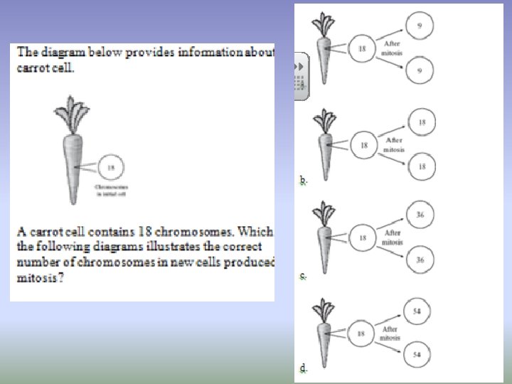
Cell Division • Humans have 46 chromosomes, 23 from each parent. • Haploid cells: only one of each chromosome (n) • Diploid cells: two of each chromosome (2 n)
 G 1 phase M phase S phase G")
Cell Cycle (click for video clip) G 1 phase M phase S phase G 2 phase

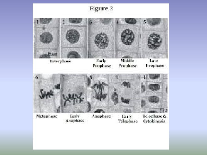
Interphase- longest • G 1 Phase • Cell growth • S Phase • DNA replication • G 2 Phase • preparation for mitosis • M Phase • mitosis and cytokinesis Allium G 1 M phase S G 2

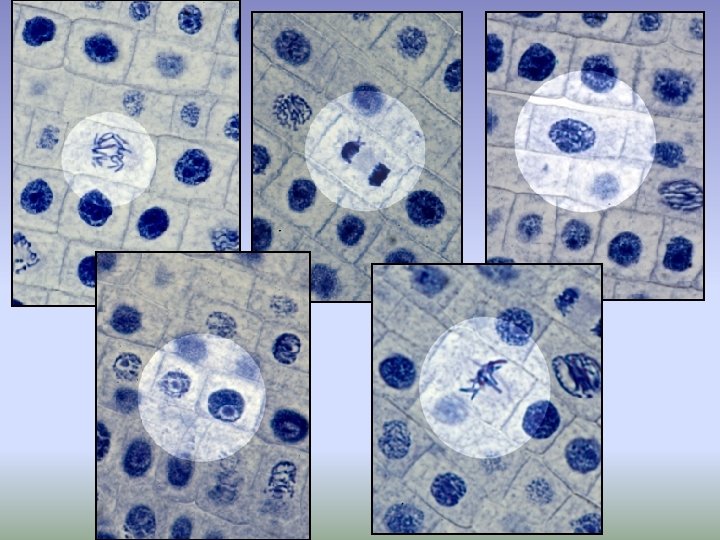
Interphase • Nucleus and nuclear envelope are visible. • One or more nucleoli are visible. • The rest of the nucleus is filled with chromatin.

Figure 10 -5 Interphase Prophase Cytokinesis Telophase Metaphase Anaphase

Prophase pro- = first; first stage of mitosis • Nuclear envelope breaks down • Chromatin condenses into chromosomes • Centrioles move to opposite poles of the cell • Spindle fibers attach to chromosomes at the centromere.

Metaphase • Chromosome line up in the Middle of the cell • Chromosome centromeres are attached to the spindle at the poles of the cell

Anaphase • Chromosome centromeres separate and sister chromatids move Apart to the opposite ends of the cell
 • A new nuclear")
Telophase • Chromosomes precipitate back into strands of DNA (chromatin) • A new nuclear envelope begins to form around each of the two new clumps of DNA
 • Same time as Telophase • Animal Cells: • cytoplasm splits")
Cytokinesis (“cell splitting”) • Same time as Telophase • Animal Cells: • cytoplasm splits as the cell membrane draws inward (cleavage furrow) and splits the cell in two • Plant Cells: • cytoplasm splits as a cell plate forms between the two new nuclei, then a new cell membrane

Cell Cycle and Mitosis • • • I P M A T C • • • I Protect Mothers And Their Children • • • Interphase Prophase Metaphase Anaphase Telophase Cytokinesis

Cell Cycle and Mitosis Virtual Lab • Go to http: //www. biology. arizona. edu/cell_bio/act ivities/cell_cycle. html • Make a table like the one you’ll see before the activity begins. • Answer the following questions: – Which phase of the cell cycle is longest? How do you know? Which is shortest, and how do you know?

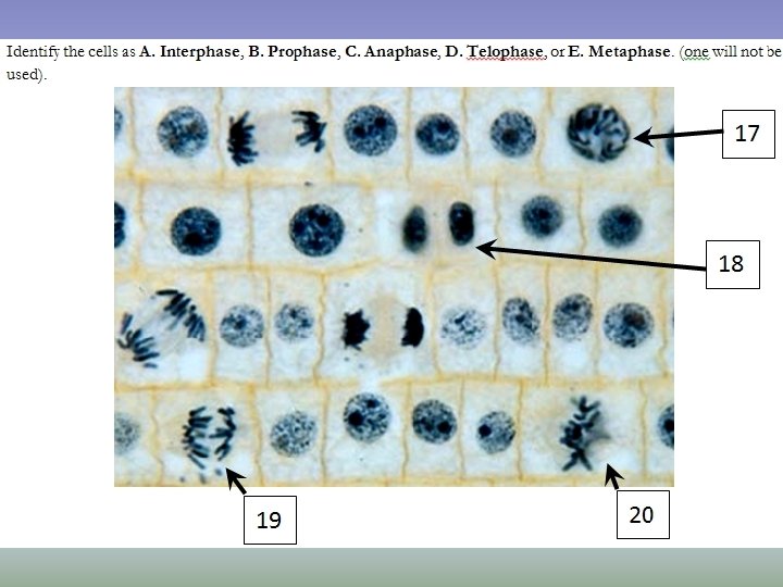
Bellwork 5/4 • What are the 4 phases of the cell cycle? • What are the 4 phases of mitosis? • What phases are shown below?

Agenda 5/4 - Cell Cycle and Cancer • • Bellwork Notes- Cell Cycle Control and Cancer Lab- Mitosis in Onion Root Tip Cells HW- finish lab questions, study for test (Fri) - DNA structure and replication, cell cycle, and mitosis
 reproduce by a type of cell division called binary")
Binary Fission • Prokaryotes (bacteria) reproduce by a type of cell division called binary fission. • In binary fission the single bacterial chromosome replicates and the two daughter chromosomes actively move apart.

G 1 phase M phase S phase G 2 phase

The Cell Cycle Control System • The events of the cell cycle are directed by a cell cycle control system, similar to a clock. G 1 checkpoint Control system G 1 M M checkpoint Figure 12. 14 G 2 checkpoint G 2 S

The Cell Cycle Control System • There are specific checkpoints where the cell cycle stops until a go-ahead signal is received. G 0 G 1 checkpoint G 1 Figure 12. 15 A, B (a) If a cell receives a go-ahead signal at the G 1 checkpoint, the cell continues on in the cell cycle. (b) If a cell does not receive a goahead signal at the G 1 checkpoint, the cell exits the cell cycle and goes into G 0, a non-dividing state.

Control of Cell Division • In density-dependent inhibition crowded cells stop dividing. • Most animal cells also exhibit anchorage dependence in which they must be attached to a substratum to divide. (a) Normal mammalian cells. The availability of nutrients, growth factors, and a substratum for attachment limits cell density to a single layer. Cells anchor to dish surface and divide (anchorage dependence). When cells have formed a complete single layer, they stop dividing (density-dependent inhibition). If some cells are scraped away, the remaining cells divide to fill the gap and then stop (density-dependent inhibition). Figure 12. 18 A 25 µm

Control of Cell Division • Cancer cells: no density-dependent inhibition or anchorage dependence. • Do not respond normally to the body’s control mechanisms and form tumors. • Loss of the cell cycle controls. Cancer cells do not exhibit anchorage dependence or density-dependent inhibition. (b) Cancer cells usually continue to divide well beyond a single layer, forming a clump of overlapping cells. Figure 12. 18 B 25 µm

Control of Cell Division • Malignant tumors invade surrounding tissues and can metastasize, exporting cancer cells to other parts of the body where they may form secondary tumors.

Cancer Risk Factors Quiz • http: //www. webmd. com/cancer/rm-quizcancer-myths-facts

Lab Objectives • To list the phases of the cell cycle in the order in which they occur. • To list the phases of the M-phase of the cell cycle (Mitosis) and explain what happens during each. • To illustrate/draw what happens during each phase of Mitosis. • To identify the role of cell structures during cell division (centrioles, chromatin, chromosomes, nucleus). • To learn the importance of cell division for organisms (asexual reproduction, growth, repair).

Mitosis in Allium Root Interphase Prophase Metaphase

Mitosis in Allium Root Anaphase Telophase

Prophase Telophase Interphase Metaphase Anaphase

Allium Lab- Mitosis Y X • X = the area just behind the root cap • Y = the area behind region X (fewer cells, fewer nuclei visible)

Bellwork 5/6 The illustration below represents a cell that is entering mitosis. 1. Identify one function of mitosis. The cell shown in the illustration has recently completed the synthesis phase (S phase) of the cell cycle and is in prophase of mitosis. 2. What happened in this cell during the S phase in preparation for division? Describe the evidence that supports your answer. 3. In your Student Answer Booklet, draw the end products that will be formed when this cell completes mitosis.

Agenda 5/6 - Mitotic Cell Division • Bellwork • Discuss lab, start lab report • Review for Monday’s test

Agenda 5/9 - Sexual vs. Asexual Reproduction • Cell Cycle, Mitosis, and DNA Replication Test • New information: Sexual vs. Asexual Reproduction • Video- Life’s Greatest Miracle • HW- – Due Tues: Lab Report – Due Wed: Vocabulary (on next slide)

HW- vocabulary- use book or internet to define these terms (max 7 words per definition, in your OWN WORDS) and draw a picture for each. • Homologous chromosomes • Tetrad • Crossing over • Trait • Gene • Allele • • • Hybrid Homozygous Heterozygous Phenotype Genotype Probability

Eukaryotic Cell Division • Mitosis- normal cell division – Parent cell divides into two identical daughter cells (diploid = 2 n) • n = number of types of chromosomes • Meiosis- cell division resulting in reproductive cells (gametes) – Parent cell divides into 4 daughter cells, each with half the number of chromosomes (haploid = 1 n)

Mitosis • 2 diploid daughter cells formed, just like parent cell (two of each chromosome) • Preceded by interphase (G 1, S, G 2 phase) • Mitosis: – Prophase – Metaphase – Anaphase – Telophase

Figure 10 -5 Interphase Prophase Cytokinesis Telophase Metaphase Anaphase

Why don’t you look exactly like your parents or siblings? • A different form of cell division: Meiosis! • Sexual reproduction increases genetic variation in a population, which increases a species’ ability to adapt to changes in their environment.
 and female gamete (egg) are haploid (1 n)")
Sexual Reproduction • Male gamete (sperm) and female gamete (egg) are haploid (1 n) • Fertilization occurs when an egg and a sperm fuse to form a zygote (2 n). • Meiosis is the form of cell division that results in gametes.
 cells to produce reproductive cells (gametes) • 4 haploid")
Meiosis occurs in somatic (body) cells to produce reproductive cells (gametes) • 4 haploid daughter cells formed (only one of each chromosome) • Preceded by interphase and DNA replication • Two Divisions: – Meiosis II

Bellwork 5/10

Agenda 5/10 - Sexual vs Asexual Reproduction • Bellwork • New Information: Sexual vs. Asexual Reproduction • Video- finish and discuss • HW- vocabulary

Why Sex? The Evolution of Sexual Reproduction Asexual Reproduction • offspring are identical to parents • little variety in population • binary fission, budding Sexual Reproduction • offspring are a combination of parents • introduces variety in population

The Evolution of Sexual Reproduction
 The Evolution of Sexual Reproduction")
The Red Queen Hypothesis (start at 3: 00 min) The Evolution of Sexual Reproduction §The leading hypothesis for why sexual reproduction has persisted §Refers to Lewis Carroll’s Through the Looking Glass The Red Queen says to Alice: “It takes all the running you can do, to stay in the same place. ”

The Red Queen Hypothesis The Evolution of Sexual Reproduction The Red Queen Hypothesis: As species that live at each other's expense co-evolve, they are engaged in a constant evolutionary struggle for a survival advantage. They need "all the running they can do" because the landscape around them is constantly changing. (predator/prey, disease/host) In other words, hosts change the locks, and parasites, etc invent new keys. Sexual reproduction is necessary to fight disease, avoid predation, get food, etc.

Why Sex? Advantages and Disadvantages of Asexual Reproduction Sexual Reproduction Advantages: § Variation § More rapid rate of adaptation in a changing environment Disadvantages: § Costs of mating behavior: competition, no mating § Only pass on half your genes Asexual Reproduction Advantages: § Pass on all of your genes § No mating behavior costs (don’t have to spend time or energy looking for a mate, no energy wasted on showy displays) Disadvantages: § Little or no variation § More susceptible to parasites, viruses, etc. http: //www. pbs. org/wgbh/evolution/library/01/5/l_015_03. html

Costs of Sexual Reproduction: Displays, Competition for Mates

Life’s Greatest Miracle • Video with worksheet. • Answer all questions- classwork grade.

Life’s Greatest Miracle • Video link • Answer the questions on the worksheet

Bellwork 5/11
 •")
Agenda 5/11 - Meiosis • Bellwork • New Information: Meiotic Cell Division (Meiosis) • Activities: – Modeling Crossing Over – Mitosis vs. Meiosis Card Sort – Cell Division Vocabulary Squares
 • 4 haploid daughter")
Meiosis occurs in germ cells to produce reproductive cells (gametes) • 4 haploid daughter cells formed (only one of each chromosome) • Preceded by interphase and DNA replication • Two Divisions: – Meiosis II

The Human Karyotype Homologous Chromosomes chromosome pair, one from each parent, that are similar in length, gene position and centromere location

Meiosis- click for video clip • Meiosis I- reduction division; like mitosis but only one copy of each homologous chromosome goes to each new cell; two new cells formed – Prophase I – Metaphase I- homologous chromosomes line up on equator to form a tetrad; crossing over occurs – Anaphase I- homologous chromosomes separate – Telophase I- two new cells, each with half the genetic material • Meiosis II- the two cells formed in Meiosis I divide again, with sister chromatids separating – Basically like Mitosis

Meiosis I - Reduction Division Interphase I Cells undergo a round of DNA replication, forming duplicate Chromosomes. Prophase I Each chromosome pairs with its corresponding homologous chromosome to form a tetrad. Metaphase I Homologous chromosomes line up along the middle of the cell. Spindle fibers attach to the chromosomes. Anaphase I The fibers pull the homologous chromosomes toward the opposite ends of the cell.

Meiosis I - Reduction Division Interphase I Cells undergo a round of DNA replication, forming duplicate Chromosomes. Prophase I Crossing over- Metaphase I Homologous chromosomes line up along the middle of the cell. Anaphase I The fibers pull the homologous chromosomes toward the opposite ends of the cell. exchange of genes between. Spindle fibers attach to the chromosomes. homologous This reduces the chromosomesnumber in each increases daughter cell by half. genetic variation among offspring

Meiosis II - Similar to Mitosis Prophase II Meiosis I results in two haploid (N) daughter cells, each with half the number of chromosomes as the original. Metaphase II The chromosomes line up in a similar way to the metaphase stage of mitosis. Anaphase II The sister chromatids separate and move toward opposite ends of the cell. Telophase II Meiosis II results in four haploid (N) daughter cells.

Modeling Crossing Over • Use the paper to create a model of crossing over • First, draw a pair of duplicated homologous chromosomes using two different colors. • Model how the homologous chromosomes separate during Meiosis I • Model how the sister chromatids separate during Meiosis II

Mitosis vs. Meiosis • • Animation Mitosis- 2 diploid cells produced Meiosis- 4 haploid cells produced Why?

Comparing Mitosis and Meiosis • Place the cards in order • How are the two processes similar? How are they different?

Meiosis Using two successive divisions to reduce the number of chromosomes by half and create gametes. Homologous chromosomes: a pair of chromosomes with the same genes (but may have different alleles); one from each parent.

Mitosis vs. Meiosis Mitosis Meiosis # of divisions 1 2 # of daughter cells produced 2 4 # chromosomes in parent cell 46 (diploid) # chromosomes in daughter cell 46 (diploid) 23 (haploid) Type of cells produced Somatic (body) cells Gametes +* (sex cells)
- Slides: 70