Basis of the MEEG signal Jordi Costa Faidella

Basis of the M/EEG signal Jordi Costa Faidella & Tal Machover 1
 Special thanks to Randal Pinto")
Electroencephalography (EEG) Special thanks to Randal Pinto

History 1929: Hans Berger developed the electroencephalography (=graphic representation of the difference in voltage between two different cerebral locations plotted over time) following the studies of Richard Caton in nonhuman animal species. He described the human alpha and beta rhythms

EEG Acquisition Electrodes: Ag/Ag. Cl, tin… Active electrodes: Attached to the scalp Reference electrode: Mastoid, nose, ear lobe. . . The EEG records differences in voltage – difference in electrical potential from one electrode to another!! Amplification

Continuous EEG recording

EEG Rhythms

What does the EEG record? Mainly NOISE!! Volume Conduction The electrical activity flows through the tissue between the electrical generator and the recording electrode. Thus, the EEG is a 2 -D representation of a 3 -D reality, which poses a problem in localizing the sources of the electrical activity Inverse problem
 To pick up the electrical activity of the")
Neural basis of the EEG (1) To pick up the electrical activity of the brain from scalp recordings, it must be of sufficient strength and duration… Action Potentials…? Rapid, transient, all-or-none nerve impulses of 100 m. V aprox. and a duration of 1 ms that flow from the body to the axon terminal of a neuron. They don’t create a dipole …NO!
 Post-synaptic potentials… …fullfill the requirements to be recorded")
Neural basis of the EEG (2) Post-synaptic potentials… …fullfill the requirements to be recorded by EEG!! 1) When an Action Potential reaches the axon terminal the neuron releases a neurotransmitter. 2) The neurotransmitter binds to the receptor. 3) The postsynaptic neuron gets depolarized (Na+ inward currents – excitatory - EPSP) or hyperpolarized (Cl- inward currents – inhibitory -IPSP). 4) EPSP and IPSP summate temporally and spatially. 5) If the postsynaptic neuron reaches a given depolarization threshold, an action potential is generated.
 When an EPSP is generated in the dendrites")
Neural basis of the EEG (3) When an EPSP is generated in the dendrites of a neuron an extracellular electrode detects a negative voltage difference, resulting from Na+ currents flowing inside the neuron’s cytoplasm. The current completes a loop further away the excitatory input (Na+ flows outside the cell), being recorded as a positive voltage difference by an extracellular electrode. This process can last hundreds of milliseconds. Thus, a small dipole is generated!!
 Pyramidal neurons are spatially aligned and perpendicular to")
Neural basis of the EEG (5) Pyramidal neurons are spatially aligned and perpendicular to the cortical surface. Thus, EEG represents mainly the postsynaptic potentials of pyramidal neurons close to the recording electrode. The electrical activity from deeper generators gets dispersed and attenuated by volume conduction effects.
")
Neural basis of the EEG (4)
 EEG averaging technique used to study the electrical activity time -locked")
Event-related potentials (1) EEG averaging technique used to study the electrical activity time -locked to an event. - Needs a considerable amount of trials - Comprises a mixture of different brain rhythms, depending on the filters applied. - Only about 20% of the evoked activity is shown - Other approaches to study electrical brain activity: Timefrequency analysis
")
Event-related potentials (2)
 Electrophysiological response to an acoustic stimulus (simple tone). Reference set at")
Event-related potentials (3) Electrophysiological response to an acoustic stimulus (simple tone). Reference set at the tip of the nose. 0. 3 -30 Hz Band-pass filter Negative response at frontocentral electrodes with polarity reversal at mastoid electrodes, indicative of its probable origin in the temporal lobe (silvian fissure, auditory areas).

Magnetoencephalography

• The connection between electricity and magnetism was first discovered by Hans Christian Orsted in 1819. • He demonstrated that a magnetic compass needle was affected by a current passing through a circuit. • You can try this at home.

• An electrical dipole is always surrounded by a corresponding magnetic field. • The polarity of the field is determined by the direction of the current. • Magnetic fields summate in the same way as voltages.

• The magnetic field is perpendicular to the current. • If the current is running parallel to the scalp the magnetic field exits the head from one side of the dipole and re-enters on the other side and so can be measured. • But if the current is perpendicular to the scalp the magnetic field does not leave the scalp and cannot be measured.

• This means that MEG is more sensitive to the activity of the pyramidal cells in the walls of the sulci. • An EEG signal could come from either a sulcus or a gyrus or both, making it even more difficult to localize its origin. • MEG registers less of the brain’s activity as it contains no information from radially aligned axons. • But this does mean that we can make stronger inferences about the origin of the signals.

Bone is transparent to magnetism and magnetic fields are not smeared by the resistance of the skull.

• Though EEG does give better spatial resolution it is not in itself a solution to the ‘inverse problem’. • The MEG signal can be time-locked to a stimulus to produce Event Related data.

• The magnetic fields generated by the brain are minute: 100 million times weaker than the earth’s magnetic field, one million times weaker than the magnetic fields generated by the urban environment. • By way of contrast, MRIs generate a magnetic field of between 3 to 3. 5 tesla.

• Since bio-magnetic fields are so small we need very sensitive equipment to detect them.

• In 1963 Gerhard Baule and richard Mcfee made the first recording of the biomagnetic field generated by the human hart. • They used two copper pick-up coils twisted round a core of ferrite material with 2 million turns. • The two coils were connected in opposite directions so as to cancel out the background fluctuations. Never the less, they had to conduct their experiment in the middle of a field because the signal was still very noisy. • A group working in the Soviet Union (Safonev et al, 1967) produced similar results but in a shielded room which reduced background noise by a factor of 10. • However thermal noise set the limit to the use of copper.

- In 1911, the Dutch physicist Heike Onnes discovered that, when cooled to 269 C, solid mercury suddenly lost all resistance to the flow of electric current. - This phenomenon – "superconductivity" – was later found in other materials, such as tin and metal alloys. _ When two superconducting materials are separated by a thin insulating layer a ‘tunnel effect’ is produced which enables the flow of electrons - even in the absence of any external voltage. This is a Josephine Junction.

There are two types of superconduction, one that completely rejects magnetic fields and the other where superconductivity and magnetism can coexist. It is this latter, type II supercoductivity, that is deployed in SQUIDs. When cooled to very low temperatures, superconductors conduct electricity without resistance. This lack of resistance allows a SQUID to measure the interference of quantum-mechanical electron waves circulating in its superconducting loop as the magnetic flux enclosed by the loop changes. A SQUID can measure magnetic fields as small as 1 femtotesla. The SQIUDs are connected to sensors arranged in a helmet shape that fit round the subjects head.

• In the late 1960’s David Cohen, at MIT, Boston used SQUIDs to record a clean MCG in an urban environment. • They conducted their experiment in a magnetically sheilded room.

The sensitivity of the SQUID to magnetic fields may be enhanced by coupling it to a superconducting pickup coil (“flux transformer”) which has greater area and number of turns than the SQUID inductor alone. made of superconducting wire and is sensitive to very small changes in the magnitude of the impinging magnetic flux. The magnetic fields from the brain Magnetometers causes a supercurrent to flow. First Order Gradiometer

- Subjects can perform tasks in a sitting position. The testing environment is quiet.

• The sensors do not need to come into direct contact with the scalp. Unlike EEG, MEG does not mess up your hair! • This cuts down preparation time and makes MEG more child-friendly. • Helmets used today have as many as 300 sensors.
 http: //imaging.")
MEG data brain activation film (recorded during comprehension of a spoken word) http: //imaging. mrccbu. cam. ac. uk/meg/

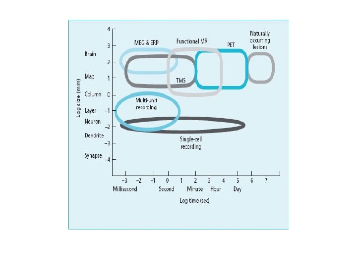
Though MEG has better spatial resolution than EEG (1 mm as opposed to 1 cm) it does not provide anatomical information Combining MEG data with FMRI results in a hybrid image which has both good temporal and spatial resolution.

Multimodal Imaging • • Combining MEG data with FMRI results in a hybrid image which has both good temporal and spatial resolution.
 • Signal")
EEG vs. MEG EEG • Cheap • Large Signal (10 m. V) • Signal distorted by skull/scalp • Spatial localization ~1 cm • Sensitive to tangential and radial dipoles (neurons in sulci & on gyri) • Allows subjects to move • Sensors attached directly to head • Extracellular secondary (volume) currents MEG • Good temporal resolution (~1 ms) • Problematic spatial resolution (forward & inverse problems) • Expensive • Tiny Signal(10 f. T) • Signal unaffected by skull/scalp • Spatial localization ~1 mm • Sensitive mostly to tangential dipoles (neurons in sulci) • Subjects must remain still • Sensors in helmet • Requires special laboratory • Intracellular primary currents’ magnetic fields Thanks to last year’s slides & wikipedia
. Can be used")
ADVANTAGES OF M/EEG Non-invasive (records electromagnetic activity, does not modify it). Can be used with adults, children, infants, newborns, clinical population. High temporal resolution (up to 1 millisecond or less, around 1000 x better than f. MRI) => ERPs study dynamic aspects of cognition. Allow quiet environments. Subjects can perform tasks sitting up- more natural than in MRI. DISADVANTAGES OF M/EEG Problematic spatial resolution (forward & inverse problems) Anatomical information not provided.
 (2007) Magnetism in Medicine. Wiley")
References/suggested reading • Andro, W. and Nowak, H, (eds) (2007) Magnetism in Medicine. Wiley - VCN • Handy, T. C. (2005). Event-related potentials. A methods handbook. Cambridge, MA: The MIT Press. • Luck, S. J. (2005). An introduction to the event-related potential technique. Cambridge, Massachussets: The MIT Press • Rugg, M. D. , & Coles, M. G. H. (1995). Electrophysiology of mind: Event-related brain potentials and cognition. New York, NY: Oxford University Press. • Hamalainen, M. , Hari, R. , Ilmoniemi, J. , Knuutila, J. & Lounasmaa, O. V. (1993). MEG: Theory, Instrumentation and Applications to Noninvasive Studies of the Working Human Brain. Rev. Mod. Phys. Vol. 65, No. 2, pp 413 -497. • Kandel, E. R. , (2006) In Search of Memory. W. W. Norton (Chapters 5 and 6) • Kandel, E. R. , (2000). Principles of neural science (pp 776 -777). Mc. Graw-Hill. NY, London. • Olejnickzac, P. , (2006). Neurophysiologic basis of EEG. Journal of Clinical Neurophysiology, 23, 186 -189. • Silver, A. H. (2006). How the SQUID was born. Superconductor Science and Technology. Vol. 19, Issue 5 , pp 173178. • Sylvain Baillet, John C. Mosher & Richard M. Leahy (2001). Electromagnetic Brain Mapping. IEEE Signal Processing Magazine. Vol. 18, No 6, pp 14 -30. • Basic MEG info: • http: //www 1. aston. ac. uk/lhs/research/facilities/meg/introduction/ • http: //web. mit. edu/kitmitmeg/whatis. html • http: //www. nmr. mgh. harvard. edu/martinos/research/technologies. MEG. php

Thanks to James Kilner for making the time to help us. And to the squid for its contribution to the march of science.
- Slides: 39