Basics of Malignant Melanoma Management Dr Malek Alhamidi

Basics of Malignant Melanoma Management Dr Malek Alhamidi Mar 5, 2020

More melanoma patients will be diagnosed in the future • Melanoma incidence is increasing in men more rapidly than any other malignancy and in women more rapidly than any other malignancy except lung cancer • Between 2002 and 2006 the incidence of melanoma increased at an overall rate of 33% for men and 23% for women • The guidelines of melanoma management continue to change dramatically NCCN guidelines version 1. 2020

Melanoma Features • The criteria are asymmetry, borders, color, diameter ≥ 5 mm, and evolving over time (ABCDE)1, 25 • The criteria were developed for easy identification of the physical characteristics of melanoma 25 – Less common features include itching, bleeding, skin breakdown, pain, or lump 26 • Feature development in melanoma is generally over months 27 Symptom (N=369) Frequency (%) Change in size 51 Change in color 40 Bleeding 26 Itch 22 Skin breakdown 18 Pain 7 Lump 23 Common melanoma symptoms. 26 The ABCDE criteria for evaluating suspicious lesions (adapted from Garbe C, et al and Abasi NR, et al)1, 25 Asymmetry of lesion Irregular Border Variability in color Diameter ≥ 5 mm Evolving over time ABCDE = asymmetry, borders, color, diameter ≥ 5 mm, and evolving over time. 1. Garbe C, et al. Eur J Cancer 2010; 46: 270– 83. 25. Abbasi NR, et al. JAMA 2004; 292: 2771– 6. 26. Negin BP, et al. Cancer 2003; 98: 344– 8. 27. Clinical Practice Guidelines for the Management of Melanoma in Australia and New Zealand. Available from www. nhmrc. gov. au (last accessed February 11, 2011). 3

Dysplastic Nevus Syndrome 4

Histological Growth Patterns • Melanomas are categorized anatomically and histologically – The subtypes of melanoma have varying incidence and prognosis Cutaneous Risk Factor Nodular Superficial Spreading Lentigo Maligna Acral Lentiginous Mucosal Uveal Anatomic location Any skin site Head/neck Palms, soles, subungual Sinus, nasal, oral, Iris, ciliary body, anal, vulvar, vaginal, choroid urethral mucosa Incidence Increasing Stable Trend in ethnic incidence Highest in C, equal in No trend AA and As No trend Highest in C; As higher than AA Clinical features Typically diagnosed at later stage Typically diagnosed in the intraocularonly stage Preferentially metastasize to liver Typically diagnosed at earlier stage Low metastatic potential Typically diagnosed at later stage Abbreviations: AA = African American; As = Asian; C = Caucasian. Anatomical and histological melanoma categorization. 5– 8 5. Woodman SE, et al. Biochem Pharmacol 2010; 80: 568– 74. 6. Four types of melanoma. Available from www. skincarephysicians. com (last accessed April 20, 2011). 7. Seetharamu N, et al. Oncologist 2010; 15: 772– 81. 8. The Liverpool Ocular Oncology Centre, Royal Liverpool University Hospital. Available from www. eyetumour. co. uk (last accessed March 2, 2011). 5

Principles of biopsy of a suspicious pigmented lesion • Excisional biopsy with 1 to 3 mm margins is preferred. Avoid wider margins to permit accurate subsequent lymphatic mapping • Full thickness incisional or punch biopsy of clinically thickest or most atypical portion of lesion is acceptable in certain anatomic areas (eg: sole/palm, digit face ear) or for very large lesions

Stage III with clinically positive LN/M 0 • Wide excision of the primary site with therapeutic lymph node dissection TLND In the setting of inguinal lymphadenopathy a pelvic dissection if • PET-CT or pelvic CT-scan reveals iliac and/or obturator lymph node involvement • A positive Cloquet′s LN is found on intra-operative frozen section • 3 or more inguino-femoral lymph node involved

Stage III with clinically positive LN/M 0 • Adjuvant nivolumab significantly improve RFS but its impact on OS has not been yet reported • Adjuvant pembrolizumab significantly improve RFS but its impact on OS has not been yet reported • Adjuvant dabrafenib/trametinib for patients with BRAF V 600 E mutation • Adjuvant radiotherapy

Stage III with clinical or microscopic satellite/in-transit metastasis: Limited resectable disease • Wide excision of the primary site with therapeutic lymph node dissection TLND • Adjuvant nivolumab significantly improve RFS but its impact on OS has not been yet reported • Adjuvant pembrolizumab significantly improve RFS but its impact on OS has not been yet reported • Adjuvant dabrafenib/trametinib for patients with BRAF V 600 E mutation • Adjuvant radiotherapy

Stage III with clinical or macroscopic satellite/in-transit metastasis: Unresectable disease Use systemic therapy
 melanoma 2 options • Resection then systemic therapy • Systemic therapy")
Metastatic limited (resectable) melanoma 2 options • Resection then systemic therapy • Systemic therapy

Metastatic melanoma: Systemic therapy
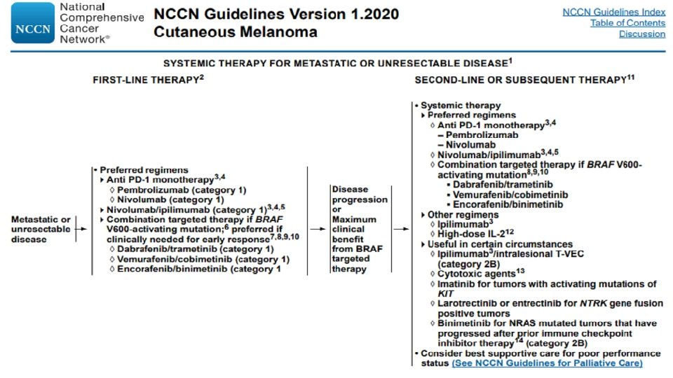

Systemic therapy for metastatic melanoma BRAF/MEK inhibitors: for BRAF V 600 mutation: • Dabrafenib/trametinib • Vemurafenib/cobimetinib • Oncorafenib/binimetinib Checkpoint inhibitors: effective regardless of BRAF mutation status • Anti PD-1: nivoluman, Pembrolizumab • Anti TCLA-4: ipilimumab Imatinib: for activating KIT mutations Cytotoxic chemotherapy

BRAF/MEK inhibitors for metastatic melanoma • BRAF/MEK inhibitor combination therapy was associated with improved response rate, PFS, and OS compared to BRAF inhibitor monotherapy, as well as a similar or better toxicity profile • If BRAF/MEK inhibitor combination is contraindicated, BRAF inhibitor monotherapy with vemurafenib or dabrafenib are recommended options, especially in patients who are not appropriate candidates for checkpoint immunotherapies Patients treated with BRAF targeted therapy as first line therapy who achieved maximum clinical benefit (but not complete remission), a switch to immune check inhibitor may be considered

Toxicities associated with targeted therapy Dermatologic: BRAF inhibitors are associated with: • Squamous cell carcinoma • Extreme photosensitivity Occur much less often with BRAF/MEK inhibitors

Toxicities associated with targeted therapy Pyrexia: • 55% of BRAF/MEK combination patients • 20% of BRAF inhibitors monotherapy patients • Episodic • The onset is often 2 to 4 weeks after start of treatment • Median duration 9 days • May be associated with chills, night seats, dehydration, electrolyte abnormalities, and hypotension

Toxicities associated with targeted therapy Pyrexia: • Stopping or holding BRAF/MEK inhibitors will often interrupt the episode • Treatment can be resumed with full dose upon cessation of pyrexia and pyrexia related symptoms • Upon re-exposure to BRAF/MEK inhibitors repeat pyrexia events can occur but grade> 3 events are uncommon (21%) • If prolonged or severe pyrexia not responsive to BRAF/MEK inhibitors discontinuation, low dose steroid (prednisolone 10 mg) can be used • Antipyretics as needed and increase fluid intake

Chekpoint inhibitors for metastatic melanoma • Anti PD-1: nivolumab, Pembrolizumab • Anti TCLA-4: ipilimumab

Selection of subsequent systemic therapy Factors affecting selection of subsequent systemic therapy: • Response and toxicities on prior therapies • Rate of progression of the underling disease (symptomatic or not) • Presence or absence of CNS progression • Performance status • Patient preference and compliance

Selection of subsequent systemic therapy • Anti PD-1 and anti TCLA-4 are not considered the same class of agent because they target different molecules • Therefor patient who received first line ipilimumab, subsequent treatment with anti PD-1 therapy is a recommended option, and vice versa • Patient who experience disease control (CR, PR, SD) and have no residual toxicity, but subsequently experience disease progression/relapse > 3 months after treatment discontinuation, reinduction with the same agent or same class of agent may be considered

Metastatic melanoma with activating KIT mutations • Mucosal melanoma • Acral melanoma • Chronically skin damaged melanoma

Metastatic Melanoma: Cytotoxic Therapy • Dacarbazin • Paclitaxel/carboplatin • Paclitaxel • Albumin bound paclitaxel • Temozolamide • Cisplatin/vinblastine/dacarbazin

Metastatic melanoma with brain metastases • Priority to prevent or delay intratumoral hemorrhage, seizures, and neurologic dysfunction • Treatment is based on symptoms, number of lesions, location of lesions, presence of extracranial metastases……. • WBRT • Resection may be un option in highly selected patients • After treatment of the brain, options for manaegement of extracranial sites are the same as for patients without brain metastases • HD interlukin-2 is contraindicated: low efficacy, worsen edema • Caution: concurrent or sequential BRAF targeted therapy and radiation: potential for increased toxicity
- Slides: 24