Basic Principles of Protein Structures Proteins The Molecule

Basic Principles of Protein Structures

Proteins: The Molecule of Life Proteins: Building Blocks Proteins: Secondary Structures Proteins: Tertiary and Quartenary Structure Proteins: Geometry

Proteins: The Molecule of Life Proteins: Building Blocks Proteins: Secondary Structures Proteins: Tertiary and Quartenary Structure Proteins: Geometry

Why Proteins? Architecture: Structural proteins Cytoskeletal proteins Coat proteins Metabolism Energy and Synthesis: Catalytic enzymes Sensory and Response Locomotion Flagella, cilia, Myosin, actin Function and Role of Proteins Transport and Storage: Porins, transporters , Hemoglobin, transferrin, ferritin Defence and Immunity Regulation And Signaling: Transcription factors Growth, Development and Reproduction

The Protein Cycle Structure Function Sequence KKAVINGEQIRSISDLHQTLKK WELALPEYYGENLDALWDCLTG VEYPLVLEWRQFEQSKQLTENG AESVLQVFREAKAEGCDITI Evolution ligand

Protein Structure Diversity 1 CTF 1 TIM 1 A 1 O 1 K 3 R 1 NIK 1 AON

Protein Structure Primary structure Ala Secondary Structure Tertiary Structure Gln Glu Leu Lys Leu Ile Thr Ala Thr Sequence of Amino acids Local interactions Native protein

Proteins: The Molecule of Life Proteins: Building Blocks Proteins: Secondary Structures Proteins: Tertiary and Quartenary Structure Proteins: Geometry

Review of Acid-Base Chemistry What is an acid or a base? An acid is a material that can release a proton (or hydrogen ion, H+), and a base is a material that can donate a hydroxide ion (OH-) (Arhennius definition), or accept a proton (Lowry Bronsted definition). Note: It is important to notice that just because a compound has a hydrogen or an OH group does not mean that it can be an acid or a base!! - The hydrogen of methane (CH 4) and usually of methyl groups (-CH 3) are all strongly attached to the carbon atom - Glycerol has three OH groups (CH 2 OH – CH 2 OH) and all 3 are alcoholic groups.

Review of Acid-Base Chemistry Acid plus base makes water plus a salt: AH + BOH (HCL + Na. OH AB + H 2 O Na. Cl + H 2 O) The chemical dissociation of nitric acid is: HNO 3 (NO 3)- + H+ Which can be rewritten as: HNO 3 + H 2 O (NO 3)- + H 3 O+ acid base conjugate base acid

Review of Acid-Base Chemistry p. H is a measure of how acidic or alkaline (basic) a solution is. The p. H of a solution is the negative log of the hydrogen ion concentration. [H+] p. H p. OH [OH-] Strong base 10 -14 14 0 0 Base 10 -12 12 2 10 -2 Weak base 10 -9 9 5 10 -5 Neutral 10 -7 7 7 10 -7 Weak acid 10 -4 4 10 10 -10 Acid 10 -2 2 12 10 -12 Strong acid 0 0 14 10 -14

Review of Acid-Base Chemistry Dissociation of a weak acid: HA A- + H + Dissociation of a weak base: BOH B+ + OH- For an (acid, base) pair: Equilibrium constant:

Review of Acid-Base Chemistry Titration:

The Basic Block: Amino Acid Sidechain R H + N 8. 9 < p. Ka < 10. 8 Ca OC H H H Amino group “zwitterion” O Carboxyl group 1. 7 < p. Ka <2. 6
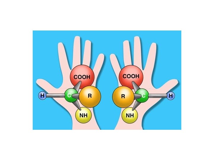

Amino Acid Chirality R R CA CA H CO N L-form H N CO D-form (CORN rule) Amino acids in proteins are in the L-form Threonine and Isoleucine have a second optical center which is also identical in all natural amino acids.

The 20 amino acids 1 -letter 3 -letter Amino acid 1 -letter 3 -letter Amino Acid A Alanine M Methionin C Cysteine N Asn Asparagine D Aspartic Acid P Proline E Glutamic Acid Q Gln Glutamine F Phenylalanine R Arginine G Glycine S Serine H Histidine T Threonin I Ile Isoleucine V Valine K Lysine W Trp Tryptophan L Leucine Y Tyrosine

Amino Acids: Usage

The 20 amino acids Hydrophobic Polar, neutral Acidic Basic

Polar Amino acids: Cysteine SG CB Names: Cys, C S CH 2 Occurrence: 1. 8 % C p. Ka sidechain: 8. 3 CA SG 1 Can form disulphide bridges in proteins CB 2 CB 1 CA 1 SG 2 CA 2

Polar Amino acids: Histidine NE 2 CD 2 CB CE 1 ND 1 CG N CH CH N C Name: His, H H Occurrence: 2. 2 % CH 2 CA C p. Ka sidechain: 6. 04

Different ionic states of Histidine H + N CH CH N H C H N CH 2 CH CH N CH C CH 2 C CH N CH 2 CH + CH N C CH 2 C C H H

Charged Amino acids: Aspartic Acid OD 1 CG OD 2 CB O- O Names: Asp, D C CH 2 CA C Occurrence: 5. 2 % p. Ka sidechain: 3. 9

Charged Amino acids: Glutamic Acid OE 1 OE 2 CG CD CB O O C Names: Glu, E CH 2 CA - C Occurrence: 6. 3 % p. Ka sidechain: 4. 25

Charged Amino acids: Lysine NZ NH 3+ CE CD CH 2 Names: Lys, K CH 2 CG CB CH 2 CA C Occurrence: 5. 8 % p. Ka sidechain: 9. 2

Charged Amino acids: Arginine NH 2+ NH 2 NH 1 NH 2 CZ CZ NE CD NE CG CB CA CH 2 Names: Arg, R Occurrence: 5. 2 % CH 2 C p. Ka sidechain: 12. 5

Unusual Amino Acids: Cyclosporin Where is the error? CH 3 http: //purefixion. com/attention/2006_03_26_archive. html

Unusual Amino Acids: Cyclosporin Correct!! http: //www. cellsignal. com/products/9973. html

Structural Bioinformatics: Proteins: The Molecule of Life Proteins: Building Blocks Proteins: Secondary Structures Proteins: Tertiary and Quartenary Structure Proteins: Geometry

The Protein: A polymer of Amino acids Peptide bond H N H Ca H H Nter N C Ca O R n+1 O Rn C Cter

The Peptide Bond Peptide bond H H N Ca N O Rn C Ca O R n+1 C The peptide bond is planar H H Ca N C Ca O Conformation “Trans” O N C Ca Conformation “Cis” Ca
 <-> N (i+4)")
Helices Cter Nter Hydrogen bonds: O (i) <-> N (i+4)
 p-helix (516)")
Helices 310 helix a-helix (413) p-helix (516)

Helices 310 helix “Thin”; 3. 0 residues /turn; ~ 4 % of all helices p-helix (516) “Fat”; 4. 2 residues /turn; instable a-helix (413) “Right”; 3. 6 residues /turn; 5. 4 Å /turn; most helices

Identify Helix Type 1. Find one hydrogen bond loop 11 10 4 9 12 2. Count number of residues (by number of C atoms in the loop). Here : 13 3 8 7 1 5 6 2 4 4 2 1 3 3. Count number of atoms in the loop (including first O and last H). Here: 13 413 helix = a-helix

The b-strand N-H---O-C Hydrogen bonds Extended chain is flat “Real b-strand is twisted”

Two types of b-sheets Parallel Anti-parallel

b-turns Type II O is down 3 O is up 2 1 3 2 4 1 The chain changes direction by 180 degrees 4

Favorable /Unfavorable Residues In Turns Turn 1 2 3 4 I Asp, Asn, Ser, Cys Pro Gly II Asp, Asn, Ser, Cys Pro Gly, Asn Gly

The b-hairpin

Structural Bioinformatics: Proteins: The Molecule of Life Proteins: Building Blocks Proteins: Secondary Structures Proteins: Tertiary and Quartenary Structure Proteins: Geometry

Protein Tertiary Structure • All a proteins • All b proteins • Alpha and beta proteins: - a/b proteins (alternating a and b) - a + b proteins
 PDB")
All-Alpha topologies • The lone helix Glucagon (hormone involved Is regulating sugar metabolism) PDB code : 1 GCN • The helix-turn-helix motif ROP: RNA-binding Protein PDB code: 1 ROP The 2 helices are twisted

All Beta Topology Beta sandwiches: Fatty acid binding protein PDB code: 1 IFB

Closed Beta Barrel PDB file: 2 POR

The Greek Key Topology Folds including the Greek key topology include 4 to 13 strands.

The Jellyroll Topology A Greek key with an extra swirl PDB code 2 BUK (coat protein of a virus)

The Beta Propellor Eight-plated propellor: Each plate is a four-stranded anti-parallel sheet PDB code 4 AAH

The Beta Helix PDB code 2 PEC

Alpha- Beta Topology The Rossman fold: Alternate beta / alpha motif Always right handed

The Horseshoe PDB code: 2 BNH

The alpha/beta barrel In a succession of alpha/beta motifs, if the first strand connects to the last, then the structure resembles a Barrel. PDB code : 1 TIM

Quaternary Structures Assemblies of Protein Chains Hemoglobin - 4 chains: 2 -a chain, 2 -b chain (Heme- four iron groups)

Structural Bioinformatics: Proteins: The Molecule of Life Proteins: Building Blocks Proteins: Secondary Structures Proteins: Tertiary and Quartenary Structure Proteins: Geometry

Protein Structure Representation CPK: hard sphere model Ball-and-stick Cartoon

Degrees of Freedom in Proteins Bond length 1 2 Dihedral angle 3 1 2 Bond angle + 4

Protein Structure: Variables Backbone: 3 angles per residue : j, f and w Sidechain: 1 to 7 angles, c; each c has 3 favored values: 60 o, -60 o, 180 o.
.")
Ramachandran Plots y y f All residues, but glycine f Glycine Acta Cryst. (2002). D 58, 768 -776

What have we learnt? • All proteins are polymers built up from 20 amino acids. • All 20 amino acids have a similar structure: they all have a main-chain, consisting of an amino group and an acidic group, attached to a central carbon, named CA; the remaining atoms form the side-chain, that can be hydrophobic, polar or charged (acid or basic). • The conformation of the backbone of amino acids is restricted, except for glycine that does not have a sidechain. • There are 3 main graphical representations of proteins: space-filling, wireframe and cartoon.

What have we learnt? • There are 3 major types of secondary structures: a-helices, b-sheets and b-turns. • Most helices are a-helices, stabilized through a network of CO (i) --- HN (i+4) hydrogen bonds • There are two types of b-sheets: parallel and anti-parallel • b-turns correspond to 180 change in the backbone direction.

What have we learnt? • There are three main classes of proteins: all Alpha, all Beta and Alpha + Beta. The latter can be divided in two, considering the alternating alpha/beta proteins as defining their own class. • Bundles are common alpha-proteins • Common beta folds include the greek key and the sandwiches. Immunoglobulins adopt a beta fold. • The Rossman fold (alternating alpha/beta) is a common motif in proteins. It is found in the horseshoe, as well as in the TIM fold.
- Slides: 61