Basic Implant Surgery Richard Palmer Paul Palmer and

Basic Implant Surgery Richard Palmer, Paul Palmer, and Peter Floyd

Introduction • Successful implant surgery required good planning (appreciation of the restorative requirement and visualization of the desired end result of the treatment), and meticulous technique (adequate surgical training and experience of the selected implant system).

Pre-requisites before surgery 1/ The patient medical history should be checked with particular relevance to: - poorly controlled diabetes, blood dyscrasias, medications, irradiation to jaws, mucosal diseases, substances abuse including tobacco and alcohol, and need for antibiotics. 2/ The patient should understand the procedure and be warned of any complications.

3/ The diagnostic set-up, surgical stent and relevant radiographs should be available. 4/ The surgeon should have a clear idea of the number, size, and planned location of the implant.

Operative requirements Ø Good operating light, foot adjusted dental chair, sterile drapes, gowns, gloves, high volume suction, and suction tube. Ø A high speed surgical drill unit ( upto 3000 rpm) and low drilling speeds (down to 10 rpm), with irrigation system for keeping bone cool during the drilling process.

Ø Appropriate surgical instrumentation for the implant system being used. Ø Appropriate number and design of implants planned plus an adequate stock to meet unexpected eventualities during surgery. Ø A trained assistance, and a third person to act as a runner between the sterile and non-sterile environment.

Anaesthesia and Analgesia • Most implant surgery can be carried out under local anaesthesia, although sometimes we need sedation or general anaesthesia. In complex cases (2 -3 hrs) its essential to use regional block anaesthesia ( infraorbital, palatal. . . etc), and supplemented by local infiltration to improve anaesthesia and hemorrhage control. • Its good idea to give analgesics immediately before surgery

Sterile technique • Every effort should be made to conduct implant surgery under sterile operating condition. • Chlorhexidine 0. 2% is used as a pre-operative mouth wash and skin preparation cicumorally.

Surgical Techniques for Implant Installation • Anatomical considerations • Flap design • Surgical preparation of the bone • Implant placement • Implant insertion

Anatomical Considerations • The anatomic structures that will affect implant placement include: o In maxilla : air sinuses, nasopalatine canal, floor of the nose and nasal spine, palatine and pterygoid vessels. o In mandible : sublingual vessels, mental nerve, inferior dental nerve, incisive branch of IDN, genial tubercles.

o Teeth : position, length and angulations of roots adjacent to implant site. o Available bone : ridge morphology, bone density (cortical, medullary), localized deformities (tooth sockets, residual cysts or granuloma).

Flap Design • A full thickness mucoperiosteal flap is elevated to completely exposed the alveolar ridge to determine its shape and whether any concavities are present. • The flap has vertical labial relieving incisions, a crestal incision mesiodistally, and has included elevation of the papilla to give good access to the narrow edentulous space……. fig 1 a, b
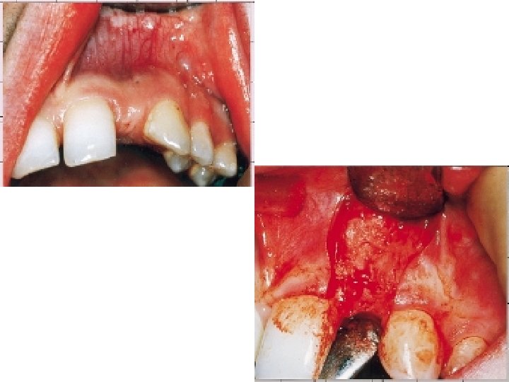

• However some surgeons prefer to avoid raising the papilla in case future aesthetics are compromised. • Care should be taken to ensure that incision lines are not placed over structures such as mental nerve and palatine arteries.

Surgical Preparation of the Bone • Heating the bone above 47 c should be avoided as this will causes bone cells death and prevent osseointegration. This problem may be avoided by : v Using sharp drills, with incremental drilling procedure with increasing diameter drills, and frequent withdrawal of the drill to allow bone swarf to clear particularly in dense/hard bone.
 and excessive pressure on the")
v Avoidance of excessive speed(no more than 3000 rpm) and excessive pressure on the drill. v Using copious sterile saline irrigation. The drill can be cooled by spraying the external surface of the drill, however some systems use internally irrigated drills

A clear blow-down stent has been placed over the adjacent teeth and provides the surgeon with the position of the labial face and gingival margin of the tooth which is to be replaced. The initial preparation of the implant site is with a round bur which readily penetrates the cortex, and this is followed by a twist drill (illustrated) which determines the initial angulation and depth of the implant Steps of preparation: -

A guide or indicator post is placed in the site to check on the position and angulation of the implant. Small adjustments can be made at this stage

The site is enlarged by a pilot drill. This has a noncutting end of the same diameter as the previous twist drill and a wider cutting portion. It should therefore maintain the line of the previous drill

The site is enlarged with the final twist drill which is slightly narrower than the implant itself. The drill has marks on it corresponding to the available implant lengths. All drilling to this stage is carried out at moderate speeds (about 1, 500 rpm) with copious saline irrigation

Submerge implant system The surface of the bone is countersunk to accept the head of the implant. The depth of countersinking is determined by the need to sink the head of the implant to allow a good emergence profile of the restoration. The head of the implant should therefore be about 2 to 3 mm apical to the labial cementenamel junction on the adjacent teeth or apical to the planned gingival margin determined by the stent

The depth of the implant site is verified with a measuring guage

Implant placement • Based on a properly designed stent produced from the diagnostic wax-up, an implant should be placed such that : 1. Its within the bone along its entire length, limited areas of bone defects(dehiscence or fenestrations) may be acceptable, but larger ones may required augmentation.

2. It does not damage adjacent structures such as teeth, nerves, nasal or sinus cavity. Its acceptable to engage the nasal or sinus floor with small degree of penetration(eg 1 -2 mm). An adequate safety margin of about 2 mm above the inferior dental canal is recommended. 3. Its located directly apical to the tooth its replacing and not in an embrasure space.

4. The angulation of the implant is consistent with the design of the restoration, particularly with screw retained restoration. Multiple implants are placed in a fairly parallel arrangement to facilitate seating of the restoration. However most implant systems allow convergence/divergence of upto 30 degrees without the use of angled/customized abutements
 when used to replace a")
5. The top of a standard diameter implant(4 mm) when used to replace a single upper incisor tooth, should be 2 -3 mm apical to the labial cementenamel junction of the adjacent natural tooth, to allow good emergency profile of the prosthesis. This is often achieved by countersinking the head of the implant

6. The implant should be immobile at placement. A loose implant at this stage will fail to osseointegrate 7. Adequate bone, about 3 mm between adjacent implants, and 1 -1. 5 mm between implant and adjacent tooth.

Insertion Technique • In general the final bone preparation site diameter is slightly smaller than the implant, it depend on bone quality or density; o In poor quality bone the site can be made smaller to produce compression of the surrounding bone to improve the initial stability.

o In dense bone the site has to be more closely match the size of the implant. o In bone with poor medullary quality, where initial stability is difficult to achieved, the implant is secured at each end in cortical bone (bicortical stabilization)

• Screw shaped implants are either : q Self tapped into the prepared site q Or inserted following tapping of the bone with a screw tap

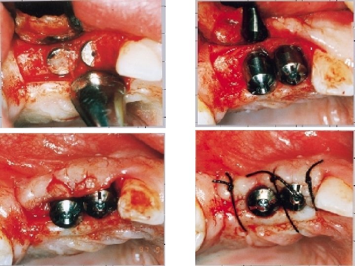
• A self tapping implant is inserted into the prepared site at slow speed and with irrigation • An implant is being driven into the site. It is connected to a slowly rotating handpiece by an implant mount and handpiece connector

• The drilling sequence has been completed and a tap is being used to cut a thread in the bone. The colored marks refer to the length of standard implants • The solid threaded implant is being screwed into the site using a hand ratchet. This is done slowly with irrigation

• cylindrical implants are either pushed or knocked into place. • following placement the head and inner screw thread of the implant is protected with a cover or closure screw.

The mucoperiosteal flaps are carefully closed with multiple sutures either to: Bury the implant completely. Or around the neck of the implant in non-submerged systems

Post-operative care • After surgery patients should be warned to expect : 1. Swelling, bruising, and discomfort, which can be controlled by oral analgesics and ice packs. 2. A transitory disturbance in sensation, if surgery has been closed to a nerve.

• They should be advised : ü Not to wear a dentures over the surgical area for at least 1 wk to void loading the implant and disrupting the suture. ü To keep the area clean by using Chlorhexidine mw 0. 2% for one min twice a day. ü Not to smoke, which will compromises healing of soft tissue and bone. Ideally patients should stop smoking for some wks before surgery and for as long as possible thereafter.

Ø The original protocols recommended an antibiotic such as amoxicillin 250 mg 8 hourly for 5 -7 days. Ø Alternative regimes include administration of 3 gm amoxicillin 1 hr before surgery or 500 mg every 8 hrs for 48 hr. Ø A provisional bridges have an advantages in that they do not load the implant sites, providing some allowance has been made to accommodate some swelling.

Ø All patients should be seen after 1 wk for review, suture removal and adjustment and refitting of the denture with adequate soft linings. Ø In some cases the tissues will not be able to accept a denture for 2 wks, particularly in edentulous mandible , and with surgical flap designs which affect the sulcus shape.
 • In non-submerge systems a second surgical")
Surgery for abutment connection (second stage surgery) • In non-submerge systems a second surgical stage is not required and abutments are simply exchanged for the closure screw. • In general implants placed in the mandible are exposed and load earlier than those in the maxilla (around 3 months compared with 6 months).

• Exposure of the implant at stage 2 surgery can be achieved with minimal flap reflection, or making small incision just to allow removal of the cover screw and attachment of the healing abutment, which has to be about 1 mm above the mucosa

• In some cases bone grows over the cover screw and this necessitates greater soft tissue reflection to allow bone removal with hand instruments or burs prior to abutment connection. • Its important to check that the abutments are fully seated on the implant. This can be achieved during surgical exposure by direct vision and radiographically at later stages.

• Soft tissue handling at abutment connection surgery should aim to preserve keratinized and attached mucosa on the labial and lingual aspects. • The position of the crestal exposing incision should serve to achieve this goal. e. g, keratinized tissue overlying the implant or towards the palatal aspect can be repositioned to the buccal aspect….

• The patient should expect some post-operative discomfort and advised to take analgesics. They should use Chlorhexidine mouth wash until adequate oral hygiene can be re-established. • The soft tissue healing will allow the restorative phase to commence in 3 -4 wk.

conclusion § Implant surgery is highly technique sensitive and required adequate training and an understanding of the restorative requirement of the proposed treatment. § However, control of these factors can produce a highly predictable, aesthetic and long-lasting result.

§ In instances where there is insufficient bone and/or soft tissue to allow successful implant placement and subsequent restoration, grafting and augmentation procedure will be required, and these are dealt with in next part.

THANKS
- Slides: 47