
Basic Hematology Rasoul Azarfarin MD FACC Professor of
Basic Hematology Rasoul Azarfarin MD, FACC Professor of Anesthesiology Fellowship of Cardiac Anesthesia Rajaie Cardiovascular Medical & Research Center
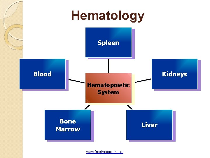
Hematology Spleen Blood Kidneys Hematopoietic System Bone Marrow Liver www. freelivedoctor. com
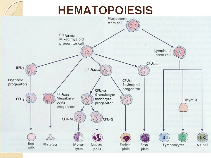
HEMATOPOIESIS
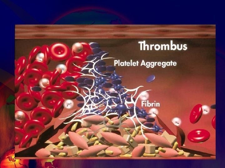
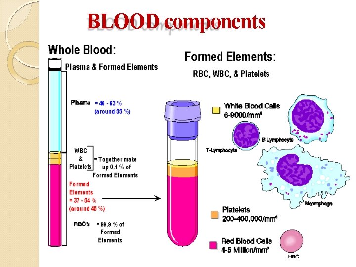
BLOOD components
COMPLETE BLOOD PICTURE

RED BLOOD CELLS also known as erythrocytes. mature red blood cells are flexible biconcave disks. has a disk diameter of 6– 8 µm and a thickness of 2 µm. Male: 4. 50– 6. 50 x 106/µL Female: 3. 80– 4. 80 x 106/µL Life span: - 120 days.
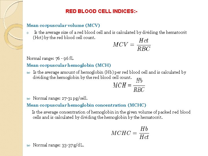
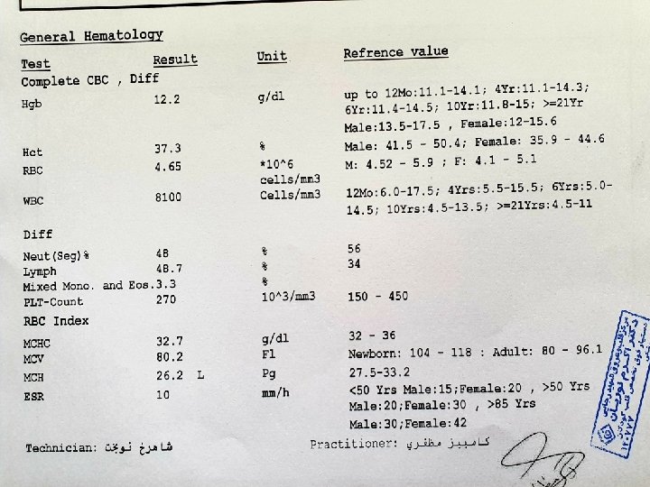
RED BLOOD CELL INDICES: Mean corpuscular volume (MCV) o Is the average size of a red blood cell and is calculated by dividing the hematocrit (Hct) by the red blood cell count. Normal range: 76 - 96 f. L Mean corpuscular hemoglobin (MCH) Is the average amount of hemoglobin (Hb) per red blood cell and is calculated by dividing the hemoglobin by the red blood cell count. Normal range: 27 -31 pg/cell. Mean corpuscular hemoglobin concentration (MCHC) Is the average concentration of hemoglobin in the given volume of packed red blood cells and is calculated by dividing the hemoglobin by the hematocrit. Normal range: 33 -37 g/d. L.
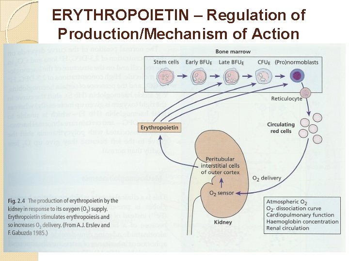
ERYTHROPOIETIN – Regulation of Production/Mechanism of Action
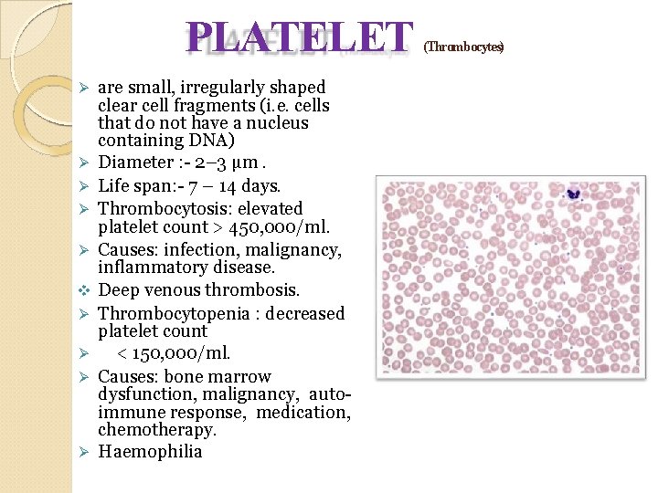
PLATELET are small, irregularly shaped clear cell fragments (i. e. cells that do not have a nucleus containing DNA) Diameter : - 2– 3 µm. Life span: - 7 – 14 days. Thrombocytosis: elevated platelet count > 450, 000/ml. Causes: infection, malignancy, inflammatory disease. Deep venous thrombosis. Thrombocytopenia : decreased platelet count < 150, 000/ml. Causes: bone marrow dysfunction, malignancy, autoimmune response, medication, chemotherapy. Haemophilia (Thrombocytes)
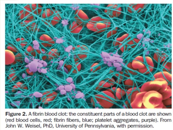
HEMOGLOBIN Is the iron-containing oxygen-transport metalloprotein in the red blood cells. Male: - 13. 0 – 17. 0 g/d. L Female: - 12. 0 – 15. 0 g/d. L Childerns: - 11. 5 – 15. 5 g/d. L Increased Hemoglobin : -Polycythemia Decreased Hemoglobin : - Anemia Hematocrit: is the percentage (%) of the concentration of red blood cells in blood. Male: - 42. 0 – 52. 0%, Female : -37. 0 – 47. 0 %
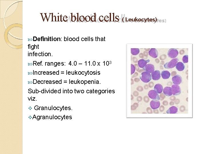
White blood cells ( Leukocytes) Definition: blood cells that fight infection. Ref. ranges: 4. 0 – 11. 0 x 103 Increased = leukocytosis Decreased = leukopenia. Sub-divided into two categories viz. Granulocytes. Agranulocytes
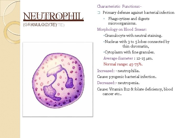
Characteristic Functions: - NEUTROPHIL (GRANULOCYTE) � Primary defense against bacterial infection ◦ Phagocytizes and digests microorganisms. Morphology on Blood Smear: ◦Granulocyte with neutral staining. ◦Nucleus with 3 to 5 lobes connected by thin chromatin, ◦Cytoplasm with fine granules. Average diameter : 12 -15 µm. Normal range: 45 -75%. Increased: - neutrophilia. Cause: pyogenic bacterial infection. Decreased: - neutropenia. Cause: Vitamin B 12 & folate deficiency, blood cancer etc. .
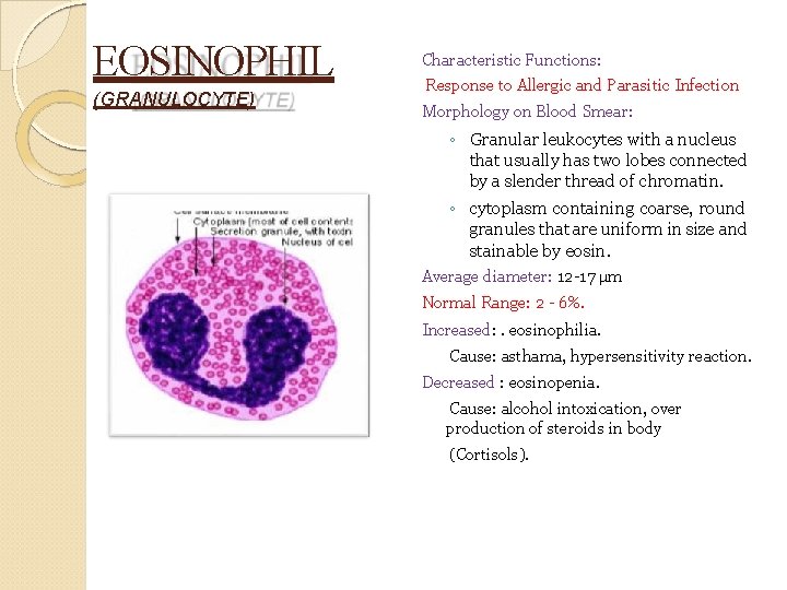
EOSINOPHIL (GRANULOCYTE) Characteristic Functions: Response to Allergic and Parasitic Infection Morphology on Blood Smear: ◦ Granular leukocytes with a nucleus that usually has two lobes connected by a slender thread of chromatin. ◦ cytoplasm containing coarse, round granules that are uniform in size and stainable by eosin. Average diameter: 12 -17 µm Normal Range: 2 - 6%. Increased: . eosinophilia. Cause: asthama, hypersensitivity reaction. Decreased : eosinopenia. Cause: alcohol intoxication, over production of steroids in body (Cortisols).
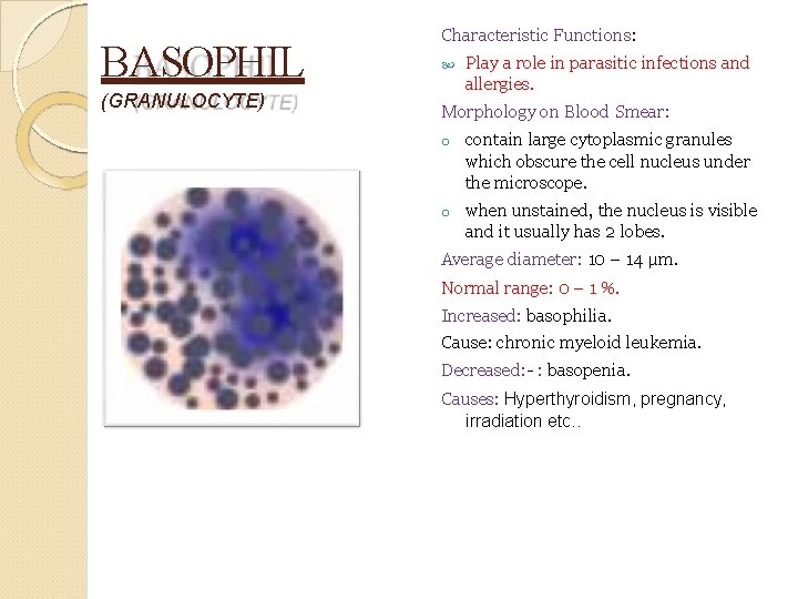
BASOPHIL (GRANULOCYTE) Characteristic Functions: Play a role in parasitic infections and allergies. Morphology on Blood Smear: o contain large cytoplasmic granules which obscure the cell nucleus under the microscope. o when unstained, the nucleus is visible and it usually has 2 lobes. Average diameter: 10 – 14 µm. Normal range: 0 – 1 %. Increased: basophilia. Cause: chronic myeloid leukemia. Decreased: - : basopenia. Causes: Hyperthyroidism, pregnancy, irradiation etc. .
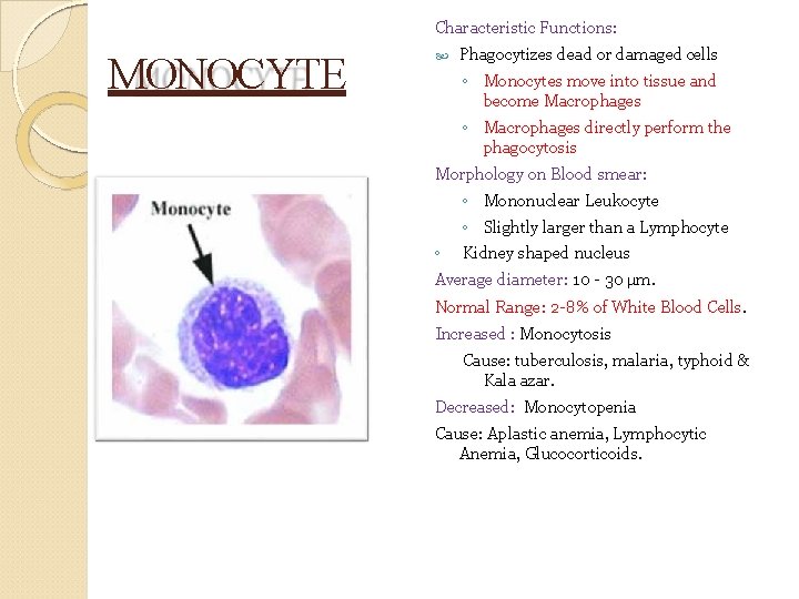
Characteristic Functions: MONOCYTE Phagocytizes dead or damaged cells ◦ Monocytes move into tissue and become Macrophages ◦ Macrophages directly perform the phagocytosis Morphology on Blood smear: ◦ Mononuclear Leukocyte ◦ ◦ Slightly larger than a Lymphocyte Kidney shaped nucleus Average diameter: 10 - 30 µm. Normal Range: 2 -8% of White Blood Cells. Increased : Monocytosis Cause: tuberculosis, malaria, typhoid & Kala azar. Decreased: Monocytopenia Cause: Aplastic anemia, Lymphocytic Anemia, Glucocorticoids.
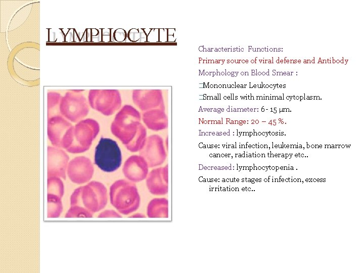
LYMPHOCYTE Characteristic Functions: Primary source of viral defense and Antibody Morphology on Blood Smear : �Mononuclear Leukocytes �Small cells with minimal cytoplasm. Average diameter: 6 - 15 µm. Normal Range: 20 – 45 %. Increased : lymphocytosis. Cause: viral infection, leukemia, bone marrow cancer, radiation therapy etc. . Decreased: lymphocytopenia. Cause: acute stages of infection, excess irritation etc. .
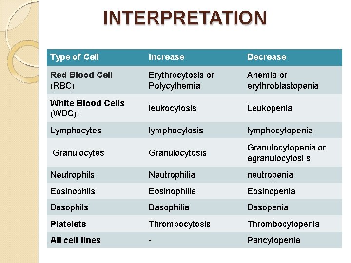
INTERPRETATION Type of Cell Increase Decrease Red Blood Cell (RBC) Erythrocytosis or Polycythemia Anemia or erythroblastopenia White Blood Cells (WBC): leukocytosis Leukopenia Lymphocytes lymphocytosis lymphocytopenia Granulocytes Granulocytosis Granulocytopenia or agranulocytosi s Neutrophilia neutropenia Eosinophils Eosinophilia Eosinopenia Basophils Basophilia Basopenia Platelets Thrombocytosis Thrombocytopenia All cell lines - Pancytopenia
Definitions 1. Define “CBC” 2. Define and explain red cell indices including derived values such as MCV, MCHC and MCH 3. Define and explain erythrocyte sedimentation rate 4. Describe the main cell types observed in peripheral blood. 5. Describe what is meant by the term “differential count”. 6. Describe the distribution & morphology of cells in certain common “Blood Pictures”
Clinical Diagnostic Laboratories offer: 1. “CBC” 2. “CBC plus Diff. ” [CBC + Differential count] 3. Above “Reticulocyte count” What do these terms mean, and how are they used?
CBC 1. White Cell count (leukocyte count) 2. Platelet count 3. Red cell count, hemoglobin concentration and red cell indices
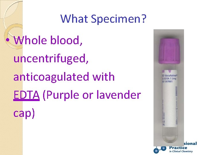
What Specimen? • Whole blood, uncentrifuged, anticoagulated with EDTA (Purple or lavender cap)
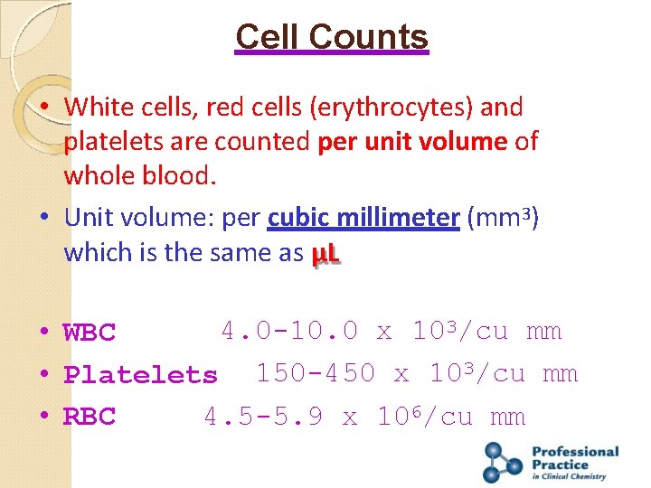
Cell Counts • White cells, red cells (erythrocytes) and platelets are counted per unit volume of whole blood. • Unit volume: per cubic millimeter (mm 3) which is the same as µL 4. 0 -10. 0 x 103/cu mm • WBC • Platelets 150 -450 x 103/cu mm • RBC 4. 5 -5. 9 x 10⁶/cu mm
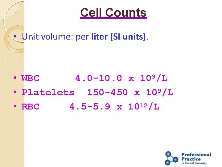
Cell Counts • Unit volume: per liter (SI units). • WBC 4. 0 -10. 0 x 109/L • Platelets 150 -450 x 109/L • RBC 4. 5 -5. 9 x 10¹²/L
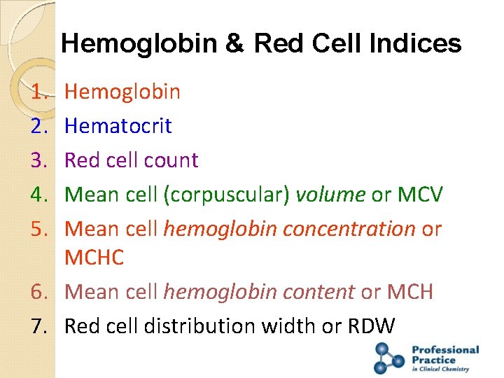
Hemoglobin & Red Cell Indices 1. 2. 3. 4. 5. Hemoglobin Hematocrit Red cell count Mean cell (corpuscular) volume or MCV Mean cell hemoglobin concentration or MCHC 6. Mean cell hemoglobin content or MCH 7. Red cell distribution width or RDW
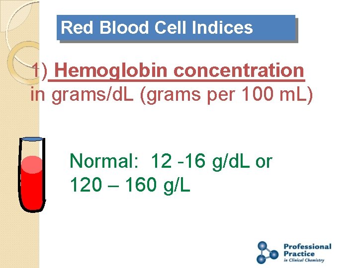
Red Blood Cell Indices 1) Hemoglobin concentration in grams/d. L (grams per 100 m. L) Normal: 12 -16 g/d. L or 120 – 160 g/L
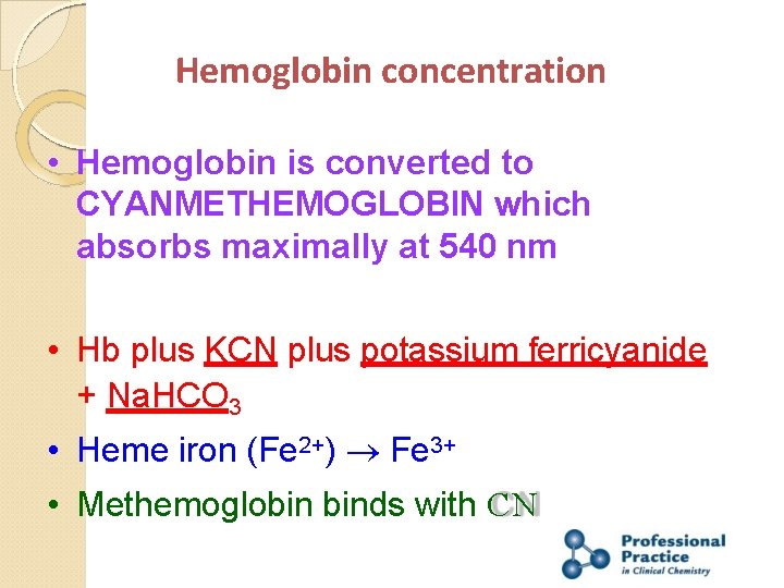
Hemoglobin concentration • Hemoglobin is converted to CYANMETHEMOGLOBIN which absorbs maximally at 540 nm • Hb plus KCN plus potassium ferricyanide + Na. HCO 3 • Heme iron (Fe 2+) Fe 3+ • Methemoglobin binds with CN
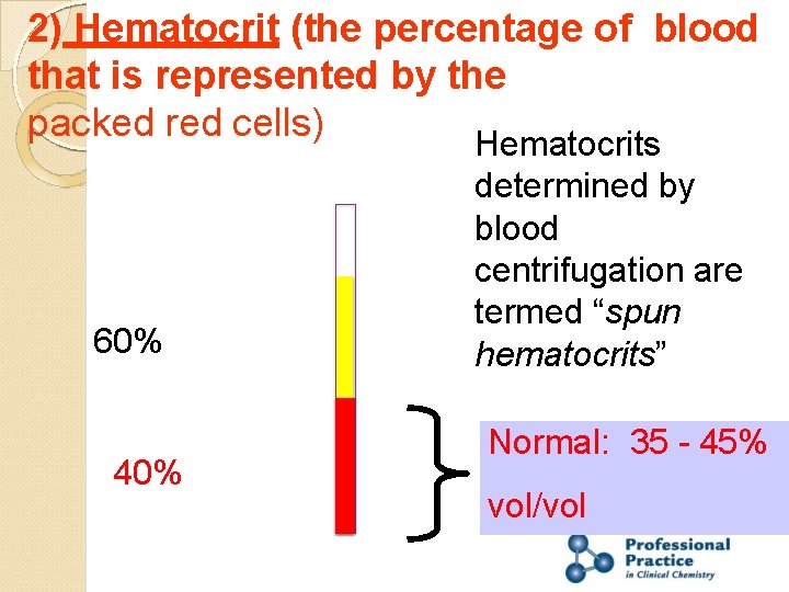
2) Hematocrit (the percentage of blood that is represented by the packed red cells) Hematocrits 60% 40% determined by blood centrifugation are termed “spun hematocrits” Normal: 35 - 45% vol/vol
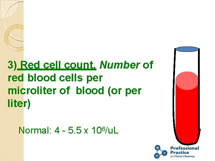
3) Red cell count. Number of red blood cells per microliter of blood (or per liter) Normal: 4 - 5. 5 x 10⁶/u. L
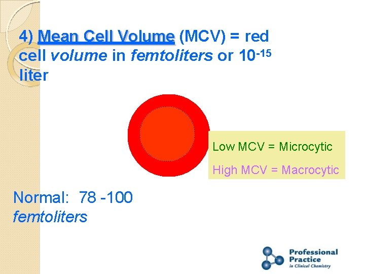
4) Mean Cell Volume (MCV) = red cell volume in femtoliters or 10 -15 liter Low MCV = Microcytic High MCV = Macrocytic Normal: 78 -100 femtoliters
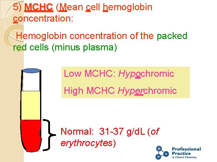
5) MCHC (Mean cell hemoglobin concentration: Hemoglobin concentration of the packed red cells (minus plasma) Low MCHC: Hypochromic High MCHC Hyperchromic Normal: 31 -37 g/d. L (of erythrocytes)
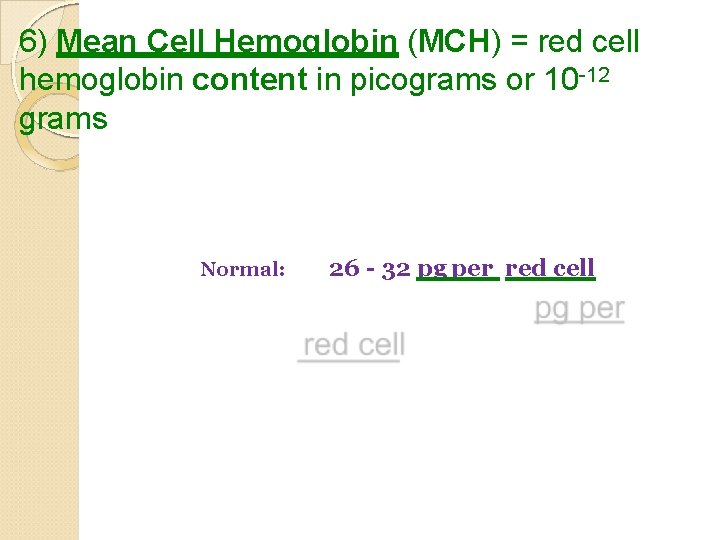
6) Mean Cell Hemoglobin (MCH) = red cell hemoglobin content in picograms or 10 -12 grams Normal: 26 - 32 pg per red cell
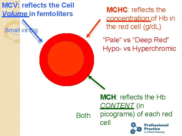
MCV: reflects the Cell Volume in femtoliters MCHC: reflects the concentration of Hb in the red cell (g/d. L) Small vs Big “Pale” vs “Deep Red” Hypo- vs Hyperchromic Both MCH: reflects the Hb CONTENT (in picograms) of each red cell
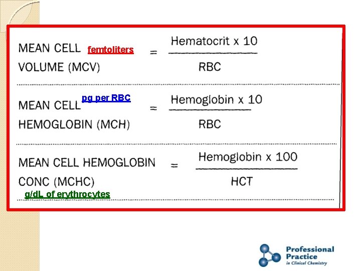
femtoliters pg per RBC g/d. L of erythrocytes
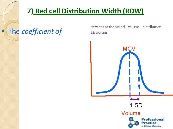
7) Red cell Distribution Width (RDW) • The coefficient of variation of the red cell volume - distribution histogram MCV 1 SD Volume
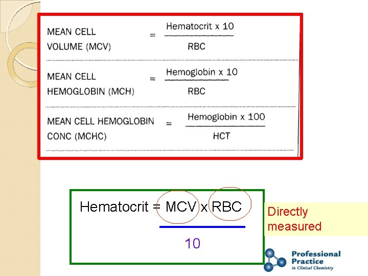
Hematocrit = MCV x RBC 10 Directly measured
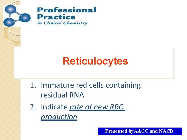
Reticulocytes 1. Immature red cells containing residual RNA 2. Indicate rate of new RBC production Presented by AACC and NACB
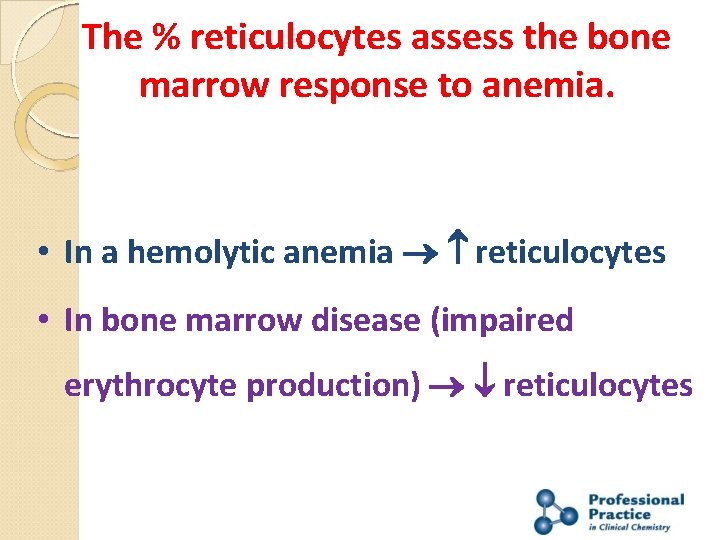
The % reticulocytes assess the bone marrow response to anemia. • In a hemolytic anemia reticulocytes • In bone marrow disease (impaired erythrocyte production) reticulocytes
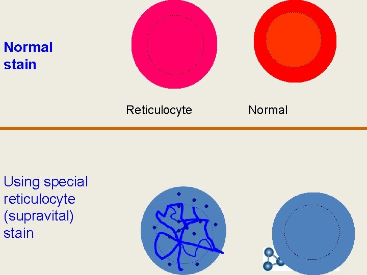
Normal stain Reticulocyte Using special reticulocyte (supravital) stain Normal
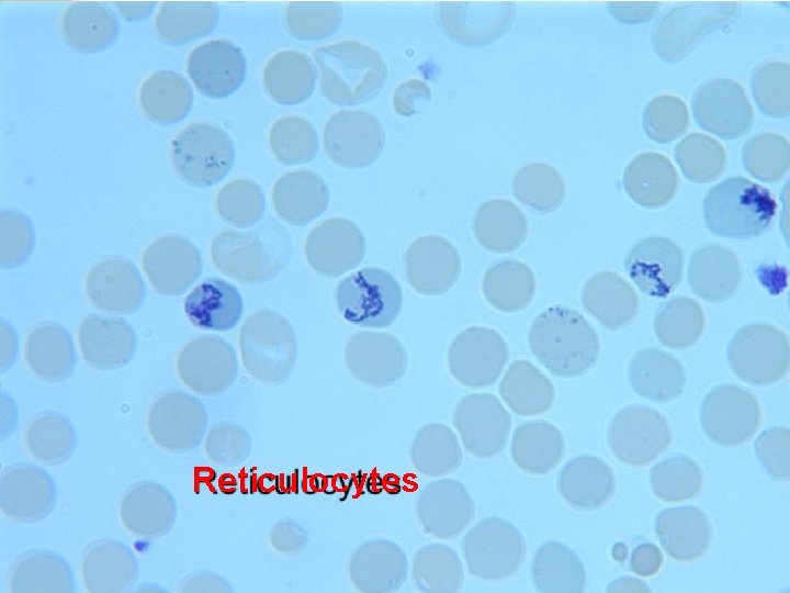
Reticulocytes
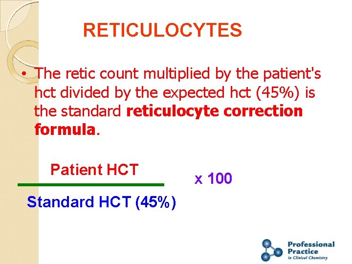
RETICULOCYTES • The retic count multiplied by the patient's hct divided by the expected hct (45%) is the standard reticulocyte correction formula. Patient HCT Standard HCT (45%) x 100
Thank You
- Slides: 44