Basic Embryology Embryology Definition the study of the

Basic Embryology

Embryology Definition: the study of the origin and development of an organism n Prenatal period: before birth n ¨ 38 weeks from conception to birth (average) “fetal” age ¨ Gynecologic timing has been from LMP therefore refers to 40 weeks “gestational” age Date of conception has been difficult to time n LMP is on average two weeks before ovulation n
 division: n “Embryonic” period: first 8 weeks ¨ All n major organs")
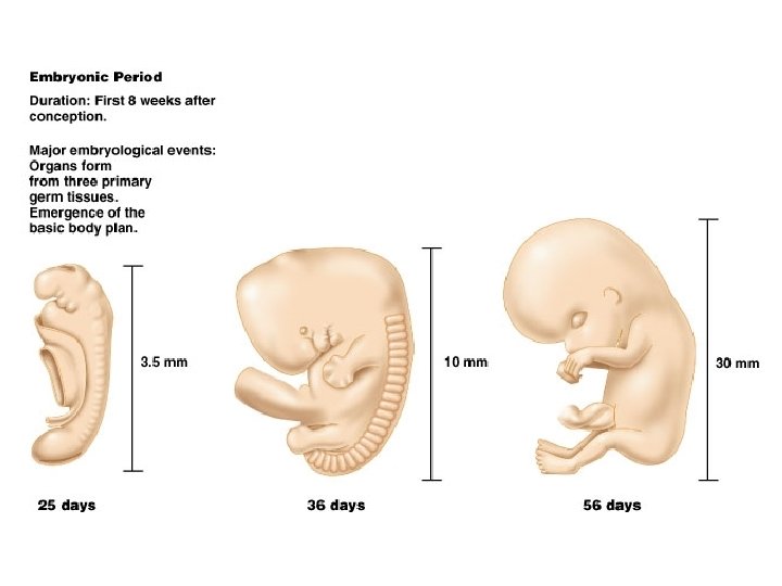
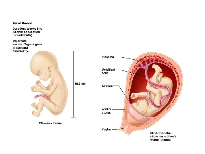
Traditional (artificial) division: n “Embryonic” period: first 8 weeks ¨ All n major organs formed “Fetal” period: remaining 30 weeks ¨ Organs grow larger and become more complex

Fertilization to Implantation

Ovulation: egg released into the peritoneal cavity n Travels down fallopian tube in which fertilization occurs n At conception in fallopian tube, maternal and paternal genetic material join to form a new human life (zygote) n Cell division occurs with travel down the tube and into the uterus n Conception (biology) or fertilisation, the fusion of gametes to produce a new organism of the same species (Wikipedia)

Week 1 post conception n n Zygote divides repeatedly moving down tube toward uterus (cleavage) The daughter cells are called blastomeres Morula: the solid cluster of 12 -16 blastomeres at about 72 hours Day 4: late 60 cell morula enters uterus, taking up fluid becoming blastocyst
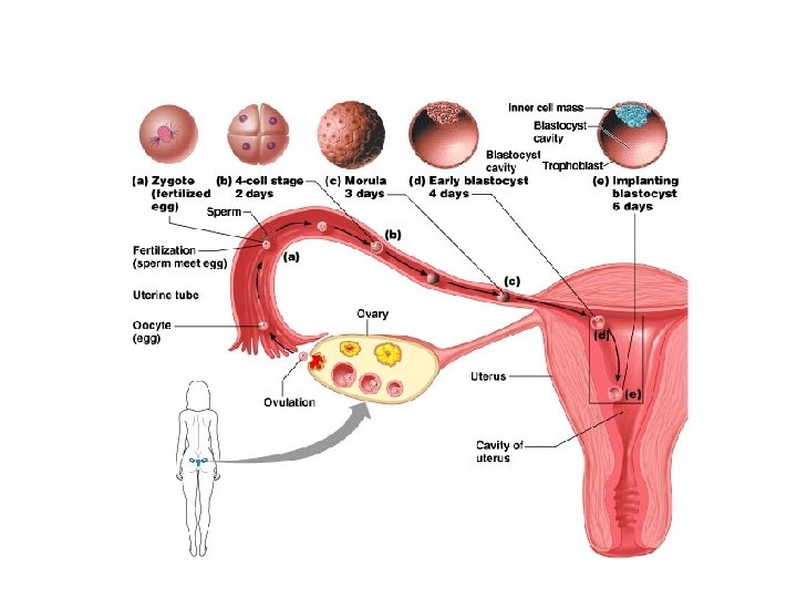

Blastocyst stage n Two distinct types of cells _____inner cell mass ______trophoblast ¨ Inner cell mass: forms the embryo ¨ Trophoblast: layer of cells surrounding the cavity which helps form the placenta n n Floats for about 3 days Implantation on about day 6 post conception Trophoblast erodes uterine wall ¨ Takes 1 week to complete ¨ n If inner cell mass of a single blastocyst divides: monozygotic (identical) twins

Week 2 n n Inner cell mass divides into epiblast and hypoblast 2 fluid filled sacs Amniotic sac from epiblast ¨ Yolk sac from hypoblast ¨ n Bilaminar embryonic disc: area of contact (gives rise to the whole body)

Blastocyst stage n Two distinct types of cells _____inner cell mass ______trophoblast ¨ Inner cell mass: forms the embryo ¨ Trophoblast: layer of cells surrounding the cavity which helps form the placenta n n Floats for about 3 days Implantation on about day 6 post conception Trophoblast erodes uterine wall ¨ Takes 1 week to complete ¨ n If inner cell mass of a single blastocyst divides: monozygotic (identical) twins

Week 3 n n Bilaminar to trilaminar disc Three primary “germ” layers: all body tissues develop from these n n n Ectoderm Endoderm Mesoderm
 on dorsal")
Formation of the 3 “germ” layers n n n Primitive streak (groove) on dorsal surface of epiblast Grastrulation: invagination of epiblast cells Days 14 -15: they replace hypoblast becoming endoderm Day 16: mesoderm (a new third layer) formed in between Epiblast cells remaining on surface: ectoderm

The three “germ” tissues n n n “Germ” as in germinate, not germs Early specialization of cells Are precursors Ectoderm and endoderm are epithelial tissue (form sheets of tissue) Mesoderm is a mesenchyme tissue ¨ Mesenchyme cells are star shaped and do not attach to one another, therefore migrate freely

Notochord n n Days 16 -18 Primitive node epiblast cells invaginate and migrate anteriorly with some endoderm cells Rod defining the body axis is formed Future site of the vertebral column
")
Neurulation n Notochord signals overlying ectoderm Formation begins of spinal cord and brain (neurulation) Neural plate to neural groove to neural tube: pinched off into body

n n n Closure of neural tube: begins at end of week 3; complete by end of week 4 (folic acid important for this step) Extends cranially (eventually brain) and caudally (spinal cord) Neural crest, lateral ectodermal cells, pulled along and form sensory nerve cells and other structures

n Mesoderm begins to differentiate ¨ Lateral to notochord, week 3 ¨ Extends cranially and caudally crown to rump) n (from head to tail or Division of mesoderm into three regions ¨ Somites: 40 pairs of body segments (repeating units, like building blocks) by end week 4 ¨ Intermediate mesoderm: just lateral to somites ¨ Lateral plate: splits to form coelom (“cavity”)

Divisions of the mesodermal lateral plate Somatic mesoderm: apposed to the ectoderm n Splanchnic mesoderm: apposed to the endoderm n Coelom in between will become the serous cavities of the ventral body cavity: n ¨ Peritoneal ¨ Pericardial ¨ Pleural

Folding begins at week 4 (main difference between the 3 week embryo and the adult body is that the embryo is still a flat disc)

24 day embryro; protrudes into amniotic cavity

Day 23, beginning to fold Lateral folds will join ventrally
 Simplified cross section through abdomen")
Cylindrical human body plan, day 28 (about ½ cm) Simplified cross section through abdomen of an adult (essentially the same as above)

Major derivatives of the embryonic germ layers

29 day embryo (this is when the heart starts pumping, about 4 weeks or 1 month, ½ cm size)
 month 3 late 5 th month (about 19 cm)")
3 month fetus (6 cm) month 3 late 5 th month (about 19 cm) month 5

By 8 weeks, about 2 months, all major organs are in place in at least a rudimentary form; this is why drugs early in pregnancy are so important to avoid – many cause birth defects; baby is a little over 1” long (below right)

- Slides: 29