BASIC ECG NITMED TUTORIALS INC GOALS OF THE

BASIC ECG NITMED TUTORIALS INC

GOALS OF THE LECTURE • At the end of this lecture, the students should be able to ; 1. Understand basic definitions and principle 2. Determine the heart rate 3. Identify common ECG abnormalities

DEFINITIONS • Electrocardiography • Electrocardiogram • Electrocardiograph

CARDIAC ELECTRICAL SYSTEM 1. Sinoatrial Node – the Pacemaker 2. Internodal fibres & intraatrial pathway 3. Atrioventricular node 4. Bundle of His 5. Bundle branches 6. Fascicles 7. Purkinje system

The Pacemaker • The Sinoatrial Node is the Pacemaker. • First described in 1907 in the countryside of Kent (UK) by Arthur Keith and his laboratory assistance; Martin Flack (a young medical student). • As such it s also called Keith –Flack Node • SAN has P cells with special property: automaticity

INTRACARDIAC ELECTRICAL SYSTEM

Ionic Basis of Cardiac Electrical Impulses • Ionic movement across the cell membrane • Major ions ; Sodium, Potassium • Cycles of depolarisation and repolarisation

THE ELECTROCARDIOGRAM PAPER

ECG Denotations • • • P wave QRS complex T wave PR interval QT interval RR interval ST segment J junction U wave
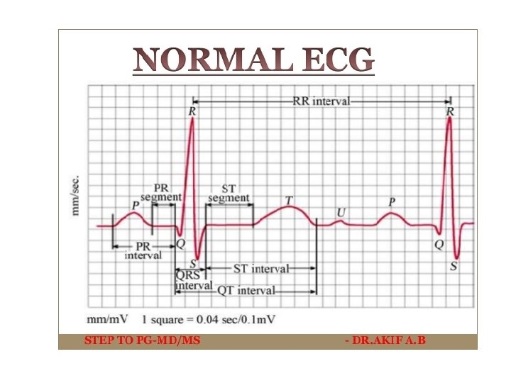

• • • P wave – 0. 12 s QRS wave – 0. 1 -0. 12 s PR interval – 0. 12 -0. 2 s QT interval : depends on age and sex RR interval : 0. 6 -1 s T wave – 0. 1 -0. 25 s
")
ELECTROCARDIOGRAPH (ECG MACHINE)

ELECTRODES • 10 Electrodes ; 4 limb and 6 chest electrodes.

Standard 12 -Lead System • Standard bipolar limb leads: I, II & III • Augmented limb leads: a. VF, a. VL & a. VR • Chest or precordial leads: V 1 -V 6

Chest Electrode Placement • • • V 1 -4 th intercostal space, right sternal edge V 2 -4 th intercostal space, left sternal edge V 3 -midway between V 2 & V 4 -5 th intercostal space, left midclavicular line V 5 -5 th intercostal space, left anterior axillary line V 6 -5 th intercostals space, left mid-axillary line

Chest Electrodes

Limb Electrodes

Heart Rate Estimation • Regular RR Interval HR = 300/x or 1500/y where x and y are no of big and small squares within RR interval respectively • Irregular RR interval Use long rhythm strip method
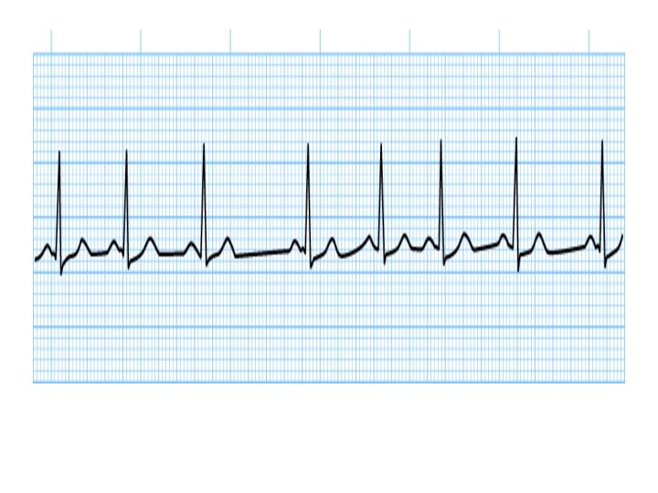

Sinus Tachycardia

Sinus Tachycardia • Sinus rhythm • Heart rate > 100 bpm • Common causes – Fever – Anaemia – Pain – Hyperthyroidism -Shock -Pulmonary Embolism -Shock -Sympathomimetics like adrenaline • Treatment: B blockers, Digoxin, CCB

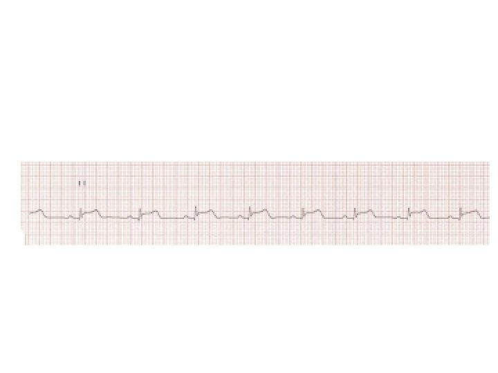
Sinus Bradycardia

Sinus Bradycardia • Sinus rhythm • Heart rate < 60 bpm • Common causes – Hypothermia – Hypothyroidism – Drugs like beta blockers – Vasovagal syncope -physical fitness • Treatment : atropine, isoprenalin, cardiac pacing

Atrial fibrillation

Atrial Fibrillation • • No discrete P wave Fibrillary waves Irregular RR interval Common causes: – Hyperthyroidism – Hypertension – Valvular Hear disease – Cardiomyopathy -Alcohol Abuse -Pulmonary embolism

Atrial fibrillation • Treatment: B blockers, digoxin, CCBs, amiodarone, cadioversion, anticoagulant

Atrial Flutter

Atrial flutter • • No discrete P waves Saw tooth appearance Causes are same as atrial fibrillation Treatment : cardioversion, B blockers

Paroxysmal Supraventricular Tachycardia

Paroxysmal Supraventricular tachycardia • • • Narrow QRS complex Occurs in paroxysms Heart rate: 150 -250 bpm P waves may not be seen Treatment : Adenosine, esmolol, digoxin, cardioversion

STEMI & NSTEMI
")
STEMI • Initial treatment: Morphine Oxygen Nitroglycerine Aspirin • Definitive treatment: PCI and thrombolysis(streptokinase)

NSTEMI • Initial Treatment: MONA • Definitive : B blockers, ace inhibitors, angiography and PCI

• Thank You
- Slides: 36