BASIC ECG INTERPRETATION RECOGNIZING STEMI OCTOBER 2020 Objectives

BASIC ECG INTERPRETATION & RECOGNIZING STEMI OCTOBER 2020

Objectives ▪ Understand the purpose of a standard 12 -lead ECG ▪ Understand how the ECG is created using electrical signals ▪ Understand what a 15 -lead and right-sided ECG are used for, and how to perform them ▪ Perform basic interpretation of ECG for emergent concerns ▪ Recognize ST elevation on an ECG ▪ Understand appropriate reporting process to initiate CODE STEMI in ER ▪ To understand nursing duties/care for patient with STEMI ▪ To understand transfer process for patient with STEMI

PRE-REQUISITES/CO-REQUISITES Refer to other PRH Critical Care modules: ▪ Prerequisite Module ▪ Cardiac Monitoring and Interpretation (rhythm interpretation is not discussed here) ▪ Co-requisite Module ▪ Performing an ECG (performance is not discussed here)

Anatomy & Physiology Refresher

Electrical Conduction Pathway During each heartbeat, a healthy heart has an orderly progression of depolarization pacemaker cells in the sinoatrial node (SA) spreads out through the atrium passes through the atrioventricular node (AV) down into the bundle of His into the Purkinje fibers spreading down and to the left throughout the ventricles This orderly pattern of depolarization gives rise to the characteristic ECG tracing.

CORONARY VESSELS Coronary vessels: A – Anterior oblique B – Left anterior oblique The major arteries are: • Left main • Left anterior descending (LAD) • Left circumflex (LCX) • Right coronary arteries (RCA)
 is the process of recording the electrical activity of the heart over")
Electrocardiography (ECG) is the process of recording the electrical activity of the heart over a period of time using electrodes placed on the skin.

A standard 12 -lead ECG
")
Why so many leads? (They all look at a different angle of the heart) Chest Leads/Precordial Leads Limb Leads

12 -lead ECG – the views ▪ This is showing that simultaneously, the 12 -lead ECG can show us a snapshot picture as if looking: ▪ Transverse ▪ Frontal ▪ Sagittal ▪ That’s a lot of information about the heart all at once!

How come on the crash cart there are only 3 wires? ▪ Eintoven’s Triangle ▪ All of this started with just 3 leads – I, III

Does electrode placement matter? Correct placement, normal ECG • Placement of the electrodes is very important to ensure consistency between tracings • What if placement is not the same? • Often, the left and right arms are mistakenly switched. RA RA LA LA LL LL

Does placement matter? Yes! Note that if RA and LA are switched, it causes major changes to the ECG tracing. This is become limb polarity is switched. Lead I is inverted. Lead II becomes lead III. Lead III becomes lead II. Lead a. VR becomes a. VL. Lead a. VL becomes a. VR.

Lead I is your major clue. All of the waveform is inverted. ▪ If you see an inverted P and an inverted T together, suspect limb (RA & LA) lead reversal ▪ Check to see that leads were applied correctly Take home message = lead placement impacts the rhythm strip! • This is true for any electrode placement – placement should be consistent from nurse to nurse • Consider marking with a dot in permanent marker

Types of ECGs

Why order an ECG? ▪ ECG is a diagnosing and investigative tool ▪ ECG can be used to show the MD or cardiologist the following: ▪ Enlargement of the heart (RA, RV, LA, LV hypertrophy) ▪ Abnormal rhythm (arrhythmia) – rapid, slow or irregular heart beats ▪ Damage to the heart such as when one of the heart’s arteries is blocked (coronary occlusion) ▪ ▪ ▪ (STEMI) Poor blood supply to the heart (ischemia) Abnormal position of the heart (axis deviation) Heart inflammation – pericarditis or myocarditis Disturbances of the heart’s conducting system (e. g. blocks) Imbalances in the blood chemicals (electrolytes) that control heart activity (e. g. hyperkalemia and others) Previous heart attacks (Q waves) ▪ Many uses… ECG expertise takes years of specialty training ▪ Here we are focusing on ST changes

3 main types: Type What is it looking for? Standard 12 -Lead Looks at ANTERIOR, INFERIOR, LATERAL portions of heart Only one lead (V 1) looks at the right side – not good for right side infarcts Does not look at the posterior 15 -lead ECG Standard 12 -lead ECG views PLUS V 4 R (right side) – now there are two leads looking at right side (V 1 and V 4 R) V 7 (optional), V 8, V 9 – looks at POSTERIOR Right-sided ECG To fully assess the RIGHT side. All chest leads are moved to right side (V 1 R-V 6 R)

Recognizing ST Segment Changes

The J-point and isoelectric line ▪ In between electrical activity, the rhythm should return to the “isoelectric line” J point ▪ Pay attention to the J point, where the S wave ends ▪ This is where you look for ST changes (elevation or depression)
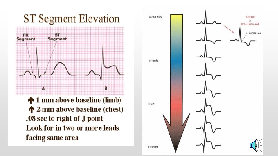

Reminder: ST Depression vs Inverted T wave inverted T wave ▪ Inverted T waves represent ischemia ▪ ST depression also represents ischemia, but may also be reciprocal changes (opposite) to the area of an ST elevation infarct. If ST depression is seen, note the area (which leads), look for ST elevation and consider the need for a 15 lead ECG or right-sided ECG

Which areas relate to which part of the heart?

12 -lead ECG

Determining Location of MI

Terms: Contiguous and Reciprocal Term Contiguous Reciprocal Definition/Meaning

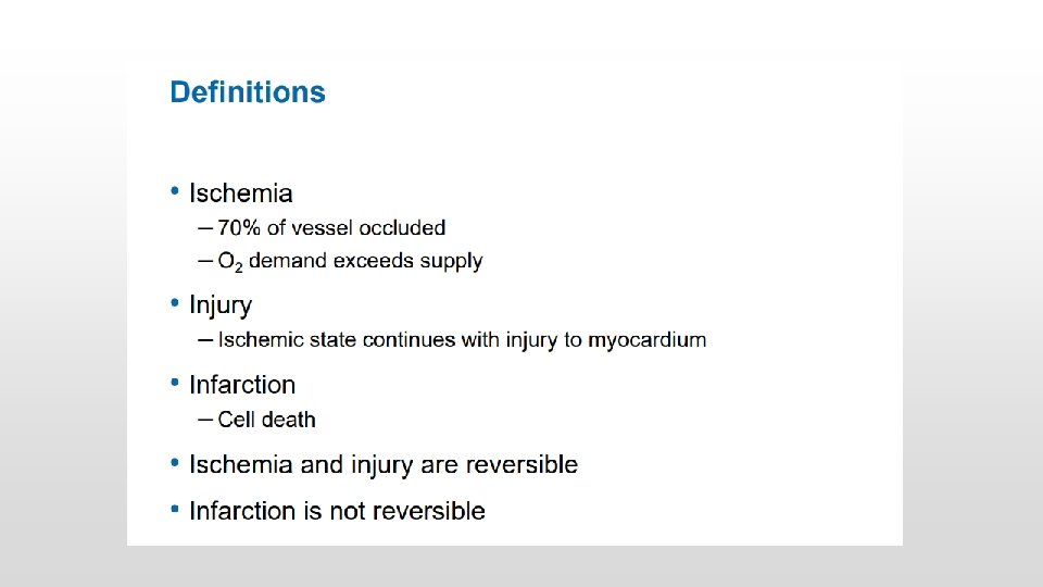
What’s the big deal with STEMI? ▪ Code STEMI = ST segment Elevation Myocardial Infarction ▪ Infarction = myocardial cellular death due to deprived of oxygen supply ▪ Myocardial cell death = decreased heart function since cells are dying ▪ Cell death is happening RIGHT NOW in a STEMI. If we can get blood flow back to the damaged area of the heart, we can decrease the damage! Time is heart!

Code STEMI timeframe? ▪ Time of onset is important to help determine if they are eligible for PCI ▪ To meet criteria, onset must be within 12 hours ▪ **In some circumstances, patients may be accepted for PCI outside of the 12 hour window**

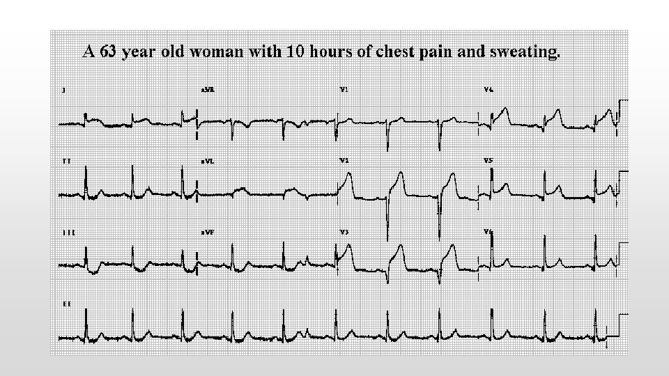
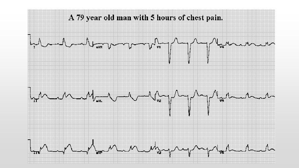
WHAT TYPE OF STEMI IS THIS? Culprit artery: LAD + LCX

LOOK AT THIS IMAGE WHILE REFERRING TO PREVIOUS ECG Look at how much of the heart is supplied by the LAD and LCX The area of the ECG tells us where the blockage is, where the damage is occurring Is this a minor or major MI?
")
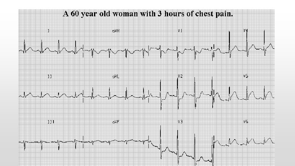
A STANDARD 12 -LEAD THAT SHOULD TRIGGER A 15 -LEAD ECG (POSTERIOR)

A 15 -LEAD ECG V 7, V 8, V 9 LOOK AT THE POSTERIOR

RIGHT SIDE INFARCTION

RIGHT-SIDED INFARCT Suspect or rule out RV infarction in all patients with inferior STEMI! Giving nitro to patients with an RV infarction is contraindicated as it can cause severe hypotension
 Can develop severe hypotension in")
Right-sided MI Preload dependent (due to poor RV contractility) Can develop severe hypotension in response to nitrates or other preloadreducing agents Severe hypotension in RVI is treated with fluid loading, and nitrates are contraindicated
 ▪ Hypotension,")
3 Clinical signs of RVI The TRIAD: ▪ Jugular vein distention (JVD) ▪ Hypotension, either presenting or following nitro administration ▪ Clear lung sounds

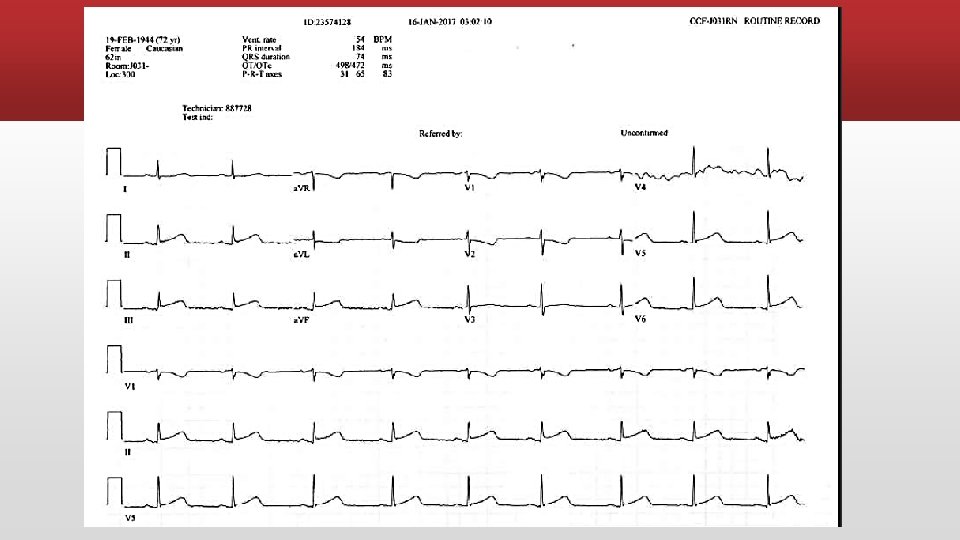
A STANDARD 12 -LEAD THAT SHOULD TRIGGER A RIGHT-SIDED ECG Look for elevation in V 1 or isolectric V 1 with ST depression V 2 ST elevation lead III more than lead II

Checkpoint! ▪ The ER RN can administer nitroglycerin without an MD order True or False

Checkpoint! ▪ Nitroglycerin is contraindicated in RV infarction True or False

Checkpoint! ▪ A patient was shown to be having an inferior infarct as per the MD. The patient’s BP pre-nitro was 125/80. The nurse administered nitroglycerin as per the MD order and the patient’s post-nitro BP was 80/40 and the patients chest pain is worse. What happened?

Checkpoint! ▪ A patient was shown to be having an inferior infarct as per the MD. The patient’s BP pre-nitro was 125/80. The nurse administered nitroglycerin as per the MD order and the patient’s post-nitro BP was 80/40 and the patients chest pain is worse. ▪ What should the nurse anticipate to improve the blood pressure? a) Wait & monitor– as the nitro wears off it will improve b) Administer another dose of nitroglycerin c) Administer IV fluid bolus d) Administer morphine

What is the preferred treatment for a right-sided infarction? ▪ IV fluids to maintain perfusion/cardiac output ▪ Avoid nitroglycerin, but nitroglycerin infusion may be used with extreme caution as it can be titrated ▪ Avoid morphine, as it is also a vasodilator and will further reduce preload ▪ Consider fentanyl for pain instead ▪ Consider inotropes (dobutamine first choice) ▪ Usual STEMI protocols: ▪ Oxygen for Sp. O 2 less than 94% ▪ ASA 180 mg chewed ▪ Thrombolytics; Anti-coagulants; Early revascularization (PCI)

CODE STEMI – ER PROCESS ENSURE THAT YOUR PRECEPTOR “RUNS THROUGH” THE STEMI PROCESS WITH YOU
")
Door-to-first ECG time = 10 MINUTES Timely diagnosis of ST‐segment elevation myocardial infarction (STEMI) in the Emergency Department (ED) is made solely by ECG Do not need to wait for BW or troponins Obtaining this test within 10 minutes of ED arrival is critical to achieving the best outcomes RN to perform if lab delay
 Pt presents to ER with chest pain; signs")
Door-to-first ECG time (goal <10 minutes) Pt presents to ER with chest pain; signs direct patient to RPN desk (nursing station) (skips triage) Pt moved into room 39, RPN or RN calls lab for STAT ECG - If not there within 5 minutes, RN to do ECG If ECG positive STEMI moved to 28 -3 (preferred) or other monitored bed, triage completed and code STEMI initiated If negative, patient returned to triage to complete triage process Review old ER chart (online) or call Health Records for old chart if any abnormalities on ECG for comparison

GOALS! Eg. TNK To qualify for PCI, the presentation must be within 12 hours of symptom onset AND Primary PCI can be delivered within 120 minutes

CODE STEMI PROCESS: When The Code is Called ▪ Once code STEMI suspected, clear 28 -3 and grab the Code STEMI box above sink by the med area in the ER ▪ Individual Code STEMI packages are in the box; if refills are needed, they are in a labelled binder in the file cabinet under the telemetry area in the ER Obtain IV access! ▪ Follow the “Nurse Checklist: Potential Code STEMI” (in package) ▪ On confirmation by MD, RN to call OHI Nursing Coordinator ▪ Code will not be called overhead ▪ Must make sure the OHI Nursing Coordinator is notified of the Code STEMI and that the patient will be enroute Get an accurate patient weight if at all possible!
 ▪ Code STEMI")
CODE STEMI ▪ Chest pain protocol implemented (ECG, BW, IV access) ▪ Code STEMI confirm by MD ▪ MD/RN call UOHI for transfer ▪ Administer upon order ▪ ▪ ▪ TNK-weight adjusted IV bolus (unless contraindicated) ASA - already given when ECG done with protocol Plavix – 300 mg (75 years or younger) Unfractionated heparin - IV bolus 60 units/kg (max 4000 units) Then UFH infusion 12 units/kg/hour

CODE STEMI ▪ Urgent transfer arranged to UOHI ▪ Send documents -- ER notes, ECG, labs if resulted, Code STEMI Transfer Checklist (in package) ▪ Need MT # (ward clerk to get) ▪ Have ward clerk prepare package, have primary RN assist with care/package/transfer ▪ Send patient with RN if going land ambulance ▪ DO NOT DELAY TRANSFER! – time is heart!

CODE STEMI PROCESS: Patient Transfer to OHI ▪ Preferred mode of patient transfer to OHI is via Air ORNGE ▪ RN escort is not needed with ORNGE ▪ Do not delay patient transport if air is not available – can’t wait for land ORNGE!!! ▪ In such a case that air is not available, emergent local land ambulance with RN escort is needed (plan accordingly for the escort with primary RN and Clinical Resource as needed) ▪ Complete “Code STEMI Transfer Checklist” to send with the patient (in Code STEMI package) ▪ When the patient leaves for OHI, update OHI of departure time and expected arrival to their facility ▪ Fax any remaining patient results to OHI if/when available (get up-to-date fax number from on-call nursing coordinator in advance)
 If you are the RN escort on a patient being transferred")
Reperfusion (particularly post-TNK) If you are the RN escort on a patient being transferred with a STEMI be aware that: ▪ This patient is UNSTABLE – high risk for cardiac arrest/other arrhythmias ▪ Be prepared with transfer medications and know how to give them ▪ Be prepared to see a lot of ectopy (ventricular irritability) in the form of PVCs, PACs, rhythm changes ▪ The patient may flip into an accelerated idioventricular rhythm (AIVR) ▪ Keep a close eye on the monitor!
")
Accelerated Idioventricular Rhythm (AIVR)

STEMI PROCESS – ICU

▪ NEW ST ELEVATION = NOTIFY MD ▪ ASSESS PATIENT (VS, CHEST PAIN, VERIFY TELEMETRY WIRE PLACEMENT) ▪ PERFORM ECG ▪ IF ST ELEVATION SEEN ON ECG, NOTIFY MD ▪ IF MD UNABLE TO COME SEE ECG, ASK ER MD TO VIEW ▪ FOLLOW MD DIRECTION ▪ (NO OFFICIAL IN-HOUSE STEMI PROTOCOL) ▪ Likely will follow ER STEMI protocol; ask ER/ICU charge RN to assist

STEMI TEST

Code STEMI test 1. Your patient with chest pain and related symptoms (suggestive of cardiac ischemia) reports timeframe of 72 hrs since symptom onset. Are Code STEMI criteria met based on this knowledge alone? A. Yes B. No

Code STEMI test 2. Your Code STEMI patient is awaiting OHI transfer via air ORNGE; ORNGE calls back and states that air is unavailable. What is your remaining transfer option? A. Awaiting land ORNGE B. Immediate local land ambulance with RN escort C. Local land ambulance without an escort D. Private transfer by patient family

Code STEMI test 3. EMS has patched in a STEMI patient. The MD confirms a Code STEMI on ambulance arrival. Prior to transferring the onto the stretcher in 28 -3, what should you do? A. Start a saline lock on the EMS stretcher B. Direct family to the family room C. Zero the stretcher scale to be able to weigh immediately D. Call for the patient’s old chart from Health Records
 Lead views that look at")
Code STEMI test 4. What does contiguous mean? a) Lead views that look at opposite areas of the heart b) Lead views that are numerically beside each other (e. g. I is next to II) c) Lead views that look at the same areas of the heart

Code STEMI test 5. You have remaining TNK leftover after the MD has given the ordered dose to your Code STEMI patient. You should promptly discard of this leftover drug into a blue pharmaceutical waste container. A. True B. False

Code STEMI test 6. A Code STEMI is called and there are no packages in the Code STEMI box. Where can you quickly find more prepared packages? A. Call the clerk to prepare one immediately B. Medication room in ICU C. Code Stroke box D. In a labelled binder in the file cabinet beside the charge nurse desk in the ER

Code STEMI test 7. The nurse recognizes that the following are signs of a STEMI: ▪ A) ST elevation in 2 contiguous leads ▪ B) New left bundle branch block ▪ C) Chest pain less than 12 hours ▪ D) All of the above

References ▪ University of Ottawa Heart Institute, Renfrew County Paramedic Services, Pembroke Regional Hospital. (n. d. ). Code STEMI protocol: Urgent transfer to UOHI for TNK facilitated PCI. ▪ PRH Clinical Skills. “Tenecteplase for acute myocardial infarction. ” ▪ PRH Policy & Procedure (policy medical) ▪ Code STEMI – Facilitated Percutaneous Coronary Intervention ▪ https: lifeinthefastlane. com/ecg ▪ https: //www. ena. org/docs/default-source/resource-library/practice-resources/tips/right-sideecg. pdf? sfvrsn=836 f 00 e 6_8 ▪ http: //cdn. esa. act. gov. au/wp-content/uploads/17 -15 -LEAD-ECG. pdf ▪ https: //www. ncbi. nlm. nih. gov/books/NBK 2214/table/A 65/? report=objectonly ▪ https: //www. ena. org/docs/default-source/resource-library/practice-resources/tips/right-sideecg. pdf? sfvrsn=836 f 00 e 6_8 ▪ https: //www. cablesandsensors. com/pages/12 -lead-ecg-placement-guide-with-illustrations ▪ http: //jaha. ahajournals. org/content/6/3/e 003528
- Slides: 68