
BASIC ECG Dr Glay LER ERDA Dersin amalar
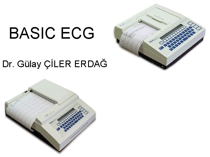
BASIC ECG Dr. Gülay ÇİLER ERDAĞ
Dersin amaçları: • Ekgnin tanıtılması • EKGnin kullanım alanları hakkında bilgi verimesi • EKGnin nasıl çekileceği hakkında bilgi verimesi • Ekg nin çalışma prensipleri hakkında bilgi verilmesi • EKG kağıdı hakında bilgi verilmesi • Kalp hızının hesplanması hakkında bilgi verilmesi • Kalp ritminin değerlendirlmesi hakkında bilgi verilmesi • P-r aralığı hesaplanması, p, ors ve r dalgalarının yapısı ve süreleri hakkında bilgi verilmesi • Aks hesaplanması hakkında bilgi verilmesi amaçlanmştır
Dersin hedefleri: • Öğrencilerin ders sonunda, çocuklarda ne zaman, ne amaçla EKG çekmeleri gerektiği konusunda bilgi sahibi olmaları • Çekilmiş olan bir EKG kağıdına bakarak kalp hızını, Kalp ritmini, kalbin aksını hesaplayabilmeleri, • Varsa p, ors , t dalgalrındaki yapısal bozuklukları farkedebilmeleri, • Varsa kalp bloklarını farkedebilmeleri • Elektrolit bozukluklarına bağlı ortaya çıkabilecek EKG değişikliklerini saptayabilmeleri

On
Learning Modules • • • ECG Basics How to Analyze a Rhythm Normal Sinus Rhythm Heart Arrhythmias Advanced 12 -Lead Interpretation
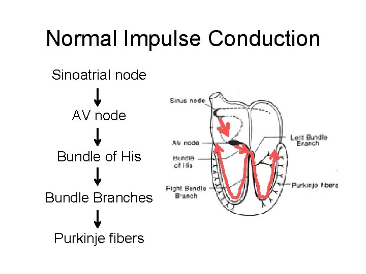
Normal Impulse Conduction Sinoatrial node AV node Bundle of His Bundle Branches Purkinje fibers
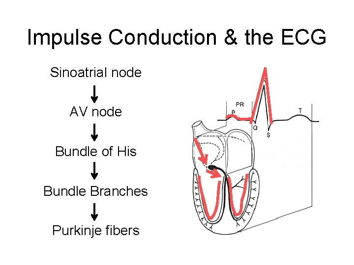
Impulse Conduction & the ECG Sinoatrial node AV node Bundle of His Bundle Branches Purkinje fibers
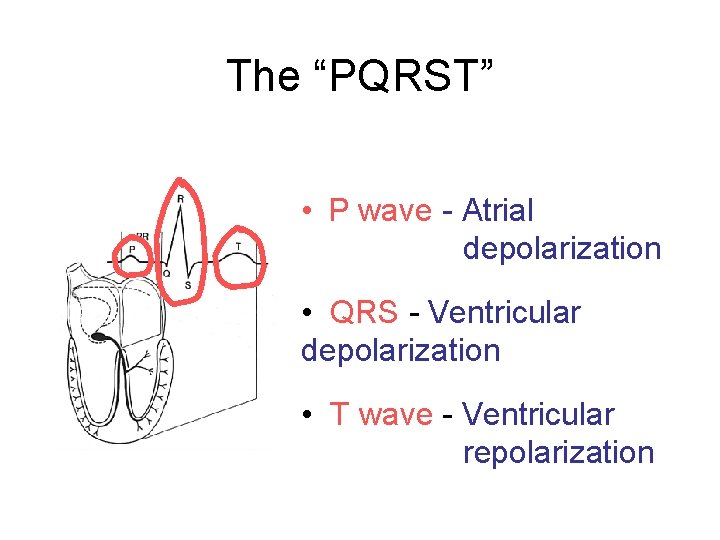
The “PQRST” • P wave - Atrial depolarization • QRS - Ventricular depolarization • T wave - Ventricular repolarization
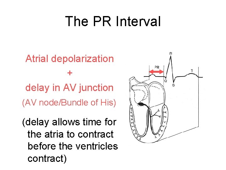
The PR Interval Atrial depolarization + delay in AV junction (AV node/Bundle of His) (delay allows time for the atria to contract before the ventricles contract)

• Atriumların kasılmasıyla oluşan dalga…P dalgası • Ventriküllerin kasılmasıyla oluşan dalga…QRS kompleksi • Ventriküllerin repolarize olmasıyla oluşan dalga…T dalgası • Uyarının sağ atriumdan çıkıp, a-v düğümden iletilmesi için geçen zaman……. . P-R aralığı
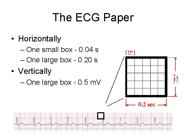
The ECG Paper • Horizontally – One small box - 0. 04 s – One large box - 0. 20 s • Vertically – One large box - 0. 5 m. V

The ECG Paper (cont) 3 sec • Every 3 seconds (15 large boxes) is marked by a vertical line. • This helps when calculating the heart rate.
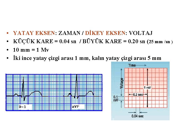
• • YATAY EKSEN: ZAMAN / DİKEY EKSEN: VOLTAJ KÜÇÜK KARE = 0. 04 sn / BÜYÜK KARE = 0. 20 sn (25 mm /sn ) 10 mm = 1 Mv İki ince yatay çizgi arası 1 mm, kalın yatay çizgi arası 5 mm
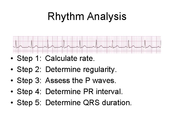
Rhythm Analysis • • • Step 1: Step 2: Step 3: Step 4: Step 5: Calculate rate. Determine regularity. Assess the P waves. Determine PR interval. Determine QRS duration.
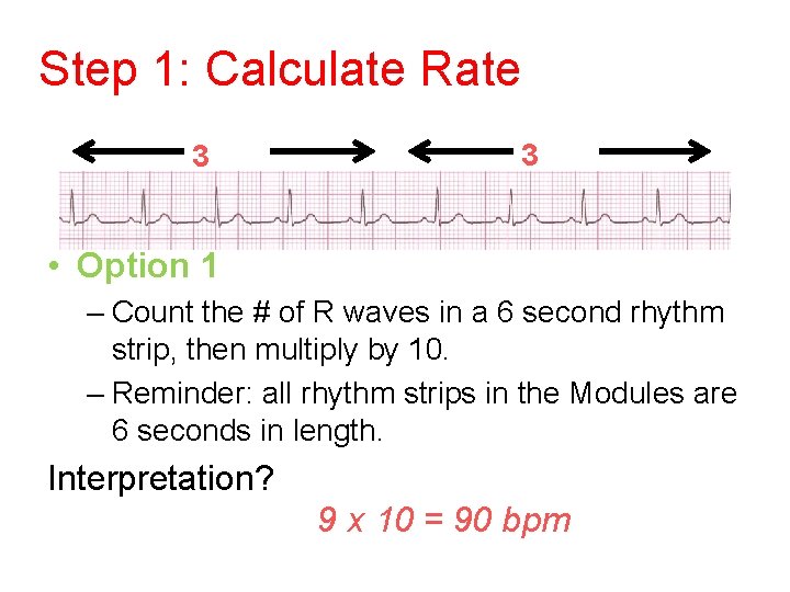
Step 1: Calculate Rate 3 sec • Option 1 – Count the # of R waves in a 6 second rhythm strip, then multiply by 10. – Reminder: all rhythm strips in the Modules are 6 seconds in length. Interpretation? 9 x 10 = 90 bpm
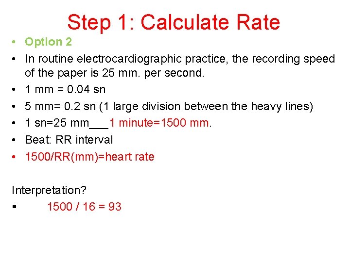
Step 1: Calculate Rate • Option 2 • In routine electrocardiographic practice, the recording speed of the paper is 25 mm. per second. • 1 mm = 0. 04 sn • 5 mm= 0. 2 sn (1 large division between the heavy lines) • 1 sn=25 mm___1 minute=1500 mm. • Beat: RR interval • 1500/RR(mm)=heart rate Interpretation? § 1500 / 16 = 93
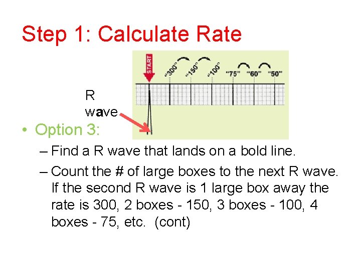
Step 1: Calculate R wave • Option 3: – Find a R wave that lands on a bold line. – Count the # of large boxes to the next R wave. If the second R wave is 1 large box away the rate is 300, 2 boxes - 150, 3 boxes - 100, 4 boxes - 75, etc. (cont)
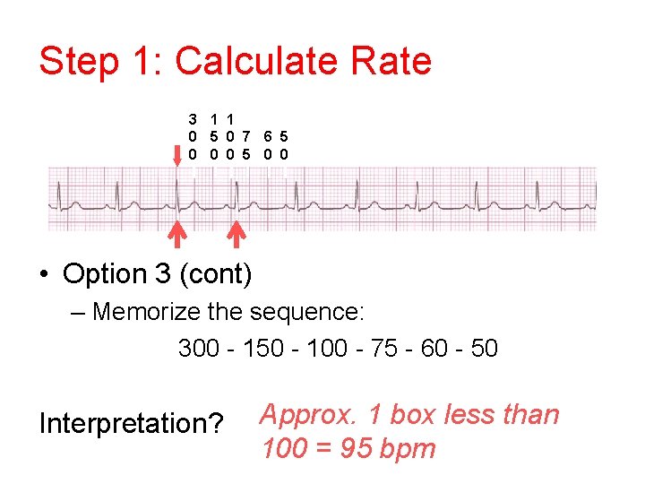
Step 1: Calculate Rate 3 1 1 0 5 0 7 6 5 0 0 0 5 0 0 • Option 3 (cont) – Memorize the sequence: 300 - 150 - 100 - 75 - 60 - 50 Interpretation? Approx. 1 box less than 100 = 95 bpm
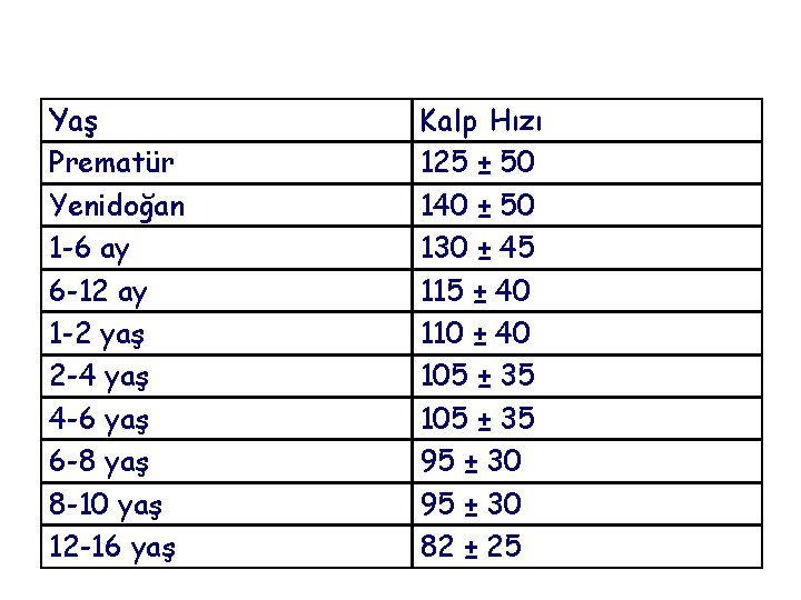
Yaş Prematür Yenidoğan 1 -6 ay 6 -12 ay 1 -2 yaş 2 -4 yaş 4 -6 yaş 6 -8 yaş 8 -10 yaş 12 -16 yaş Kalp Hızı 125 ± 50 140 ± 50 130 ± 45 115 ± 40 110 ± 40 105 ± 35 95 ± 30 82 ± 25
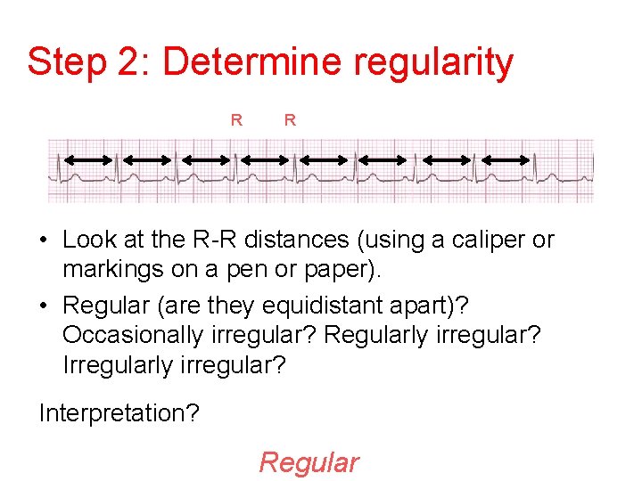
Step 2: Determine regularity R R • Look at the R-R distances (using a caliper or markings on a pen or paper). • Regular (are they equidistant apart)? Occasionally irregular? Regularly irregular? Irregularly irregular? Interpretation? Regular
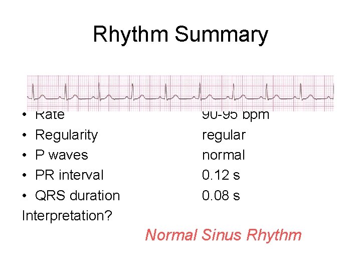
Rhythm Summary • Rate • Regularity • P waves • PR interval • QRS duration Interpretation? 90 -95 bpm regular normal 0. 12 s 0. 08 s Normal Sinus Rhythm
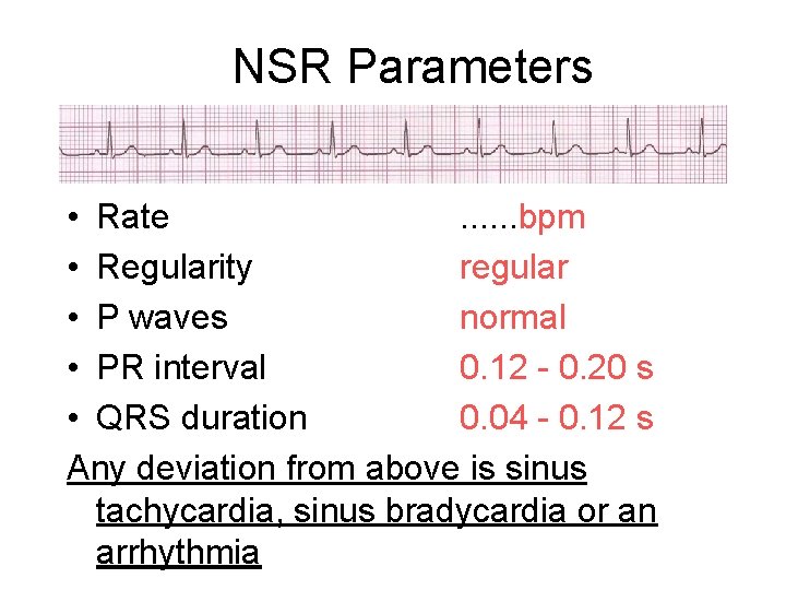
NSR Parameters • Rate. . . bpm • Regularity regular • P waves normal • PR interval 0. 12 - 0. 20 s • QRS duration 0. 04 - 0. 12 s Any deviation from above is sinus tachycardia, sinus bradycardia or an arrhythmia
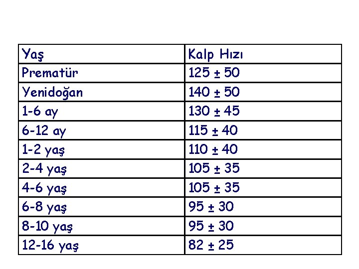
Yaş Prematür Yenidoğan 1 -6 ay 6 -12 ay 1 -2 yaş 2 -4 yaş 4 -6 yaş 6 -8 yaş 8 -10 yaş 12 -16 yaş Kalp Hızı 125 ± 50 140 ± 50 130 ± 45 115 ± 40 110 ± 40 105 ± 35 95 ± 30 82 ± 25
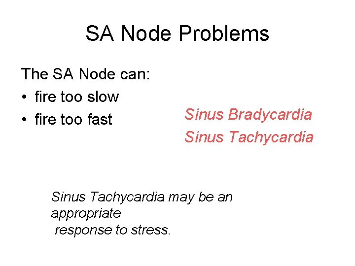
SA Node Problems The SA Node can: • fire too slow • fire too fast Sinus Bradycardia Sinus Tachycardia may be an appropriate response to stress.
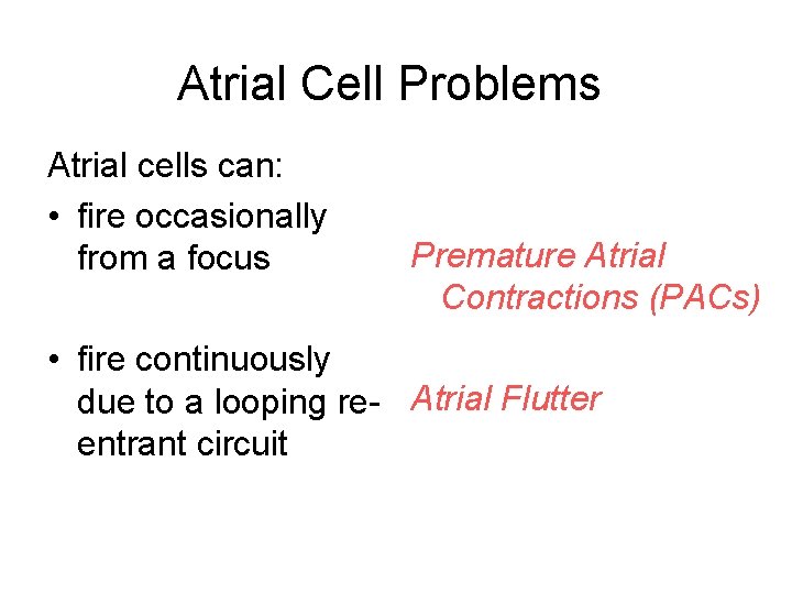
Atrial Cell Problems Atrial cells can: • fire occasionally from a focus Premature Atrial Contractions (PACs) • fire continuously due to a looping re- Atrial Flutter entrant circuit
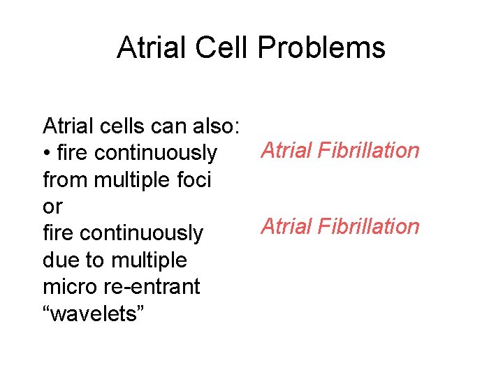
Atrial Cell Problems Atrial cells can also: Atrial Fibrillation • fire continuously from multiple foci or Atrial Fibrillation fire continuously due to multiple micro re-entrant “wavelets”
AV Junctional Problems The AV junction can: • fire continuously Paroxysmal due to a looping re Supraventricular -entrant circuit Tachycardia • block impulses AV Junctional Blocks coming from the SA Node
Ventricular Cell Problems Ventricular cells can: • fire occasionally Premature Ventricular from 1 or more foci Contractions (PVCs) • fire continuously Ventricular Fibrillation from multiple foci • fire continuously Ventricular Tachycardia due to a looping re -entrant circuit
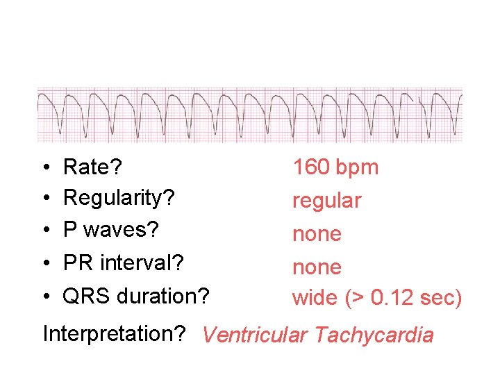
• • • Rate? Regularity? P waves? PR interval? QRS duration? 160 bpm regular none wide (> 0. 12 sec) Interpretation? Ventricular Tachycardia
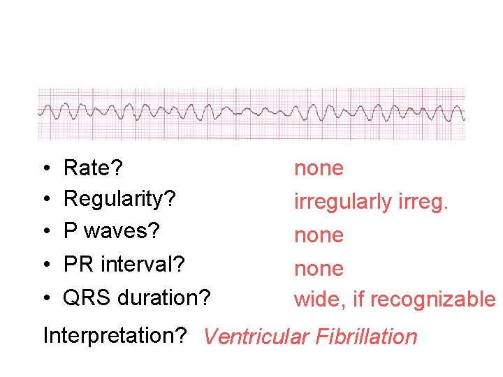
• • • Rate? Regularity? P waves? PR interval? QRS duration? none irregularly irreg. none wide, if recognizable Interpretation? Ventricular Fibrillation
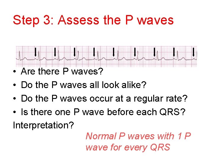
Step 3: Assess the P waves • Are there P waves? • Do the P waves all look alike? • Do the P waves occur at a regular rate? • Is there one P wave before each QRS? Interpretation? Normal P waves with 1 P wave for every QRS
P wave • P duration is measured from the onset to the end of the p wave. • The maximal p duration is 0. 10 second in normal children(2. 5 mm) • The mean p amplitude in lead II or any other lead is about 1, 5 mm. A with a maximum of 3 mm. • Tall waves are an indication of right atrial hypertrophy.
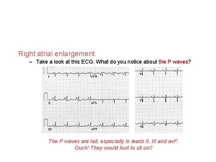
Right atrial enlargement – Take a look at this ECG. What do you notice about the P waves? The P waves are tall, especially in leads II, III and av. F. Ouch! They would hurt to sit on!!
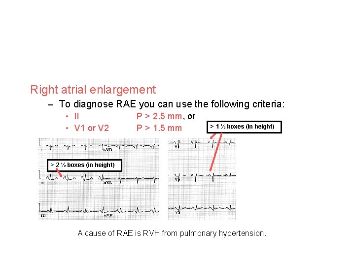
Right atrial enlargement – To diagnose RAE you can use the following criteria: • II • V 1 or V 2 > 2 ½ boxes (in height) P > 2. 5 mm, or P > 1. 5 mm > 1 ½ boxes (in height) Remember 1 small box in height = 1 mm A cause of RAE is RVH from pulmonary hypertension.
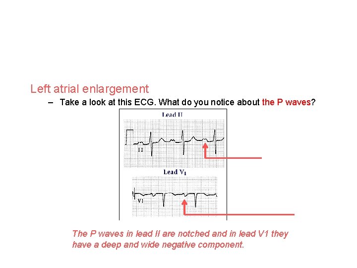
Left atrial enlargement – Take a look at this ECG. What do you notice about the P waves? Notched Negative deflection The P waves in lead II are notched and in lead V 1 they have a deep and wide negative component.
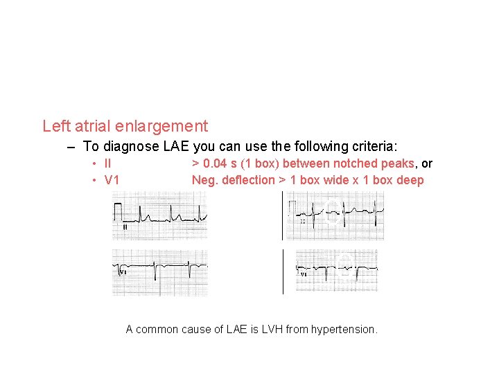
Left atrial enlargement – To diagnose LAE you can use the following criteria: • II • V 1 > 0. 04 s (1 box) between notched peaks, or Neg. deflection > 1 box wide x 1 box deep Normal LAE A common cause of LAE is LVH from hypertension.
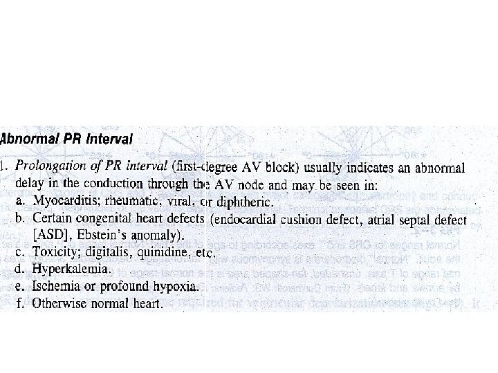
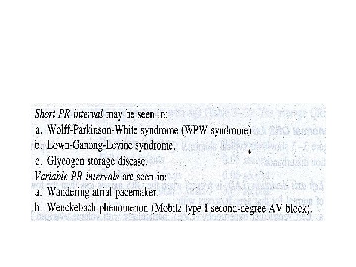
Step 4: Determine PR interval • From the onset of the p wave to the begining of the QRS complex • It is the time required for atrial depolarization and the physiologic delay of the impulse in the AV node. • The normal PR interval varies with age and heart rate. • The older the person and the slower the heart rate, the longer is the PR interval.
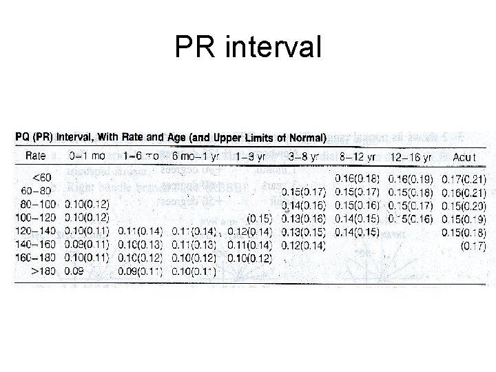
PR interval
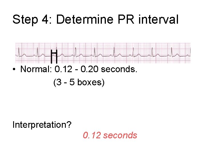
Step 4: Determine PR interval • Normal: 0. 12 - 0. 20 seconds. (3 - 5 boxes) Interpretation? 0. 12 seconds
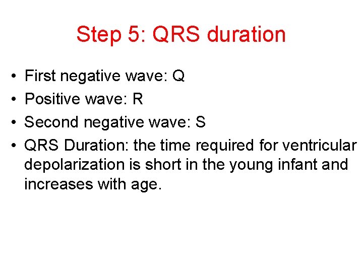
Step 5: QRS duration • • First negative wave: Q Positive wave: R Second negative wave: S QRS Duration: the time required for ventricular depolarization is short in the young infant and increases with age.
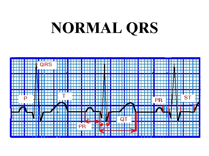
NORMAL QRS
Abnormal QRS amplitude: • Large deflexions: – Ventricular hypertrophy – Ventricular conduction disturbances • Low voltage: – Myocarditis – Pericardial effusions – Chronic constrictive pericarditis – Hypotiroidism
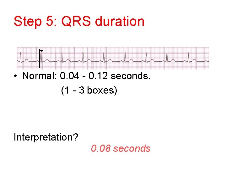
Step 5: QRS duration • Normal: 0. 04 - 0. 12 seconds. (1 - 3 boxes) Interpretation? 0. 08 seconds
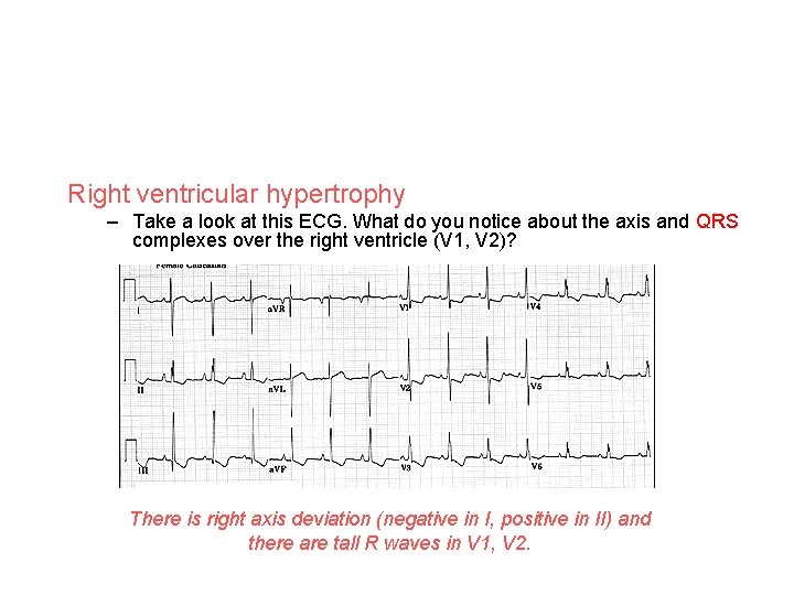
Right ventricular hypertrophy – Take a look at this ECG. What do you notice about the axis and QRS complexes over the right ventricle (V 1, V 2)? There is right axis deviation (negative in I, positive in II) and there are tall R waves in V 1, V 2.
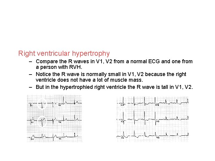
Right ventricular hypertrophy – Compare the R waves in V 1, V 2 from a normal ECG and one from a person with RVH. – Notice the R wave is normally small in V 1, V 2 because the right ventricle does not have a lot of muscle mass. – But in the hypertrophied right ventricle the R wave is tall in V 1, V 2. Normal RVH
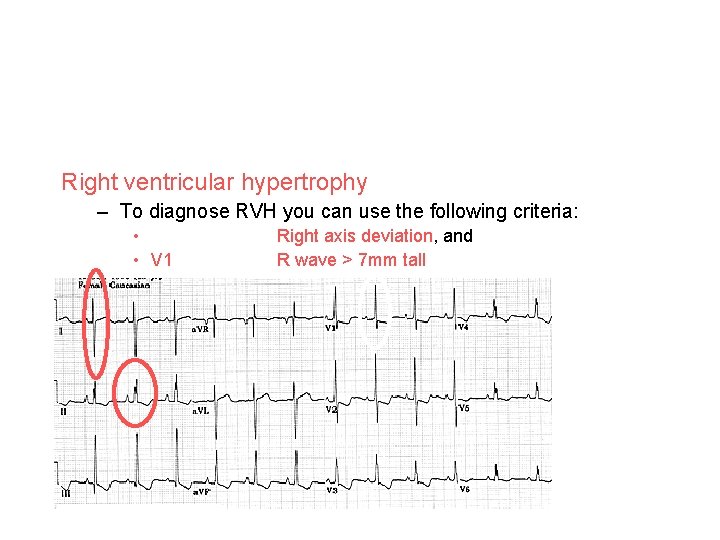
Right ventricular hypertrophy – To diagnose RVH you can use the following criteria: • • V 1 Right axis deviation, and R wave > 7 mm tall
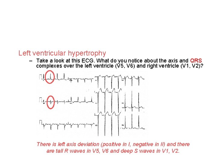
Left ventricular hypertrophy – Take a look at this ECG. What do you notice about the axis and QRS complexes over the left ventricle (V 5, V 6) and right ventricle (V 1, V 2)? The deep S waves seen in the leads over the right ventricle are created because the heart is depolarizing left, superior and posterior (away from leads V 1, V 2). There is left axis deviation (positive in I, negative in II) and there are tall R waves in V 5, V 6 and deep S waves in V 1, V 2.
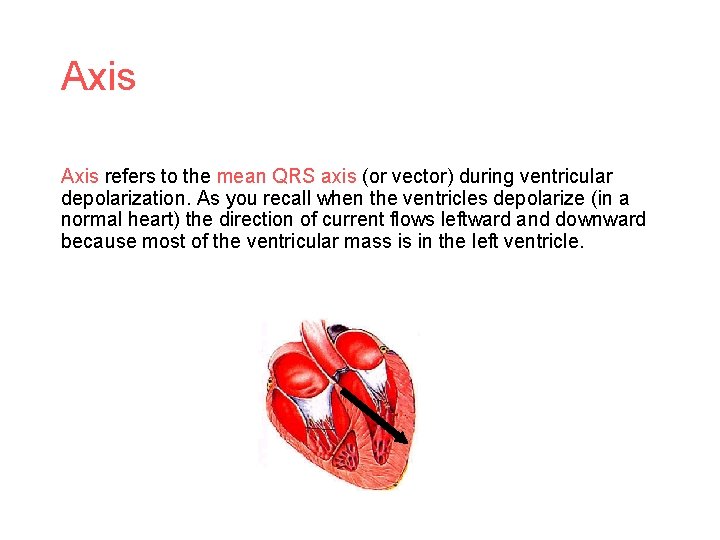
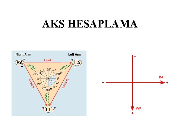
Axis refers to the mean QRS axis (or vector) during ventricular depolarization. As you recall when the ventricles depolarize (in a normal heart) the direction of current flows leftward and downward because most of the ventricular mass is in the left ventricle.
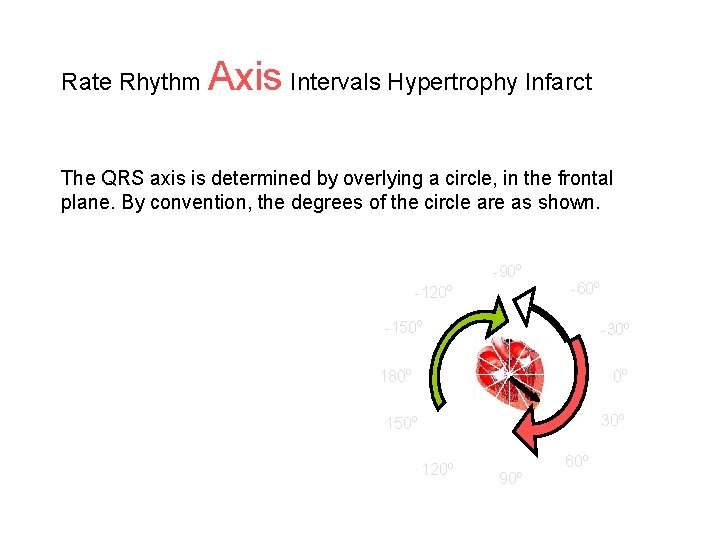
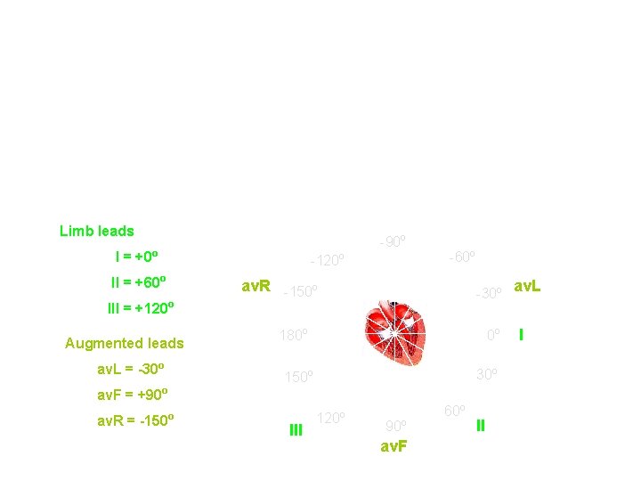
Rate Rhythm Axis Intervals Hypertrophy Infarct The QRS axis is determined by overlying a circle, in the frontal plane. By convention, the degrees of the circle are as shown. -90 o -120 o -60 o -150 o -30 o 180 o 0 o 30 o 150 o 120 o 90 o 60 o
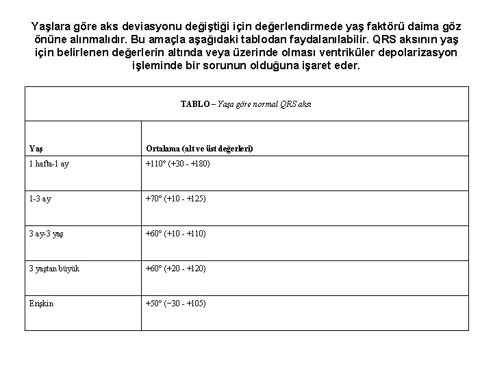
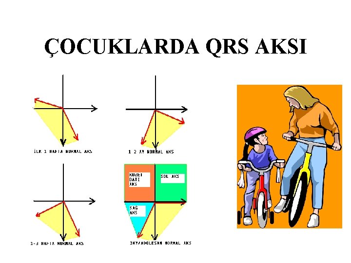
Yaşlara göre aks deviasyonu değiştiği için değerlendirmede yaş faktörü daima göz önüne alınmalıdır. Bu amaçla aşağıdaki tablodan faydalanılabilir. QRS aksının yaş için belirlenen değerlerin altında veya üzerinde olması ventriküler depolarizasyon işleminde bir sorunun olduğuna işaret eder. TABLO – Yaşa göre normal QRS aksı Yaş Ortalama (alt ve üst değerleri) 1 hafta-1 ay +110° (+30 - +180) 1 -3 ay +70° (+10 - +125) 3 ay-3 yaş +60° (+10 - +110) 3 yaştan büyük +60° (+20 - +120) Erişkin +50° (− 30 - +105)
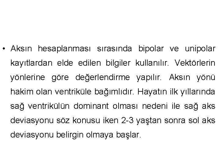
• Aksın hesaplanması sırasında bipolar ve unipolar kayıtlardan elde edilen bilgiler kullanılır. Vektörlerin yönlerine göre değerlendirme yapılır. Aksın yönü hakim olan ventriküle bağımlıdır. Hayatın ilk yıllarında sağ ventrikülün dominant olması nedeni ile sağ aks deviasyonu söz konusu iken 2 -3 yaştan sonra sol aks deviasyonu belirgin olmaya başlar.
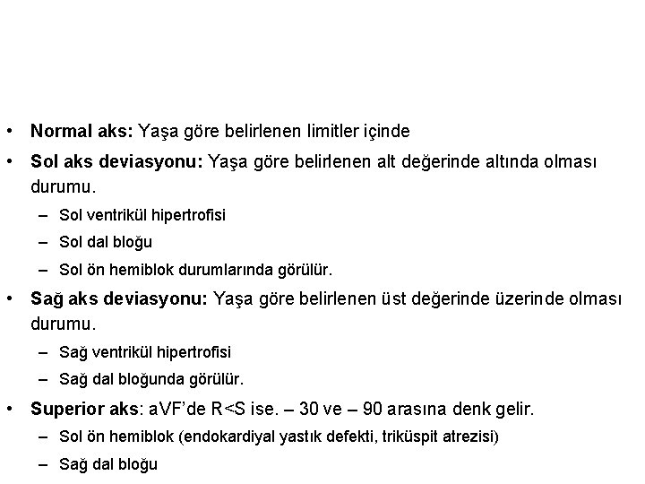
• Normal aks: Yaşa göre belirlenen limitler içinde • Sol aks deviasyonu: Yaşa göre belirlenen alt değerinde altında olması durumu. – Sol ventrikül hipertrofisi – Sol dal bloğu – Sol ön hemiblok durumlarında görülür. • Sağ aks deviasyonu: Yaşa göre belirlenen üst değerinde üzerinde olması durumu. – Sağ ventrikül hipertrofisi – Sağ dal bloğunda görülür. • Superior aks: a. VF’de R<S ise. – 30 ve – 90 arasına denk gelir. – Sol ön hemiblok (endokardiyal yastık defekti, triküspit atrezisi) – Sağ dal bloğu
AKS HESAPLAMA
P AKSI HESAPLAMA
QRS AKSI HESAPLAMA
QRS AKSI HESAPLAMA
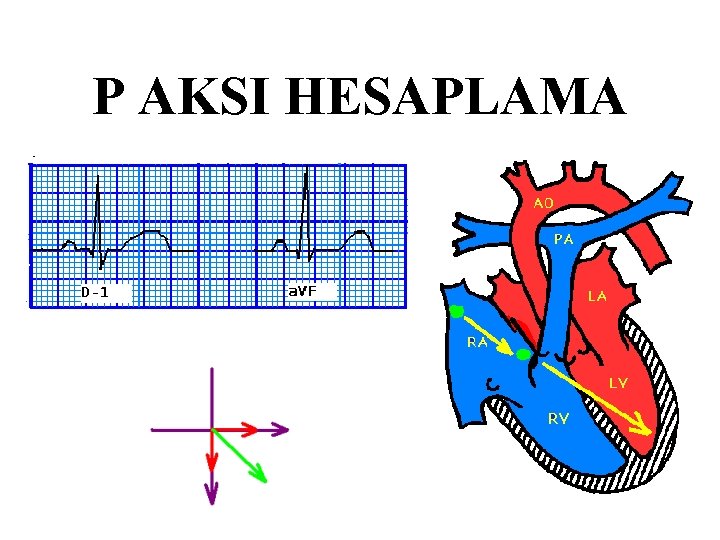
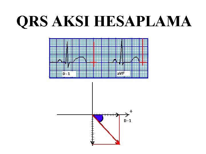
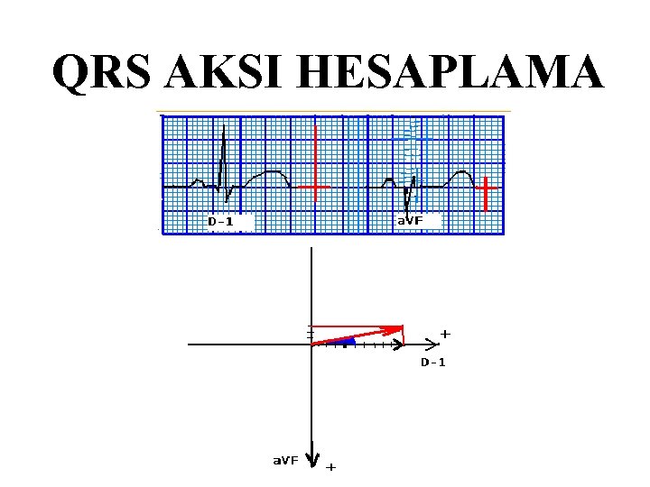
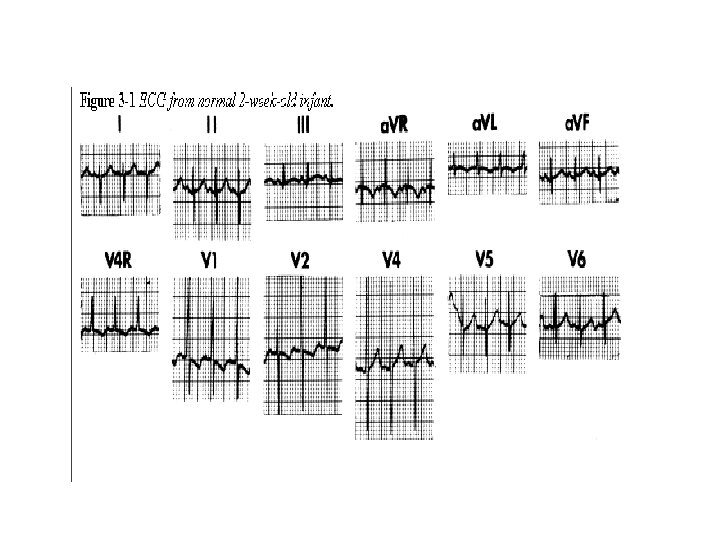
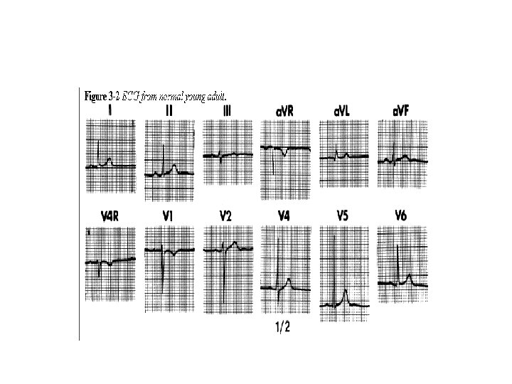
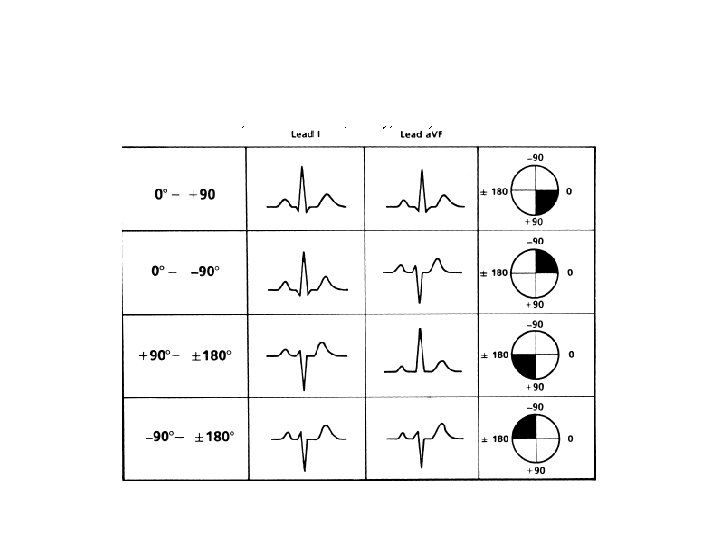
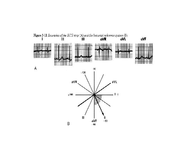
D 1 ile a. VF(veya D 3 ) gibi birbirini 90 -120 arasında kesen iki derivasyon seçilir: • Bu sistemde D I ve a. VF derivasyonlarındaki QRS defleksiyonların net yönüne bakılır. Net yönden kastedilen şudur: • Bir derivasyonda R 13 mm ve S 5 mm olsun. Bunun net yönü + 8 mm’dir. • Veya R 5 mm ve S 13 mm olsun. Bunun da net yönü -8 mm’dir. • Sadece bu derivasyonların net yönüne bakarak aksın hangi çeyrek daire içinde yer aldığı kolaylıkla saptanabilir.
Limb leads -90 o I = +0 o -120 o II = +60 o III = +120 o Augmented leads av. L = -30 o av. R -150 o -30 o 180 o 0 o 30 o 150 o av. F = +90 o av. R = -150 -60 o o III 120 o 90 o av. F 60 o II II av. L II
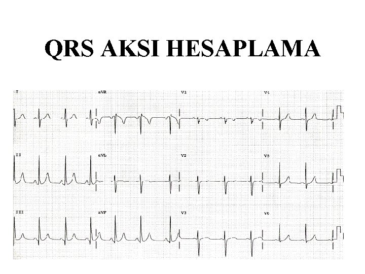
QRS AKSI HESAPLAMA
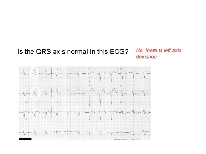
Is the QRS axis normal in this ECG? No, there is left axis deviation. The QRS is positive in I and negative in II.
ÇOCUKLARDA QRS AKSI
Digitalis:

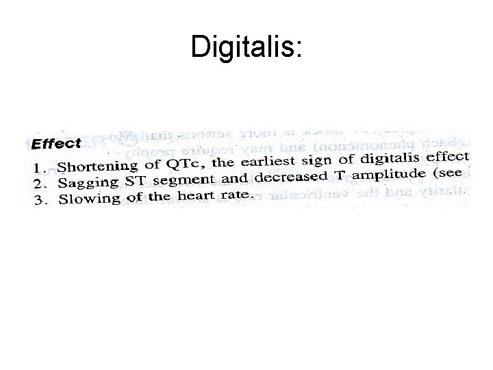
Elektrolit bozukluklarında EKG • Hiperpotasemi: • Hafif yükselme: t dalgaları sivrileşir • Orta derecede yükselme: ileti yavaşlar, P-R aralığı uzar • Çok yüksek: p amplitüdü azalır, atrial aktivite durur • Hipopotasemi: (K< 2. 7 m. Eq / lt olursa): • • S-T çökmesi T voltajında azalma T negatifliği U dalgaları • Hiperkalsemi: Q-T kısalır
AV Nodal Blocks • 1 st Degree AV Block • 2 nd Degree AV Block, Type II • 3 rd Degree AV Block
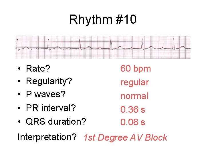
Rhythm #10 • • • Rate? Regularity? P waves? PR interval? QRS duration? 60 bpm regular normal 0. 36 s 0. 08 s Interpretation? 1 st Degree AV Block
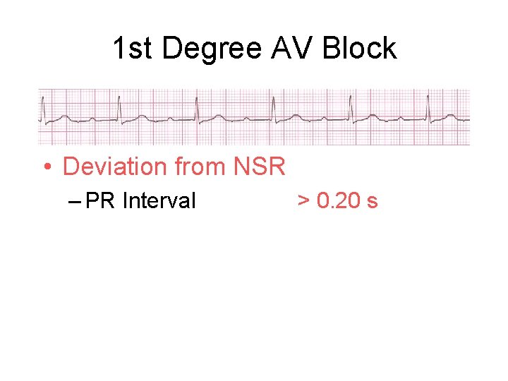
1 st Degree AV Block • Deviation from NSR – PR Interval > 0. 20 s
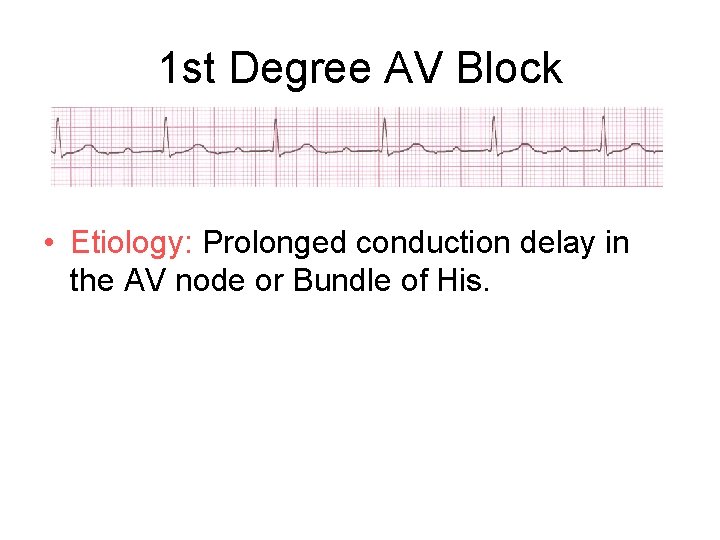
1 st Degree AV Block • Etiology: Prolonged conduction delay in the AV node or Bundle of His.
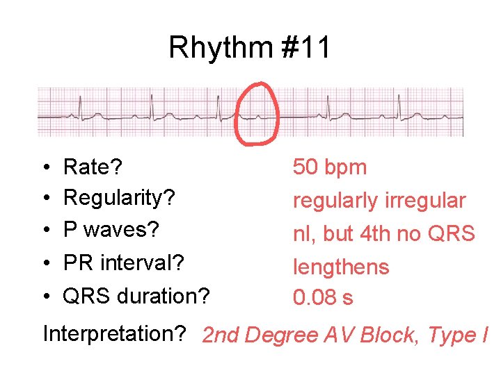
Rhythm #11 • • • Rate? Regularity? P waves? PR interval? QRS duration? 50 bpm regularly irregular nl, but 4 th no QRS lengthens 0. 08 s Interpretation? 2 nd Degree AV Block, Type I
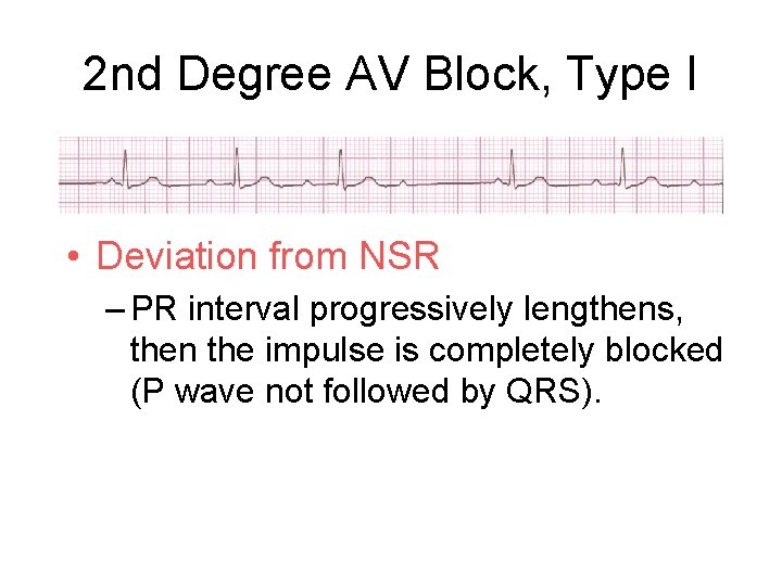
2 nd Degree AV Block, Type I • Deviation from NSR – PR interval progressively lengthens, then the impulse is completely blocked (P wave not followed by QRS).
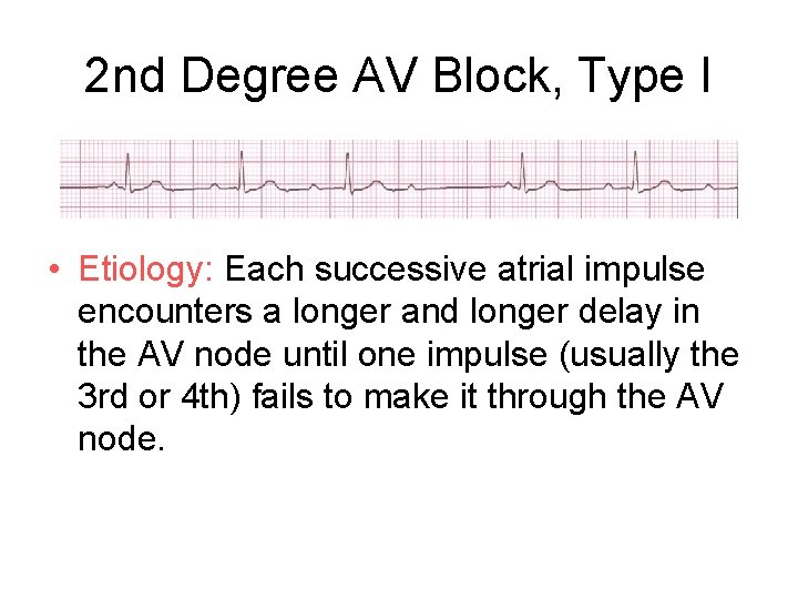
2 nd Degree AV Block, Type I • Etiology: Each successive atrial impulse encounters a longer and longer delay in the AV node until one impulse (usually the 3 rd or 4 th) fails to make it through the AV node.
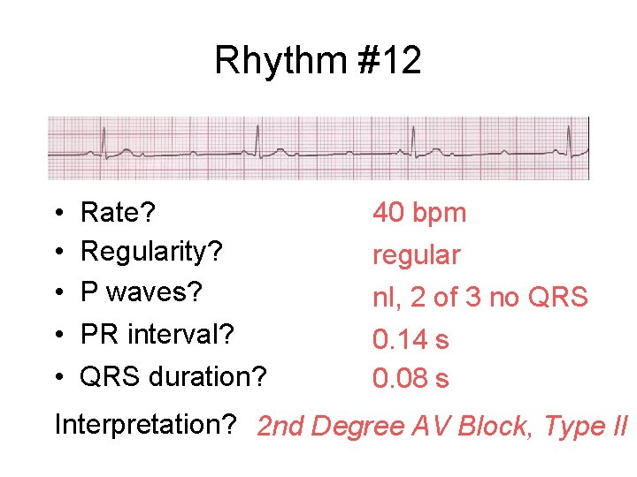
Rhythm #12 • • • Rate? Regularity? P waves? PR interval? QRS duration? 40 bpm regular nl, 2 of 3 no QRS 0. 14 s 0. 08 s Interpretation? 2 nd Degree AV Block, Type II
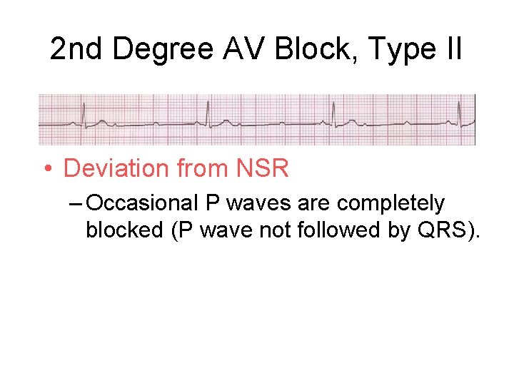
2 nd Degree AV Block, Type II • Deviation from NSR – Occasional P waves are completely blocked (P wave not followed by QRS).
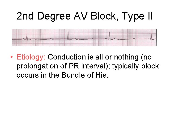
2 nd Degree AV Block, Type II • Etiology: Conduction is all or nothing (no prolongation of PR interval); typically block occurs in the Bundle of His.
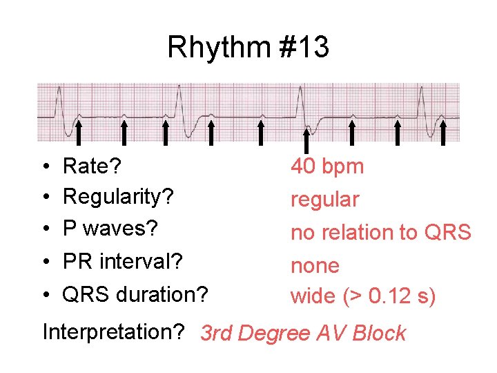
Rhythm #13 • • • Rate? Regularity? P waves? PR interval? QRS duration? 40 bpm regular no relation to QRS none wide (> 0. 12 s) Interpretation? 3 rd Degree AV Block
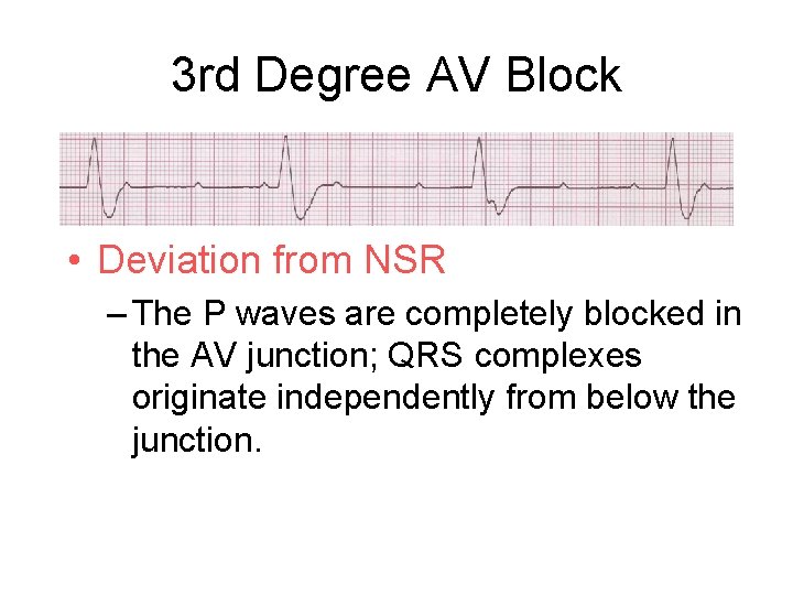
3 rd Degree AV Block • Deviation from NSR – The P waves are completely blocked in the AV junction; QRS complexes originate independently from below the junction.
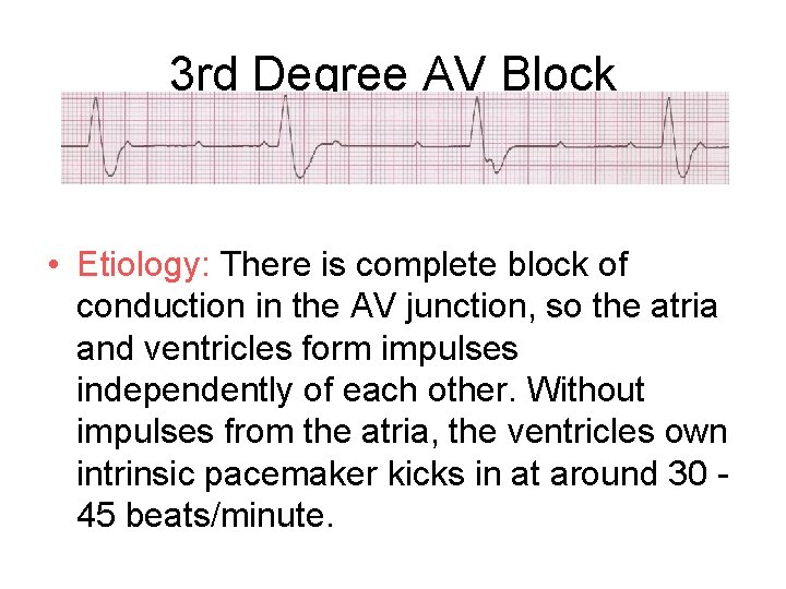
3 rd Degree AV Block • Etiology: There is complete block of conduction in the AV junction, so the atria and ventricles form impulses independently of each other. Without impulses from the atria, the ventricles own intrinsic pacemaker kicks in at around 30 45 beats/minute.
- Slides: 85