Basic Concepts of Microbiology Chapter 3 A Brief

Basic Concepts of Microbiology Chapter 3

A Brief Survey of Microorganisms Pg. 78 to 81

Prokaryotes and Eukaryotes Have Some Similar Structures. • Both have Nucleic acids – Eukaryotes has its DNA in a protective compartment called the nucleus. – Prokaryotes DNA is free in the cell. • Cell membrane (plasma membrane) composed mainly of proteins and lipids (phospholipids). – Phospholipids form two layers referred to as bilayer. • Cytoplasm is everything inside the cell membrane – Cytosol the liquid of the cytoplasm without the structures ie. water, salts, ions, and organic compounds. • Ribosomes – RNA and protein bodies. – That are used to help synthesis protein. – The prokaryotic ribosomes are smaller than the eukaryotic.

Prokaryotes and Eukaryotes Have Some Different Structures. • Eukaryotic cells have distinct compartments called organelles. • Prokaryotic cells do not have organelles. • Eukaryotic Organelles – Endoplasmic reticulum (ER). • Rough ER is specific membrane with ribosomes attached. • Smooth ER has no ribosomes attached. • Both rough and smooth ER are for protein and lipid synthesis.
 – Rough ER is specific membrane with ribosomes")
Eukaryotic Organelles • Endoplasmic reticulum (ER) – Rough ER is specific membrane with ribosomes attached. – Smooth ER has no ribosomes attached. – Both rough and smooth ER are for protein and lipid synthesis. • Golgi apparatus – Stacked flattened membranes. – This is were the proteins and lipids are processed, sorted and packaged. – • Lysosomes – Sacs containing digestive enzymes – Kills pathogens once they have been taken up by white blood cells

Eukaryotic Organelles • Mitochondria – One or more – Used to convert chemical energy to cellular energy (powerhouse of the cell). – In bacteria this occurs on their cellular membrane. • Chloroplasts – Green algae has membrane bound chloroplasts. – Used in photosynthesis converts light to chemical. – Also some bacteria can convert light to energy • Cyanobacteria • Green sulfur bacteria

Cytoskeleton • Only found in eukaryotic cells. • Made up of a complex system of proteins not attached to membranes. • Proteins form into fiber and interwoven molecules. • This helps give structure to the cell. • Also moves material through the cell.

Motility • Many prokaryotes and eukaryotes have flagella or cilia. • Flagella are long thin protein projections that extend from the cell. • In prokaryotes, the flagella rotates like the propeller of a boat. • In eukaryotes, protozoa and algae the tail whips about. • The cilia are short and more numerous. • The cilia wave together like grass in the wind and this propels the protozoa.

Cell Walls • Many microorganisms have extra structure outside of their cell membranes. • The one most commonly seen is the cell wall. • The cell wall of different microorganism differ in composition. • Fungal have chitin (polysaccharide). • Algae has cellulose. • A number of bacteria have a cell wall made up of peptidoglycan. • Cell walls provide support. • Gives shape and protects from pressure.

Taxonomy and Nomenclature

Cataloging Microorganisms Pg 92 to 100 • Taxonomy – is the systematic classification of organisms based on their categories (taxa). • Taxonomy helps arrange related microorganisms and other living organisms into logical categories. • This is accomplished by arranging organisms together on basis of similar features.

History of Classification • Aristotle In 4 BC categorized over 500 plants and animal according to appearance and habits. • Linnaeus 1735 to 1759 (Two system of classification). – Covered the globe and classified thousands of plants and animals. – He developed the kingdoms Plant and Animal. – He was not interested in microorganisms so he classified them as Vermes (vermin) in the Chaos category. – Some microorganism a few bacteria, algae and fungi were put into plants. – Protozoa in animal.
 Three System of Classification • Proposed a new system of classification. •")
Haeckel (1866) Three System of Classification • Proposed a new system of classification. • He separated the organisms into three groups not two. • Plants, Animals and Microorganisms. • Mainly due to mushrooms which did not have photosynthesis and was not an animal. • He found there was a lot of in-between organisms. • He classified these in a new group called protist. • Included protozoa, algae and bacteria
 proposed a 5 kingdom system. •")
Five Kingdom System of Classification • Whittaker (1969) proposed a 5 kingdom system. • Monera – Bacteria. • Protista – Protozoa and unicellular algae. • Fungi – Molds, Yeasts and Mushrooms. • Plants – Multicellular use photosynthesis. • Animals – Multicellular, digest food through mouth.

Whittaker’s Five Kingdom System
")
Classification Using a Hierarchical System Taxonomy Humans Escherichia coli • Kingdom Animalia Protista (Monera) • Phylum Chordata Schizophyta (Protophyta) • Class Mammalia Schizomycetes • Order Primates Eubacteriales • Family Hominidae Eubacteriaceae • Tribe None Escherichiaceae • Genus Homo Escherichia • Species sapiens coli

Taxonomy • In this classification: – A species is the most specific category, comprises all organisms with characteristics similar enough to be considered of one and the same type. – A genus comprises all related species. – A tribe comprises all related genera. – A family comprises all related tribes. – An order comprises all related families. – A class comprises all related orders. – A tribe comprises all related genera. – A phylum comprises all related classes. – A kingdom, the broadest category, comprises all related phyla.

New System Three Domain System or Super-Kingdom • Proposed by Woese 1970 • Based on new techniques in molecular biology. • His system divides the Monera into two groups. • Archaea ( archae = “ancient”) – Most have the ability to survive in very extreme environments. • Eubacteria made up of “True Bacteria” • Why two – Woese showed that these two groups of bacteria had large differences in their base sequences of the 16 S RNA in their ribosomes, and their composition of their cell walls and lipids in the cell membranes.

Domains • Eubacteria • Archaea • Eukaryotes –Protista, Fungi, Plantae and Animalia

Three Domain System

Bergey’s Bacterial Taxonomy • Bergey, 1923 one of the first systems for classification of bacteria. • The manual named after him “Bergey’s Manual of Determinative Bacteriology” is used to help researchers identify bacteria. • The first edition was released in 1984. • In 2001 a second addition was released and has more than 2, 200 new species and 390 new genera.

Bergey’s Manual • Traditional criteria used for identification of bacteria are: – Color, Shape and Size – Oxygen, p. H, temperature requirements – Staining reaction – Spore forming – Motility – Biochemical abilities photosynthesis – Ability to digest organic compounds

Molecular Taxonomy Now an Important Section in Classification • DNA sequence • RNA sequence • 16 S r. RNA sequence – Many researchers believe that the best method for classification is through the comparison of the DNA sequence of the 16 S r. RNA genes. – The reason for this is the importance of these genes in protein synthesis. – Any big changes in the DNA sequence due to mutations of the DNA will kill the bacteria so only small single base changes will occur over time. – This makes it easy to determine when species or geneses have

Nomenclature • Nomenclature is the process of assigning names to organisms following a formalized set of rules. • The names are given through set standards for each international groups committees for nomenclature, International Codes of Zoological Nomenclature, International Codes of Botanical Nomenclature and Bacteriological Code groups etc. • The names are based on identifying characteristics. • Organisms are then group into these categories based on similarities.

Naming of Microorganisms Through Binomial Nomenclature. • All organisms are named using the binomial system (binomial=“two names”) naming system. • Carolus Linnaeus was credited with the development of the binomial naming system. • The first name is the Genus to which the organism belongs. • Second name is the Species to which the organism belongs. • Genus and species names describe specific information about the organism. • The name can be derived from historical information, physical features, culture needs and diseases caused

Naming of Organisms • Name of Discoverer – Escherichia coli – Discovered by T. Escherich so genus name Escherichia – coli species name from were the bacteria inhabits large intestine, or colon. • Physical Features – Size, shape, color or odor etc. – Staphylococcus aureus genus Grape-like clusture and species golden color. – Streptococcus malodoratus organization of cocci into chains and causes a foul odor • Culture Requirements – Haemophilus aegyptius requires blood (heme) in the media to grow and was discovered in Egypt. • Disease – Propionibacterium acnes produces propionic acid and causes acne.

Naming • When a species is written only the first letter of the genus is capitalized. • The rest of the letter as well as the letters of the species name are written in lower case letters. • Both word are italicized. • Escherichia coli, Staphylococcus aureus and Streptococcus pyogenes • Some cases only the first letter of the genus name is written E. coli.

Naming Microorganisms • All the microorganisms except viruses are named by binomial naming system (genus species). • The names given are determined by set standards for each international groups committees for nomenclature, International Codes of Zoological Nomenclature, International Codes of Botanical Nomenclature and Bacteriological Code groups etc. . • Viruses have no binomial naming system. • Virus names are determined by International Committee on Taxonomy of Viruses

Micro. Focus • Read Micro. Focus 3. 4 • Pg 100

Microscopy Pg 101 - 113 • In order to determine size and shape a microscope has to be used. • Basic microscope used for microbiology is light microscope (bright-field microscope). • Read Pg 101 to 105. • Microorganisms must be stained to be viewed.
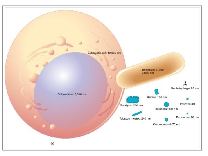

Size Comparisons Among Atoms, Molecules, and Microorganisms
 • Microorganisms need to be colored by stains to")
Staining (Pg 105 to 108) • Microorganisms need to be colored by stains to be viewed. • Stains impart color to cells or their surroundings. • Labs 2 and 3 we will stain bacteria. • There are two general staining methods: – Simple staining – Differential staining

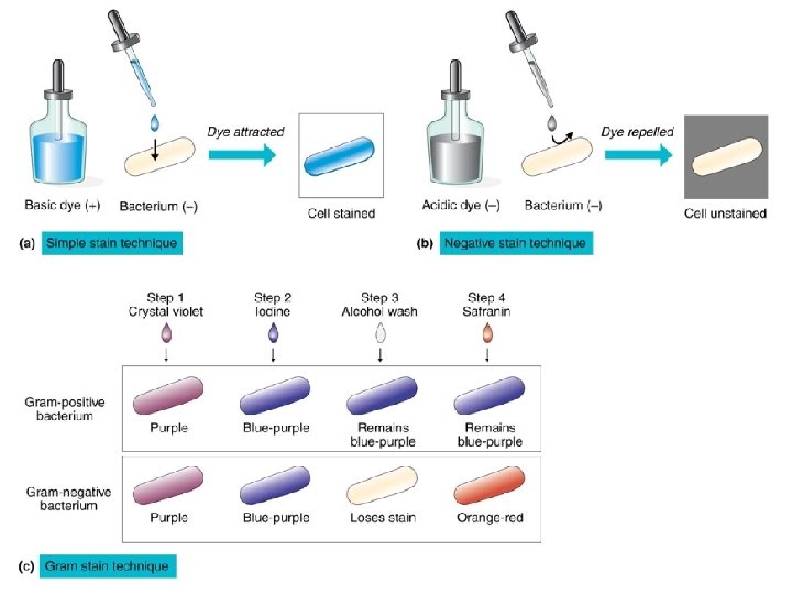
Staining • Simple staining – Uses one dye – Look at size, shape and arrangement – Two simple stains: • Positive • Negative • Differential staining – Two dyes – Differentiate types of bacteria and view special structures – Gram stain, Acid-fast stain, Flagella and Spore

Bacterial Smear Preparation • Take a little bacteria and smear on center of slide. • Allow cells to air dry. • Then briefly heat fix with Bunsen burner flame. – Three reasons for this: • Sticks cell to slide • Kills organism • Increases absorption of stain

Staining • Simple stain – a staining procedure that contrasts stained cells against bright background. • Positive Stain – negative charge of cell binds the positive charge of the dye. • Negative stain – negative charge of the cell repels the positive charge of the dye. – Stains background – No heat fixing used – Get a better idea of the shape and arrangement of cells

Differential Stains • Gram stain – Differentiates bacteria into two major groups. – Gram-negative and Gram-positive. – 4 step staining • crystal violet • iodine (mordent) • alcohol • Safranin – PPP purple, peptidoglycan, positive. – Gram negative counter stain pink with counter

Acid-fast Stain • Identify Mycobacterium. • Poorly stain with Gram stain. • Waxy substance (Mycolic acid) in the membrane prevents. • Heat carbolfuchsin into membrane. • Rinse with acid alcohol. • AF cells hold stain non-acid-fast cells lose stain. • Counter stain with blue counter stain.

• Microscopy

Other Types of Microscopes • Phase-contrast • Dark-field • Fluorescence • Electron
 • Prism splits light. • Increases contrast of images. •")
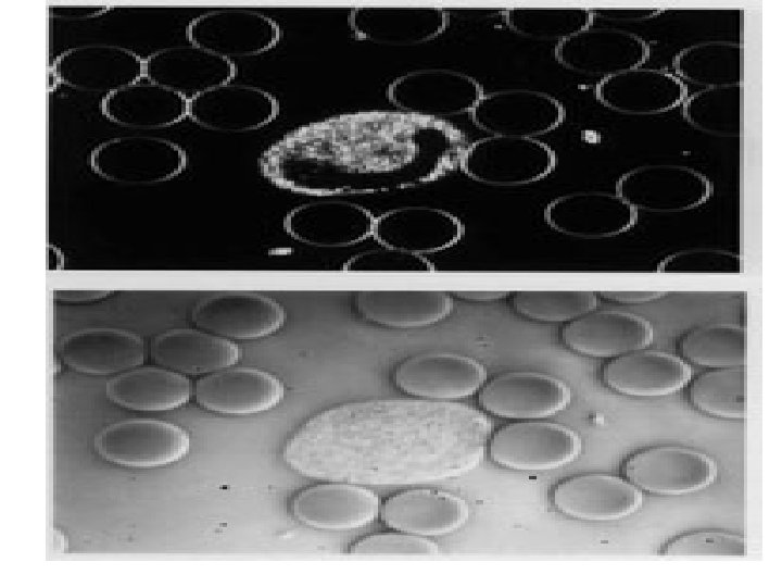
Phase-contrast Microscope (Pg 109) • Prism splits light. • Increases contrast of images. • Image looks shadowed and three-dimensional. • Look at live bacteria.
 • Background remains dark. • Light hits the side of")
Dark-field microscopy (Pg 109) • Background remains dark. • Light hits the side of the specimen. • Cause light reflection against the dark background. • Look at spiral bacteria hard to stain and at the limit of resolution 0. 2 µm. • • ie. Syphilis diameter 0. 15 µm
 • Some chemicals or object fluoresce a certain")
Fluorescence Microscopy (Pg 109 – 110) • Some chemicals or object fluoresce a certain color after light of another color is shined on them. • Microorganism is coated with fluorescent dye (fluorescein) and illuminated with UV. • Fluorescent antibody technique. • Fluorescent dye is attached to antibodies. • The antibody is specific to one bacteria. • Attaches to the bacteria of interest. • Detect with UV

Immunohistochemistry with Antibodies Against EGF-Receptor & HER-2/neu Oncoprotein Neo. Markers Ab 10 – EGF Receptor Zymed TAB 250 – HER-2/neu Oncoprotein

Electron Microscope • Electrons are used instead of light to show image. • Ruska, 1933 electrons flow in a sealed vacuum tube. • Magnets rather then lenses focus the electron beam. • Image projected on screen on the bottom of the microscope. • Resolution 2 nm. • 100 x better than a light microscope

Electron Microscope
: – Transmission electron microscope (TEM)")
Electron Microscope • Two kinds: (Pg 111 – 112): – Transmission electron microscope (TEM) • Electron beam passes through a thin sliced section of the object. – Scanning electron microscope (SEM) • Electron sweeps across the gold coat object. • As the electrons hit the gold it causes shower of electrons to be released. • Detector picks up electrons. • Edges produces less electrons so shows depth (three -dimension).
")
Transmission Electron Microscope (TEM)

Scanning Electron Microscope
- Slides: 52