Basic concepts in Lung disease SS Visser Internal

Basic concepts in Lung disease SS Visser Internal Medicine PAH and UP

Questions • • Why do we need a respiratory system? What does it consist of? How is it controlled/regulated? How is it affected by disease? How is disease recognized? How can disease be prevented or treated? Why do you have know all this?

Contents • • • Function of the respiratory system Embryology Anatomic concepts Physiologic concepts Pathology Clinical : symptoms physical signs disease patterns

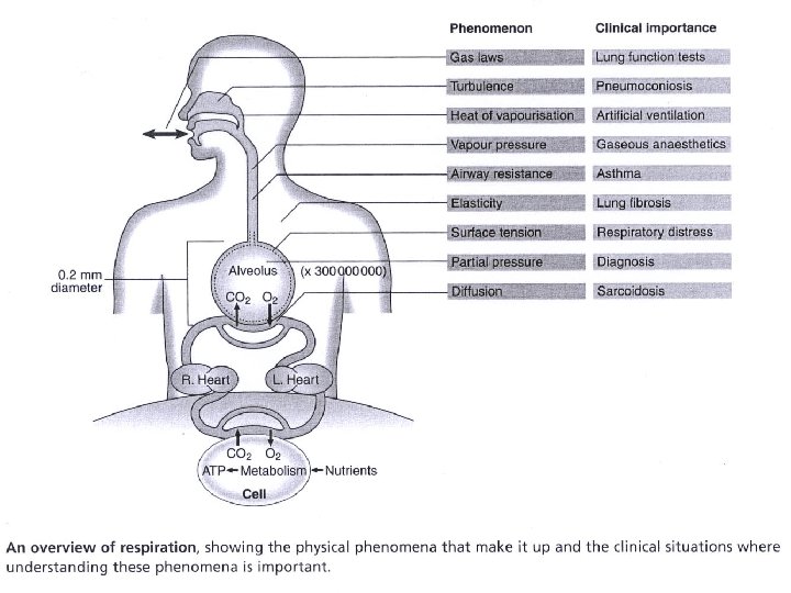
Functions of the lung Respiration: ventilation and gas exchange: O 2, CO 2, p. H, warming and humidifying Non-respiratory functions: • synthesis, activation and inactivation of vasoactive substances, hormones, neuropeptides, eicosanoids, lipoprotein complexes. • Hemostatic functions (thromboplastin, heparin) • Lung defense: complement activation, leucocyte recruitment, cytokines and growth factors • Speech, vomiting, defecation, childbirth

• What does the respiratory system consist of?

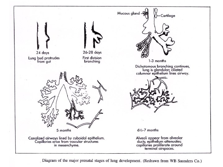
Embryology • Embryology : lung development starts from the gut 24 days after conception; diaphragm forms in cervical region at 3 -4 weeks and moves progressively downwards carrying the phrenic nerves with; lung lobes are identifiable at 12 weeks; bronchial tree is completed at 16 weeks and alveoli and capillaries appear at 24 – 28 weeks; surfactant appears at 35 weeks. • Postnatal Alveolarization: intense first 8 -10 y (alveolar buds – hyperplastic growth) and enlargement of all structures throughout adolescence and early adulthood ( hypertrophic growth)

Embryology and disease • Developmental abnormalities: tracheo-oesophageal fistula, cleft palate, cysts, agenesis, sequestration, cilia dysfunction and abnormal structure, diaphragmatic hernias. • Shared nerve supply (Vagus) between respiratory tract and GI tract – Gastro-oesophageal reflux can increase bronchial secretions (reflexively) and cause bronchial constriction ( together with oesophageal spasm). • Diaphragmatic irritation is often experienced as pain in the cervical region (referred pain) from where it evolved.

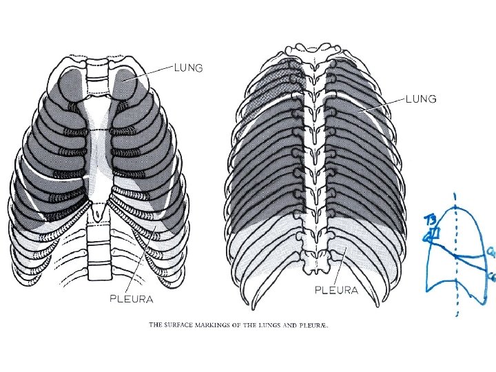
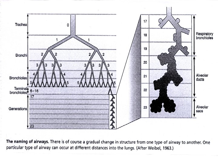
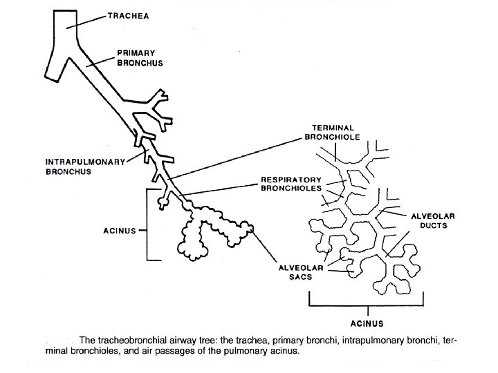
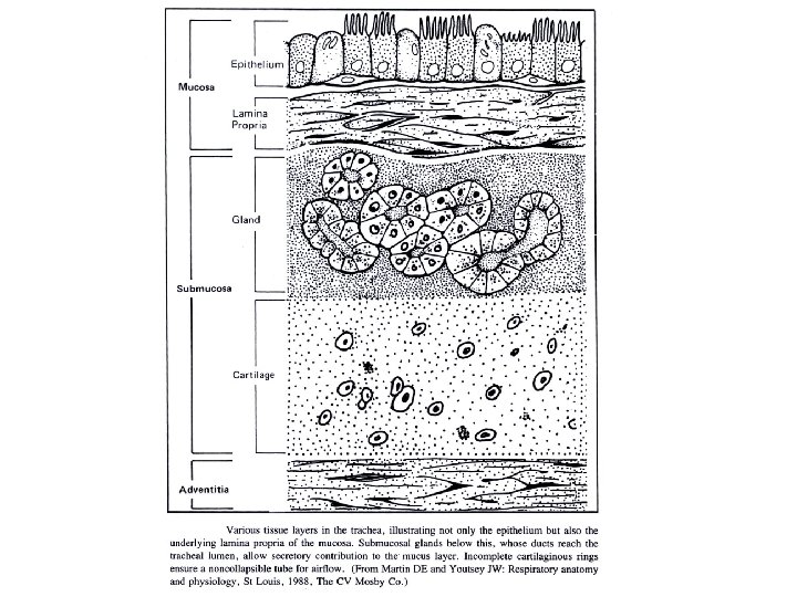
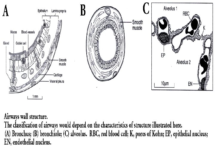
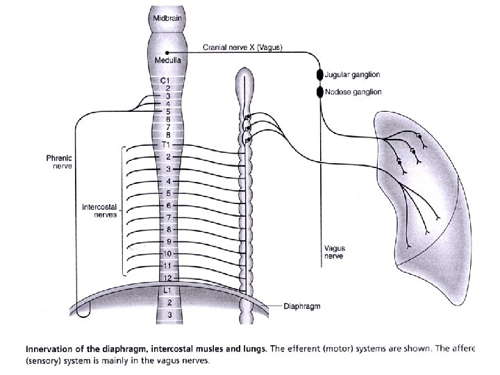
Anatomy • Surface Anatomy: • • • borders of the pleura borders of the lung fissures lung lobes Bronchial tree, vascular and nerve supply, lymphatics. Angle of Louis Histology, cilia, secretory and immunologic cells. Thoracic cage Diaphragm and accessory muscles of breathing

• How is the respiratory system controlled/regulated?

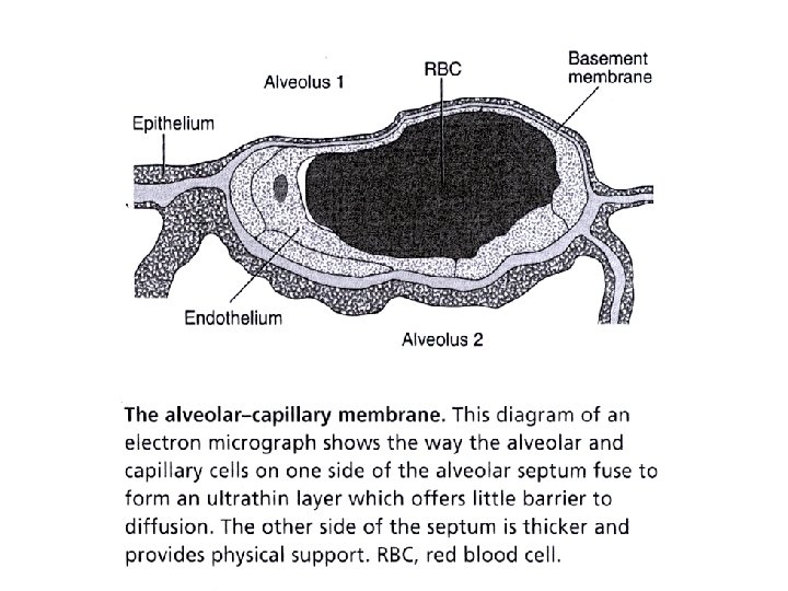
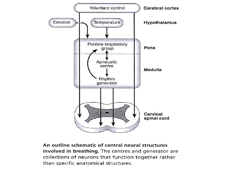
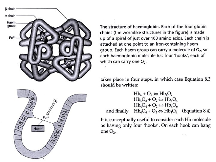
Physiology • Lung mechanics and Lung functions • Airway resistance • Diffusion : Gas laws ( Graham, alveoalar gas equation, Charles, Boyle, Dalton, Henry) • Blood gases: Pa. O 2, Pa. CO 2, p. H, HCO 3, O 2 sat • Hemoglobin, dissociation curve, 2, 3 DPG • Surfactant • Control of Breathing
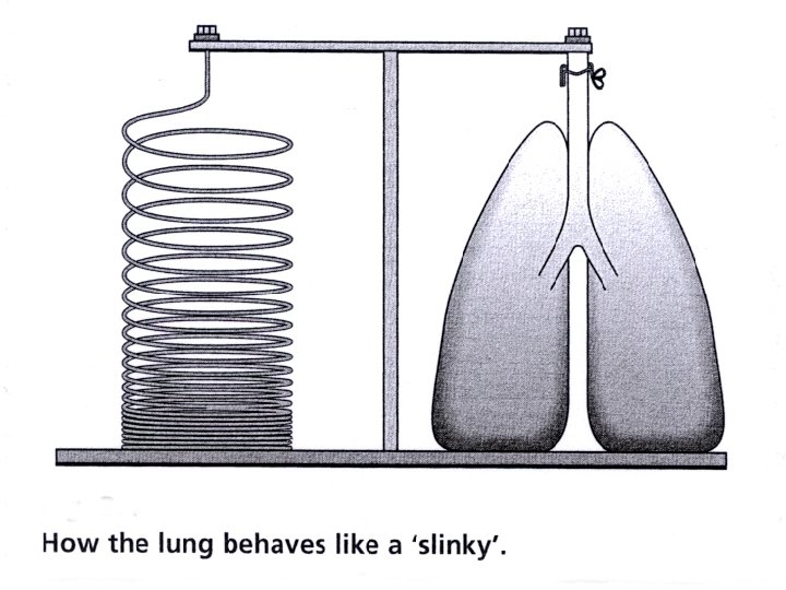

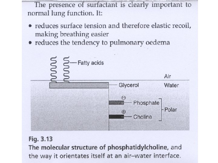
Surfactant • Reduces surface tension and therefore elastic recoil, making breathing easier • Reduces the tendency to pulmonary oedema • Equalises pressure in large and small alveoli
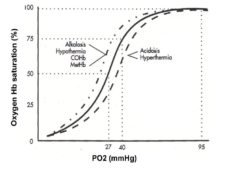

Oxyhemoglobin dissociation curve • Left shift increased HB affinty for O 2 ( release of O 2 to tissues) • Alkalosis • Hypothermia • 2, 3 DPG • COHB • Met. HB • Right shift decreased HB affinity for O 2 ( release of O 2 to tissues) • Acidosis • Hyperthermia • 2, 3 DPG

Hypoxia • Anemic hypoxia- HB • CO intoxication- HB availabilty, shifts O 2 HB dissociation curve to the left • Respiratory hypoxia-next slide • R to L extrapulmonary shunting- ASD, VSD, PDA • Circulatory hypoxiacardiac failure, shock • Ischemic hypoxia- arterial obstruction • Increased O 2 requirements- fever, exercise, thyrotoxicosis • Improper O 2 utilization- cyanide, diptheria toxin

Blood gases: PO 2 and PCO 2 • • • Hypoxemia Hypoventilation Diffusion Ventilation / perfusion inequality AV Shunt High altitude Hypercarbia • Hypoventilation • Ventilation / perfusion inequality

• How is the respiratory system affected by disease?

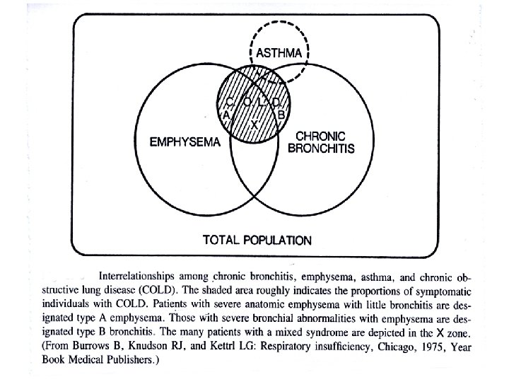
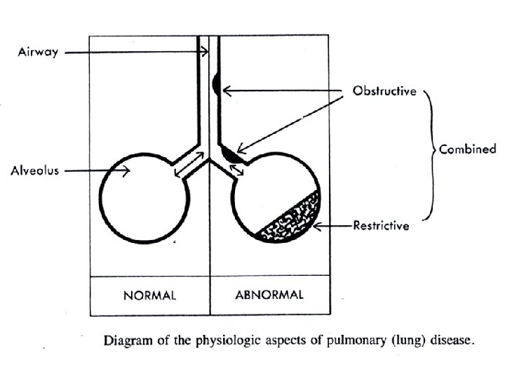
Pathology • Airway diseases: COPD, asthma, bronchiectasis, cystic fibrosis, obstructive sleep apnoea • Parenchymal disease: pneumonia, ARDS, Interstitial lung disease, pneumoconiosis • Pleural disease: pleural effusion, empyema. • Vascular disease: thrombo-embolism, primary pulmonar hypertension • Neoplastic disease: Bronchus Ca, mesothelioma, adenoma, carsinoid
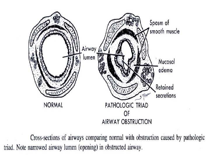
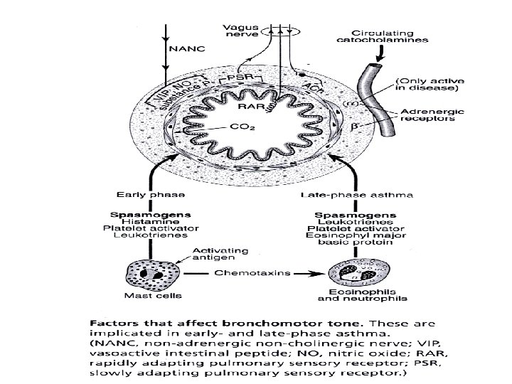

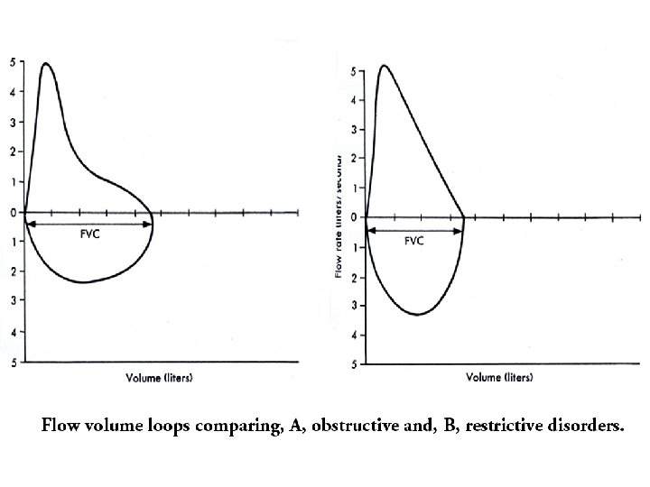
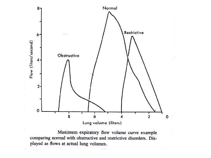
Airway diseases • Causes: atopy, cigarette smoking, infection, abnormal lung defense • Effect: obstruction to airflow • Mechanism: bronchospasm, inflammation, airway remodelling, destruction, collapsing airways • Consequences: air flow ( FEV 1, PEF); work of breathing resp muscle fatigue respiratory failure; Pa. O 2, Pa. CO 2 PHT cor pulmonale

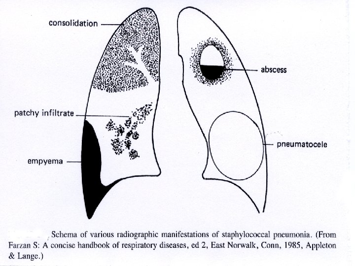
Parenchymal disease • consolidation - infection - typical/atypical • Oedema - cardiac vs non-cardiac (ARDS) • interstitial lung disease - idiopathic fibrosis, sarcoidosis, hypersensitivity pneumonitis, pneumoconiosis • Vascular – secondary/primary PHT, cor pulmonale, pulmonary thrombo-embolism (unexplained dyspnea); Virchow triade: stasis, coagulability, blood vessel abnormality, varicose veins, endothelial dysfunction DVT risk

Pleural disease • Pleural effusion: alb, LDH, pleural/serum, cholesterol, glucose, ADA, p. H. • exudate: infection, inflammation, neoplastic, blood ( permeability) • transudate: hypoproteinemia (renal, liver - oncotic pressure), systemic venous hypertension ( hydrostatic pressure - Heart failure) • Empyema • Chylothorax, pseudo-chylothorax

Neoplastic disease • Bronchus Ca: squamous, small cell ca, adeno ca, large cell ca, broncho-alveolar ca • Mesothelioma • Metastatic ca • Rare tumours: lymphoma, malt-lymphoma • Benign tumours

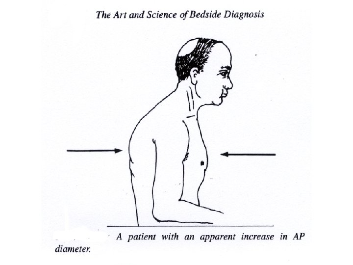
Control and Mechanism of breathing • Alveolar hypoventilation • Sleep-related: central and obstructive sleep apnoea, Ondine’s curse • Neuro-muscular diseases: polio, Guillain. Barre syndrome, myasthenia gravis, resp muscle fatigue, polimyositis • Chest wall: kyphoscoliosis, rib fractures with flail chest

Complications of Lung disease • Cor pulmonale • Respiratory failure: ventilatory failure vs oxygenation failure – hypercapnia, acidosis and hypoxaemia • Endstage lung disease • Pneumothorax

• How is disease of the respiratory system recognized?

Clinical Manifestations • Dyspnea, PND, orthopnea, trepopnea, platypnea and orthodeoxia. • Cough: productive vs non-productive, volume, character, blood, post-nasal discharge • Chest pain: ischaemic, pleuritic, chest wall, GE reflux, tearing of tissue • Constitutional: fever, night sweats, weight loss • RHF: swelling, pain R hypochondrium, abdominal distention, palpitations

Hemoptysis • Upper airway: nasopharyngeal, GIT • Tracheobronchial: neoplasm, bronchitis, bronchiectasis, trauma, foreign body • Parenchyma: pneumonia, lung abscess, TB, mycetoma, SLE, Wegeners, Goodpasture, lung contusion • Primary vascular disease: AV malformations, pulmonary embolism, pulmonary venous pressure • Others: Systemic coagulopathy, anticoagulants, pulmonary endometriosis

Massive hemoptysis • 100 – 250 ml blood per day • Causes: most frequently PTB and bronchiectasis • Rx: maintain oxygenation and prevent blood spilling into unaffected regions, avoid asphyxiation • Suppress cough • Invasive management: double lumen endotracheal tube or balloon catheter to seal off site of bleeding, mechanical ventilation, laser phototherapy, embolotherapy, resection

Respiratory system • • • signs of respiratory distress, hyperinflation, consolidation, pleural effusion, pneumothorax, sup vena cava obstruction

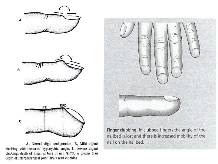
Physical signs • • 1. 2. 3. 4. General: Cyanosis, anaemia, jaundice, oedema, lymphadenopathy, clubbing Respiratory examination: Observation Palpation Percussion Auscultation

Auscultation • Intensity of breath sounds: N, or absent • Character of breath sounds: N or bronchial breathing/ amphoric breathing • Intensity of vocal sounds: (one-one, 99)N, (bronchophony) or nasal ( aegophony) • Whispering pectoriloquy ( 66) • Adventitious sounds: ronchi, creps, rubs, clicks.

APPLICATION OF ADVENTITIOUS LUNG SOUNDS Lung sounds Possible mechanism Characteristics Causes Wheezes Rapid airflow through obstructed High-pitched; most airways caused by bronchospasm, often occur during mucosal edema exhalation Asthma, congestive heart failure, bronchitis Stridor Rapid airflow through obstructed airway caused by inflammation High-pitched; often occurs during inhalation Croup, epiglottitis, postextubation Insp & exp Excess airway secretions moving with airflow Coarse and often clear with cough Bronchitis, respiratory infections Early insp Sudden opening of proximal bronchi Scanty, transmitted to mouth; not affected by cough Bronchitis, emphysema, asthma Late insp Sudden opening of peripheral airways Diffuse, fine; occur initially in dependent regions Atelectasis, pneumonia, pulmonary edema, fibrosis Crackles

Abnormality Initial impression Inspection Palpitation Percussion Ausculation Possible causes Acute airways obstruction Appears acutely ill Use of accessory muscles Reduced expansion Increased resonance Expiratory wheezing Asthma, bronchitis Chronic airways obstruction Appears chronically ill Increased antero -posterior diameter, use of accessory muscles Reduced expansion Increased resonance Diffuse reduction in breath sounds; early inspiratory crackles Chronic bronchitis, emphysema Consolidation May appear acutely ill Inspiratory lag Increased fremitus Dull note Bronchial breath sounds; crackles Pneumonia, tumor Pneumothorax May appear acutely ill Unilateral expansion Decreased fremitus Increased resonance Absent breath sounds Rib fracture, open wound Pleural effusion May appear acutely ill Unilateral expansion Absent fremitus Dull note Absent breath sounds Congestive heart failure Local bronchial obstruction Appears acutely ill Unilateral expansion Absent fremitus Dull note Absent breath sounds Mucous plug Diffuse intersitial fibrosis Often normal Rapid shallow breathing Often normal; increased fremitus Slight decrease in resonance Late inspiratory crackles Chronic exposure to inorganic dust Acute upper airway obstruction Appears acutely ill Laboured breathing Often normal Inspiratory or expiratory stridor or both Epiglottitis, croup, foreign body aspiration

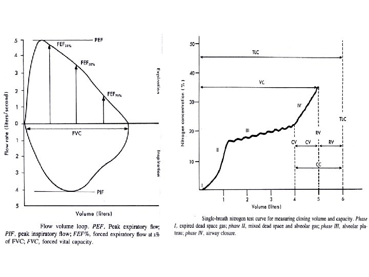
Diagnostic procedures • • XRC, CT scan, MRI scan Lung functions Blood gases Sputum, cilia function Bronchoscopy, biopsy Nuclear medicine

• How can disease of the respiratory system be treated or prevented?

Treatment/prevention • Patient education • Immunization • Medication: antibiotics, bronchodilators, anti-inflammatory drugs, diuretics, anticoagulants • Ventolators • Physiotherapy • Surgery

Why do you have to know all this? • Because so that you can one day say: “ Trust me, I am your doctor!”
- Slides: 65