BASIC ANATOMY OF THE NERVOUS SYSTEM Basic conception

BASIC ANATOMY OF THE NERVOUS SYSTEM
 CNS efferent information (nerves, axons, tracts) ascending")
Basic conception afferent information (nerves, axons, tracts) CNS efferent information (nerves, axons, tracts) ascending information (tracts) descending information (tracts)

NERVE CELL = NEURON

TYPES OF NEURONS

DIVISION OF THE NERVOUS SYSTEM CNS oligodendrocytes astrocytes PNS Schwann cells and their derivatives sensors stimulus impulse effectors Striated skeletal muscles Smooth muscles, myocardium, glands

DIVISION OF THE PNS Cranial nerves I. - XII. • run through the skull base Spinal nerves – 31 pairs • run through foramina intervertebralia
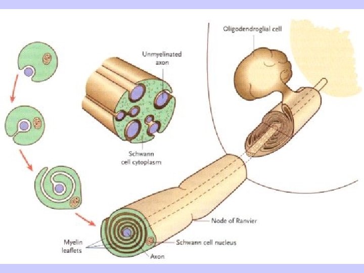

Glial cells of the CNS: astrocytes, oligodendrocytes, microglial, ependymal cells Glial cells of the PNS: myelinating and non-nemyelinating Schwann cells, satellite glial cells, terminal glial cells
 myelinated axons")
unmyelinated axons (< 1 m) myelinated axons

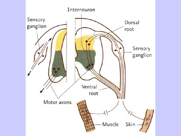
Afferent FUNCTIONAL TYPES OF AXONS IN PNS somatosensory touch, proprioception, pain viscerosensory mechanoception, pain Efferent sensory relay impulses for taste, hearing and balance somatomotor striated muscles branchiomotor striated muscles visceromotor smooth muscles sympathetic parasympathetic myocardium glands
 Spinal cord (Medulla spinalis) Brainstem (Truncus encephali) Medulla")
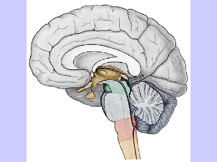
DIVISION OF THE CNS Brain (Encephalon) Spinal cord (Medulla spinalis) Brainstem (Truncus encephali) Medulla oblongata Pons Mesencephalon Cerebellum Diencephalon Telencephalon
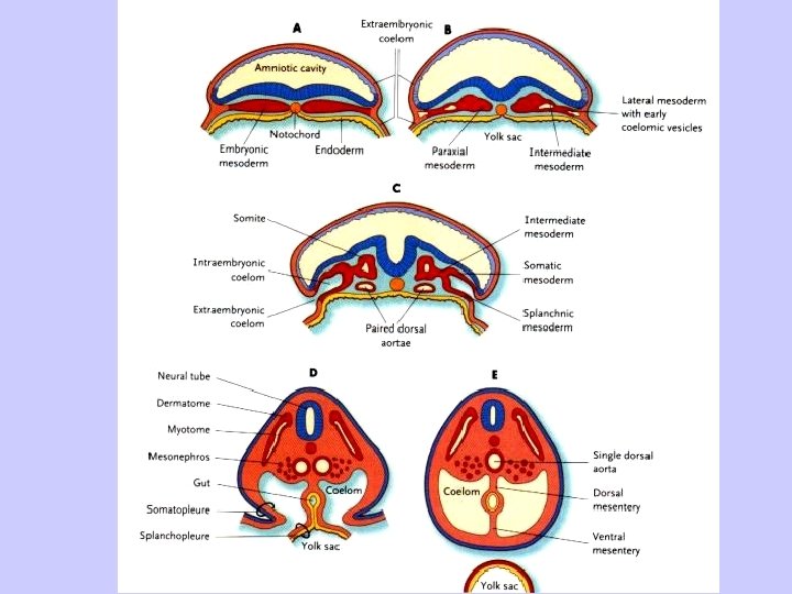

Neural crest

sulcus limitans

Primary subdivisions: prosencephalon, mesencephalon, rhombencephalon Secondary subdivision: telencephalon, diencephalon, mesencephalon, metencephalon, myelencephalon metencephalon mesencephalon medulla spinalis diencephalon telencephalon

Conus medullaris Filum terminale Cauda equina

Spinal segment Fila radicularia

Intumescentia cervicalis C 3 – T 2 Intumescentia lumbalis T 9 – T 12

Segment C 5 Segment C 1 Segment C 8 Segment C 5 Segment C 8 Segment Th 2 Segment L 4 Segment Th 10 Segment L 1 Segment S 4 Segment L 4 Segment S 4
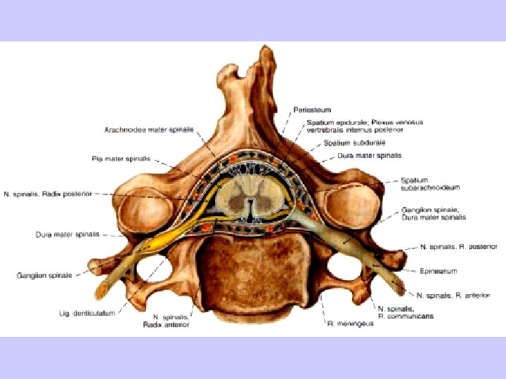
, cornu posterius (columna posterior), cornu laterale (columna")
SUBSTANTIA GRISEA – cornu anterius (columna anterior), cornu posterius (columna posterior), cornu laterale (columna lateralis), substantia intermedia, canalis centralis

SUBSTANTIA ALBA – funiculus anterior, lateralis, posterior fissura mediana ant. , sulcus medianus post. , septum medianum posterius, sulcus anterolateralis, posterolateralis

FUNCTIONAL ZONES IN THE NEURAL TUBE S SS VS cc VM SM BM

Cornu posterius FR CC Substantia intermedia Cornu anterius

Pseudounipol. neurons of the DRG Radix dorsalis Tr. dorsolateralis Lissaueri Fasc. gracilis Tr. spinobulbaris Ncl. posteromarginalis + Subst. gelatinosa Rolandi Fasc. cuneatus Ncl. proprius Ncl. thoracicus (Stilling-Clark. ) Tr. spinocerebellaris post. Ncl. intermediomedialis Tr. spinocerebellaris ant. Tr. spino-thalamicus, -reticularis, -tectalis Tr. spino-olivaris Tr. olivo-spinalis

Ncl. intermediolateralis Ncl. motorius

Tr. corticospinalis lat. Tr. rubrospinalis Tr. reticulospinalis Tr. olivospinalis Tr. tectospinalis Tr. corticospinalis ant. Tr. vestibulospinalis
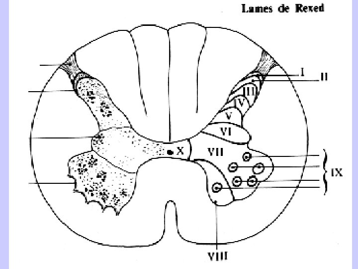
 nuclei I ncl. apicalis (ncl. posteromarginalis) II + III substantia gelatinosa")
laminae (Rexed 1952) nuclei I ncl. apicalis (ncl. posteromarginalis) II + III substantia gelatinosa Rolandi IV + V ncl. proprius VI ncl. thoracicus (Stilling - Clark) C 8 -L 3 VII group of interneurons in the anterior horn VIII medial group of motoneurons IX lateral group of motoneurons X zona centralis, gray matter around the central canal
- Slides: 31