Basal Lamina Basement Membrane Dr Mah Jabeen Muneera

Basal Lamina & Basement Membrane Dr. Mah Jabeen Muneera Associate Professor Department of Anatomy KEMU
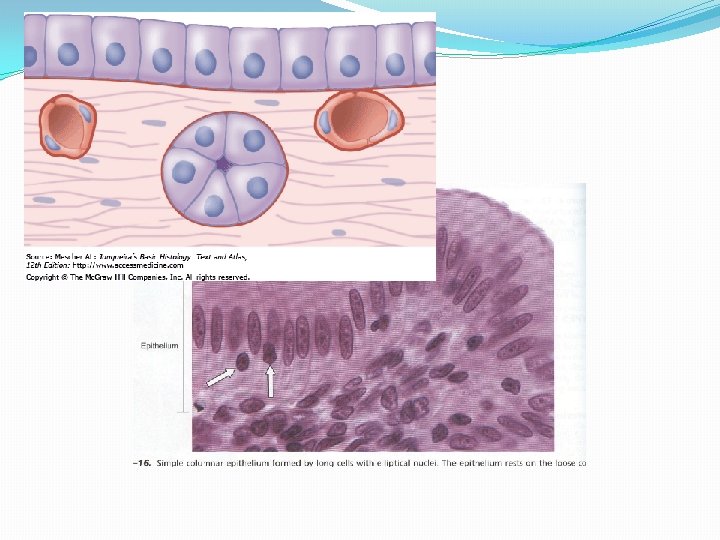

LAMINA PROPRIA �Connective tissue upon which epithelium rests �Support, provide nutrition, epithelium is avascular �Area of contact is increased by folds BASEMENT MEMBRANE �Sheet of ECM intervenes between epithelium and lamina propria �Source �Function �Special stain (PAS) �Also present around glial cells, adipocytes & muscle cells �Not present around other CT cells
 �LAMINA")
Basal lamina �Evident by routine EM preparation �Composed of upper LAMINA LUCIDA (glycoproteins) �LAMINA DENSA on lower aspect

Components �Laminin � Glycoproteins � Associated with integrins �Type IV collagen, XVIII, VII �Entactin (Nidogen) & Perlecan, Agrin � Link the first two components � Proteoglycans ; hydrated, � negative charge controls ions

Tissue preparation by HPF method �HPF- high pressure freezing �Only lamina densa is present �Lamina lucida is considered to be an artifact

External lamina �In non epithelial cells

Formation of basal lamina �Self assembly of collagen type IV & Laminin �Calcium dependent polymerization

RETICULAR LAMINA �FUNCTIONS: �Support �Barrier �Influence cellular activity e. g. proliferation, differentiation BASEMENT MEMBRANE �PAS +ve layer �Visible by Light Microscope �Two basal laminae �One basal lamina & one reticular lamina

Attachment of basal lamina to underlying tissue �Anchoring fibrils �Mutations-dystrophic epidermolysis bullosa �Fibrillin microfibrils �Marfan’s syndrome �Projections of basal lamina

Functions �Regulator of cell behavior �Organ specific molecules �Structural attachment �Compartmentalization �Filtration �Tissue Scafolding (barrier against tumor cell invasion) �Regulation & signaling
- Slides: 12