BASAL GANGLIA Dr JAMILA EL MEDANY OBJECTIVES q

BASAL GANGLIA Dr JAMILA EL MEDANY

OBJECTIVES q q q At the end of the lecture, the student should be able to: Define “basal ganglia” and enumerate its components. Enumerate parts of “Corpus Striatum” and their important relations. Describe the structure of Caudate and Lentiform (Putamen & Globus Pallidus) nuclei. Differentiate between striatum & paleostriatum in term of connections. State briefly functions & dysfunctions of Corpus Striatum.
 1. 2. Group of nerve cells deeply situated in cerebral hemispheres")
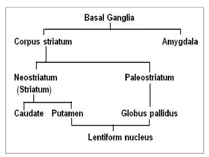
BASAL GANGLIA (NUCLEI) 1. 2. Group of nerve cells deeply situated in cerebral hemispheres Components: Caudate Nucleus Lentiform Nucleus: divided into Putamen & Globus Pallidus 3. Amygdaloid Nucleus CN LN A N

Caudate & Lentiform nuclei are functionally related to each other & called “Corpus Striatum”: Part of extrapyramidal motor system, system principally involved in the control of posture and movements (primarily by inhibiting motor functions) Amygdaloid Nucleus (part of limbic system) is only embryologically related to Corpus Striatum
 Bands of grey matter pass from lentiform nucleus across the internal")
CORPUS STRIATUM (Nomenclature) Bands of grey matter pass from lentiform nucleus across the internal capsule to the caudate nucleus, giving the striated appearance hence, the name corpus striatum

PARTS q q LENTIFORM NUCLEUS SHAPE: three sided, wedge-shaped mass of grey matter, with a convex outer surface and an apex which lies against the genu of the internal capsule (G) DIVISION: divided into 1. Larger darker lateral portion called Putamen (P) 2. Smaller, lighter medial portion called Globus Pallidus (g) G q
")
Putamen is more closely related to Caudate nucleus (regarding development, function & connections) and together constitute the Neostriatum or Striatum. Globus Pallidus is the oldest part of corpus striatum and is called Paleostriatum or Pallidum

PUTAMEN Separated from globus pallidus by a thin sheath of nerve fibers, the Lateral Medullary Lamina The white matter lateral to putamen is divided, by a sheath of grey matter, the Claustrum into two layers: § External capsule (1) between the putamen and claustrum and § Extreme capsule (2) between the claustrum and the insula Insula 2 1

GLOBUS PALLIDUS Consists of two divisions, the Lateral & the Medial segments, separated by a thin sheath of nerve fibers, the Medial Medullary lamina The medial segment is similar, in terms of cytology and connections with the pars reticulata of substantia nigra

CAUDATE NUCLEUS SHAPE: C-shaped mass of grey matter COMPONENTS: head, body & tail Head: -Rounded in shape -Lies anterior to thalamus (in frontal lobe) -Completely separated from the putamen by the internal capsule except rostrally where it is continuous with the putamen
 Tail: Tail")
CAUDATE NUCLEUS Body: -Long & narrow -Extends above thalamus (in parietal lobe) Tail: Tail -Long & tapering -Descends into temporal lobe -Continuous with Amygdaloid Nucleus
 Head of Caudate Nucleus lies: Anterior to thalamus Medial to")
CORPUS STRIATUM (Important relations) Head of Caudate Nucleus lies: Anterior to thalamus Medial to Lentiform & separated from it by anterior limb of internal capsule (A) Lentiform Nucleus: Lateral to thalamus & separated from it by posterior limb of internal capsule (P) A P
 “The input portion of Corpus striatum” Cerebral Cortex Striatu m")
STRIATUM (CAUDATE & PUTAMEN) “The input portion of Corpus striatum” Cerebral Cortex Striatu m G. P. Lateral segme nt Pars compact a Substantia Nigra Pars reticulat a G. P. Medial segme nt Thalamus (Intralamin ar nuclei) Afferents Efferents
 “The output portion of corpus striatum: medial segment of G. P.")
PALEOSTRIATUM (GLOBUS PALLIDUS) “The output portion of corpus striatum: medial segment of G. P. + Pars Reticulata of S. N. ” Thalamic fasciculus Striatu m G. P. Lateral segme nt G. P. Medial segme nt Thalamus (Ventral lateral, Ventral anterior, centromedian) Subthalamic fasciculus Afferents Efferents Subthalam ic Nucleus Pars reticulat a Substantia Nigra

CORPUS STRIATUM Function q q q The corpus striatum assists in regulation of voluntary movement and learning of motor skills as they: Facilitate behavior and movement that are required and appropriate. Inhibit unwanted or inappropriate movement.

Dysfunction Its dysfunction does NOT cause: paralysis, sensory loss or ataxia It leads to: I. Abnormal motor control: emergence of abnormal, involuntary movements (dyskinesias) II. Alteration in muscle tone: hypertonia/hypotonia
- Slides: 17