
BARRETTS ESOPHAGUS Minesh Mehta PGY4 University of Louisville

BARRETT’S ESOPHAGUS Minesh Mehta, PGY-4 University of Louisville Department of Gastroenterology
OUTLINE Importance of Barrett’s Esophagus Diagnosis Screening Surveillance Diagnosis and Management of Dysplasia Endoscopic Therapy for Barrett’s Related Neoplasia
SIGNIFICANCE OF BE Acquired condition in response to Gastro-esophageal reflux leading to columnar lined epithelium in the distal esophagus Intestinal metaplasia, characterized by goblet cells, is biologically unstable with greatest risk of neoplastic progression Making it a major risk factor in development of esophageal adenocarcinoma Incidence of EAC rising rapidly in western countries Wang KK, Sampliner RE, Practice Parameters Committee of the American College of G. Updated guidelines 2008 for the diagnosis, surveillance and therapy of Barrett's esophagus. Am J Gastroenterol 2008; 103: 788 -797.
DEFINITION OF BE Change in the distal esophageal epithelium of any length that can be recognized as columnar type mucosa at endoscopy and is confirmed to have intestinal metaplasia by biopsy of the esophagus Clearly visible endoscopically above the GEJ(>1 cm) and confirmed histo-pathologically with distal esophageal biopsies of columnar lined epithelium only
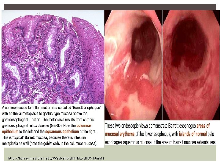
http: //library. med. utah. edu/Web. Path/GIHTML/GIIDX. html#1
ENDOSCOPIC DIAGNOSIS OF BE Must distinguish BE from Irregular Z-line Where the squamo-columnar junction appears with tongues of columnar epithelium less than 1 cm with no confluent columnar lined segments Biopsies are generally NOT RECCOMENDED for Irregular Zline If done, then label as GEJ not esophageal biopsies
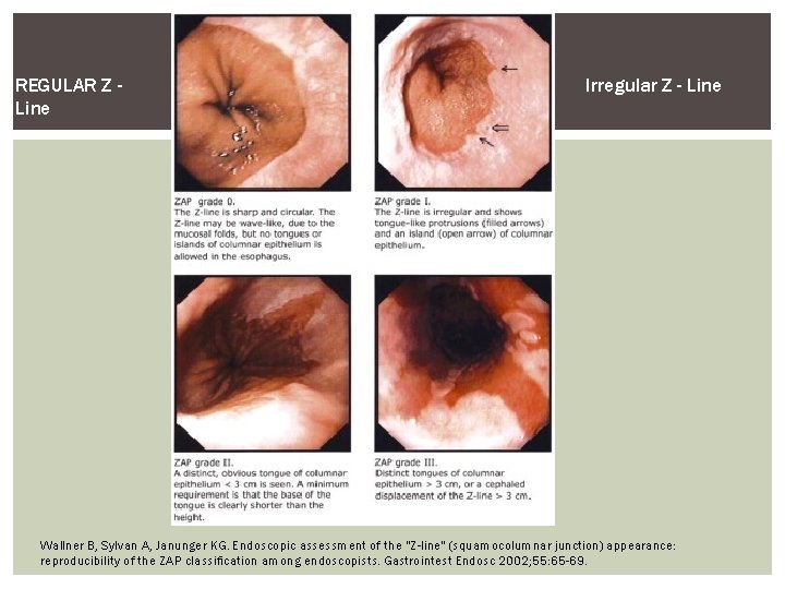
REGULAR Z Line Irregular Z - Line Wallner B, Sylvan A, Janunger KG. Endoscopic assessment of the "Z-line" (squamocolumnar junction) appearance: reproducibility of the ZAP classification among endoscopists. Gastrointest Endosc 2002; 55: 65 -69.
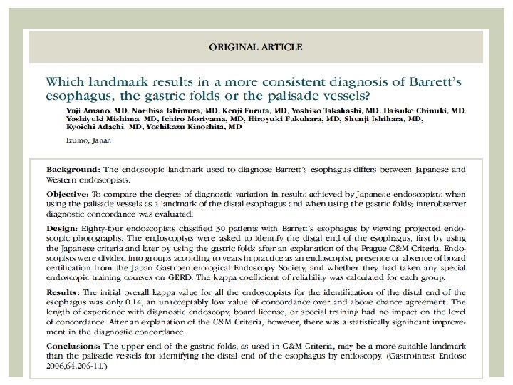
Must accurately delineate the GEJ by detecting the proximal end of the longitudinal Gastric Folds with minimal air insufflation Columnar epithelium should be visible endoscopically (minimum of 1 cm) above the GEJ to diagnose as BE
ENDOSCOPIC REPORTING OF BE Should be done using the Prague Criteria Which assesses the circumferential (C) and maximal (M) extent of endoscopically visualized Barrett’s Segment Diaphragmatic Pinch and Proximal End of the Gastric Folds The presence and location of visible lesions should be recorded according to Paris Classification
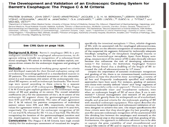
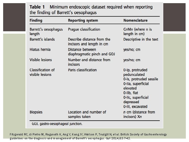
Fitzgerald RC, di Pietro M, Ragunath K, Ang Y, Kang JY, Watson P, Trudgill N, et al. British Society of Gastroenterology guidelines on the diagnosis and management of Barrett's oesophagus. Gut 2014; 63: 7 -42.
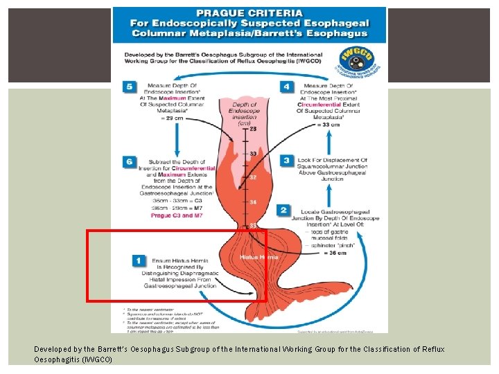
Developed by the Barrett’s Oesophagus Subgroup of the International Working Group for the Classification of Reflux Oesophagitis (IWGCO)
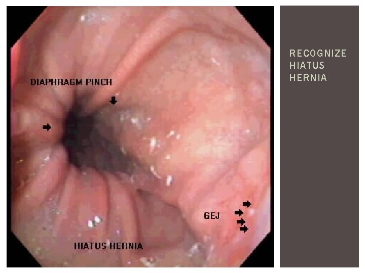
RECOGNIZE HIATUS HERNIA
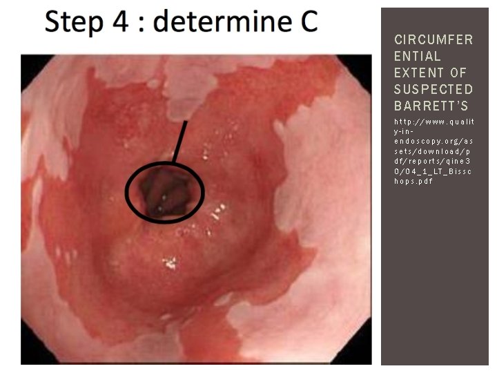
CIRCUMFER ENTIAL EXTENT OF SUSPECTED BARRETT’S http: //www. qualit y-inendoscopy. org/as sets/download/p df/reports/qine 3 0/04_1_LT_Bissc hops. pdf
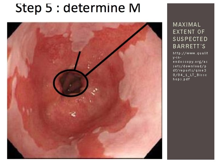
MAXIMAL EXTENT OF SUSPECTED BARRETT’S http: //www. qualit y-inendoscopy. org/as sets/download/p df/reports/qine 3 0/04_1_LT_Bissc hops. pdf
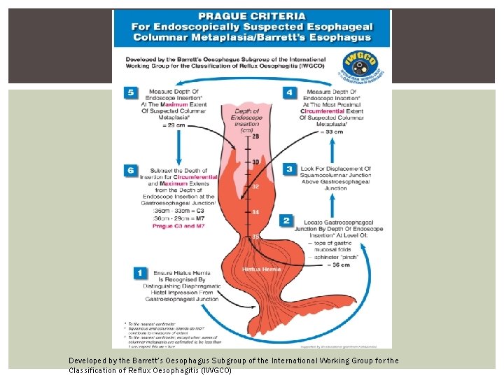
Developed by the Barrett’s Oesophagus Subgroup of the International Working Group for the Classification of Reflux Oesophagitis (IWGCO)
BIOPSY PROTOCOL Minimum of 8 biopsies provides adequate assessment of Intestinal Metaplasia Starting distally 1 -2 cm above GEJ, 4 quadrant random biopsies should be done every 2 cm while advancing proximally Targeted biopsies of visible lesions (should be done before random biopsies) Ideally, erosive esophagitis should be healed prior to biopsies for BE to avoid missing short segments of columnar epithelium Harrison R, Perry I, Haddadin W, Mc. Donald S, Bryan R, Abrams K, Sampliner R, et al. Detection of intestinal metaplasia in Barrett's esophagus: an observational comparator study suggests the need for a minimum of eight biopsies. Am J Gastroenterol 2007; 102: 1154 -1161. Hanna S, Rastogi A, Weston AP, Totta F, Schmitz R, Mathur S, Mc. Gregor D, et al. Detection of Barrett's esophagus after endoscopic healing of erosive esophagitis. Am J Gastroenterol 2006; 101: 1416 -1420.
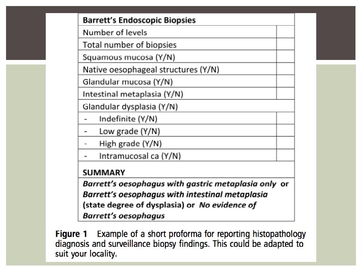
MINIMUM DATASET To improve standard of care and ease of communication between experts, the use of a minimum dataset is recommended to record histo-pathologic findings
SCREENING FOR BE Screening for BE in unselected population of patients with gastro esophageal reflux symptoms is not recommended Screening should be considered in patients with chronic GERD (> 13 years) and multiple risk factors including at least 3 of the following § § Age 50 or older White race Male sex Obesity Threshold of multiple risk factors should be lowered in those with a first degree relative with Barrett’s or esophageal adenocarcinoma
SURVEILLANCE Aim of surveillance is to detect cancer or pre-cancer at a stage when intervention may be curative Surveillance correlates with earlier staging and improved survival from cancer Endoscopic monitoring with histo-pathologic assessment of dysplasia is the only current method of surveillance Patients should have access to outpatient clinic to discuss diagnosis of BE and discussion of pros and cons of surveillance High resolution Endoscopy should be used for surveillance
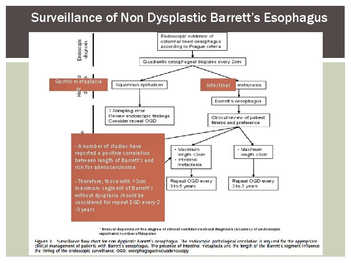
Surveillance of Non Dysplastic Barrett’s Esophagus Gastric metaplasia or - A number of studies have reported a positive correlation between length of Barrett’s and risk for adenocarcinoma - Therefore, those with >3 cm maximum segment of Barrett’s without dysplasia should be considered for repeat EGD every 2 -3 years Intestinal
PATHOLOGIC FEATURES AND REPORTING OF DYSPLASIA Dysplasia should be reported as low or high grade (controversy over indefinite) Any grade of dysplasia should be confirmed by an expert or two independent pathologist Revised Vienna Classification for GI mucosal neoplasia standardizes diagnostic terminology into biologically similar groupings with scores of 1 -5 depending on presence/absence of dysplasia or malignancy § 1. § 2. § 3. § 4. § 5. Negative for Dysplasia Indefinite for Dysplasia Low Grade Dysplasia High Grade Dysplasia Submucosal Invasion of Adenocarcinoma
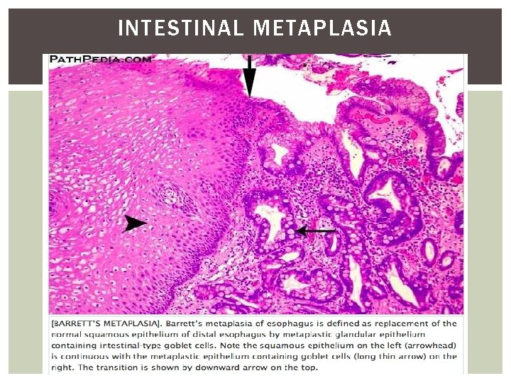
INTESTINAL METAPLASIA
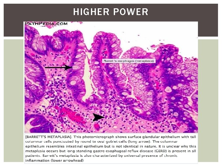
HIGHER POWER
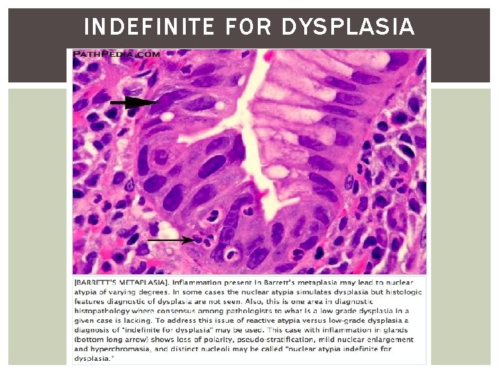
INDEFINITE FOR DYSPLASIA
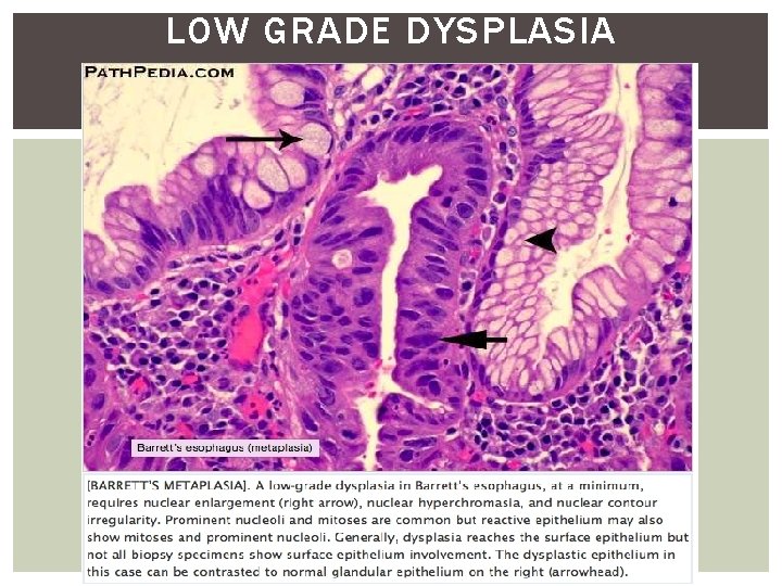
LOW GRADE DYSPLASIA
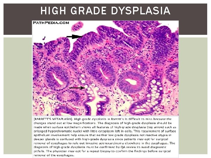
HIGH GRADE DYSPLASIA
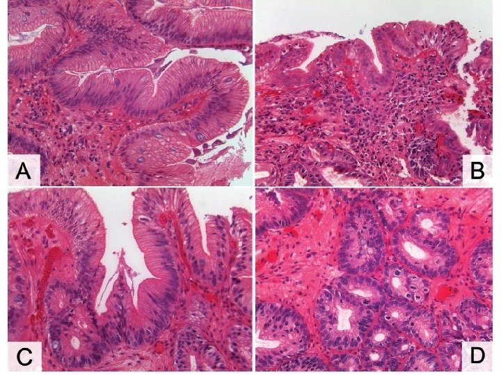
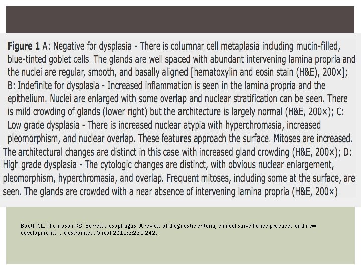
Booth CL, Thompson KS. Barrett's esophagus: A review of diagnostic criteria, clinical surveillance practices and new developments. J Gastrointest Oncol 2012; 3: 232 -242.
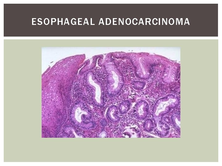
ESOPHAGEAL ADENOCARCINOMA
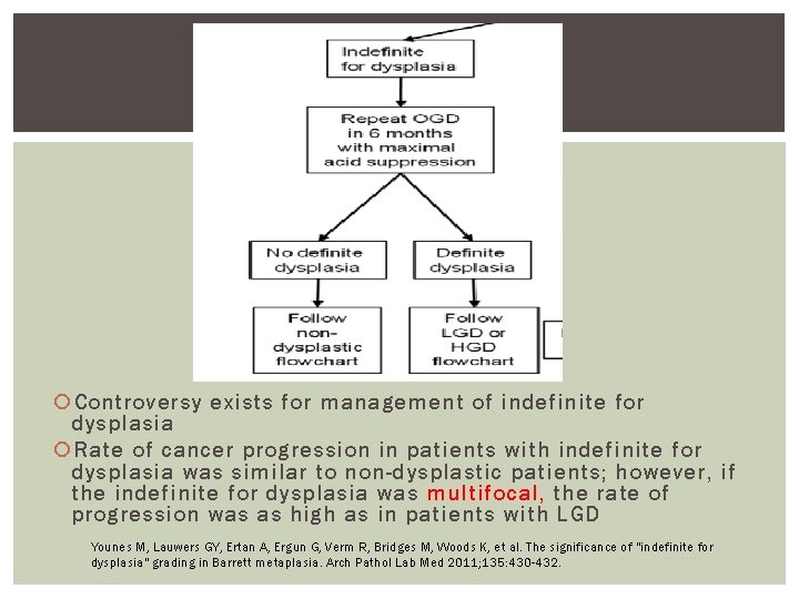
Controversy exists for management of indefinite for dysplasia Rate of cancer progression in patients with indefinite for dysplasia was similar to non-dysplastic patients; however, if the indefinite for dysplasia was multifocal, the rate of progression was as high as in patients with LGD Younes M, Lauwers GY, Ertan A, Ergun G, Verm R, Bridges M, Woods K, et al. The significance of "indefinite for dysplasia" grading in Barrett metaplasia. Arch Pathol Lab Med 2011; 135: 430 -432.
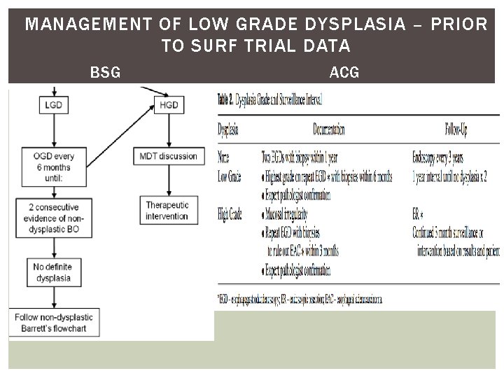
MANAGEMENT OF LOW GRADE DYSPLASIA – PRIOR TO SURF TRIAL DATA BSG ACG
BIOPSY PROTOCOL FOR KNOWN DYSPLASIA Obtain every 1 cm in patients with known dysplasia
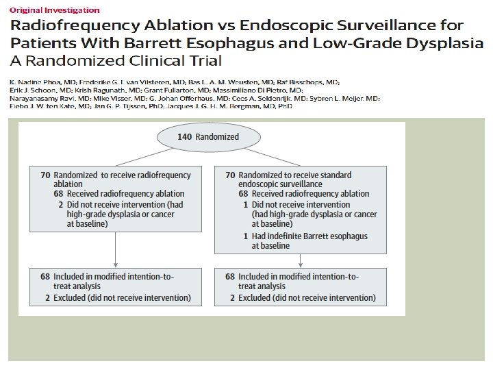
Primary outcome: occurrence of HGD or Adenocarcinoma anytime during 3 years following randomization Secondary outcomes: histological eradication of dysplasia and intestinal metaplasia, and adverse events
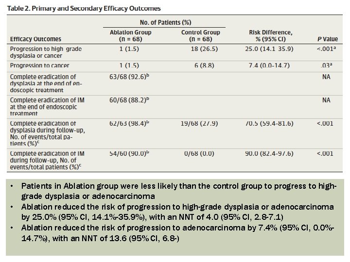
• Patients in Ablation group were less likely than the control group to progress to highgrade dysplasia or adenocarcinoma • Ablation reduced the risk of progression to high-grade dysplasia or adenocarcinoma by 25. 0% (95% CI, 14. 1%-35. 9%), with an NNT of 4. 0 (95% CI, 2. 8 -7. 1) • Ablation reduced the risk of progression to adenocarcinoma by 7. 4% (95% CI, 0. 0%14. 7%), with an NNT of 13. 6 (95% CI, 6. 8 -)
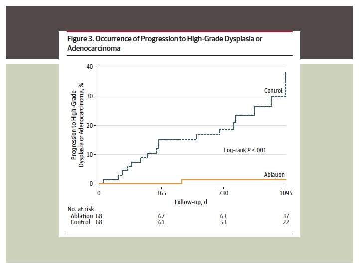
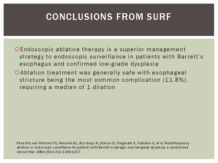
CONCLUSIONS FROM SURF Endoscopic ablative therapy is a superior management strategy to endoscopic surveillance in patients with Barrett’s esophagus and confirmed low-grade dysplasia Ablation treatment was generally safe with esophageal stricture being the most common complication (11. 8%), requiring a median of 1 dilation Phoa KN, van Vilsteren FG, Weusten BL, Bisschops R, Schoon EJ, Ragunath K, Fullarton G, et al. Radiofrequency ablation vs endoscopic surveillance for patients with Barrett esophagus and low-grade dysplasia: a randomized clinical trial. JAMA 2014; 311: 1209 -1217.
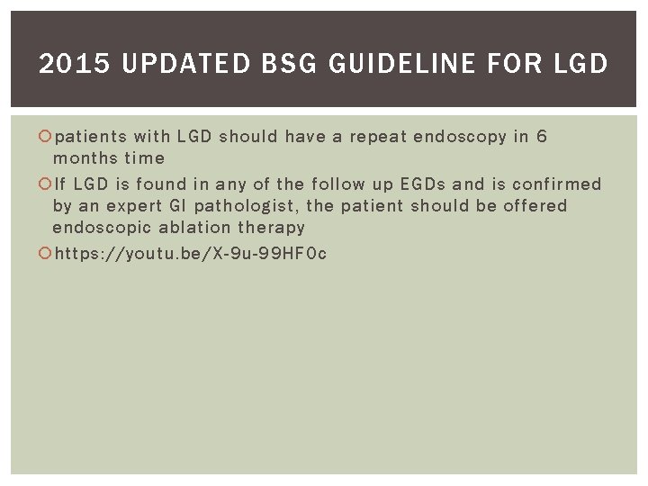
2015 UPDATED BSG GUIDELINE FOR LGD patients with LGD should have a repeat endoscopy in 6 months time If LGD is found in any of the follow up EGDs and is confirmed by an expert GI pathologist, the patient should be offered endoscopic ablation therapy https: //youtu. be/X-9 u-99 HFOc
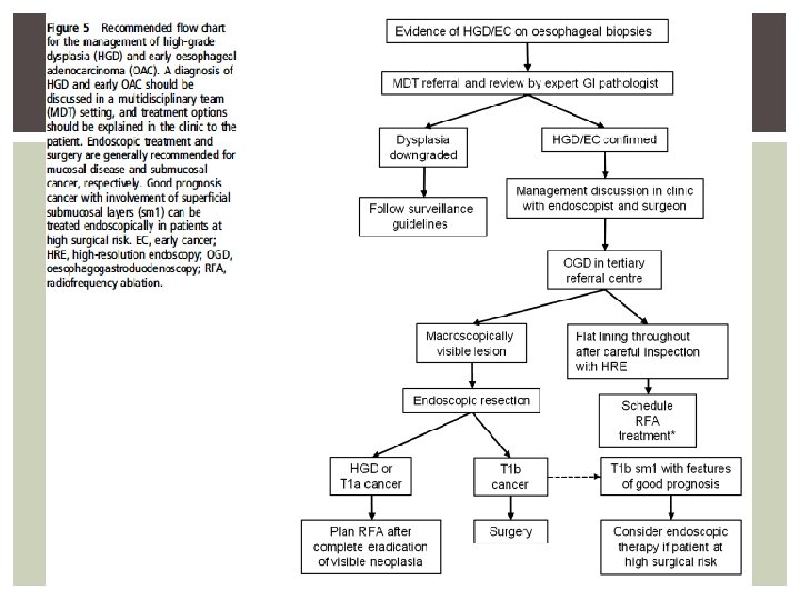
MANAGEMENT OF HIGH GRADE DYSPLASIA If the possibility of high grade dysplasia has been raised, it is critical that a high-quality baseline endoscopy is done to map out any visible lesions and extent of dysplasia Endoscopic assessment will usually identify the area with the most advanced neoplasia. EMR should aim to resect all visible abnormalities. HGD and Barrett’s-related adenocarcinoma confined to the mucosa, endoscopic therapy is preferred over esophagectomy or endoscopic surveillance
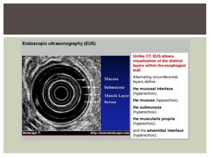
ESOPHAGEAL WALL
ESOPHAGEAL CANCER STAGING
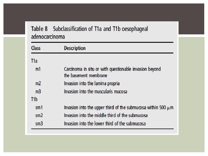
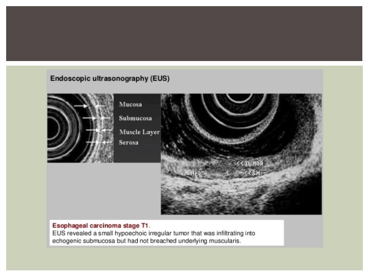
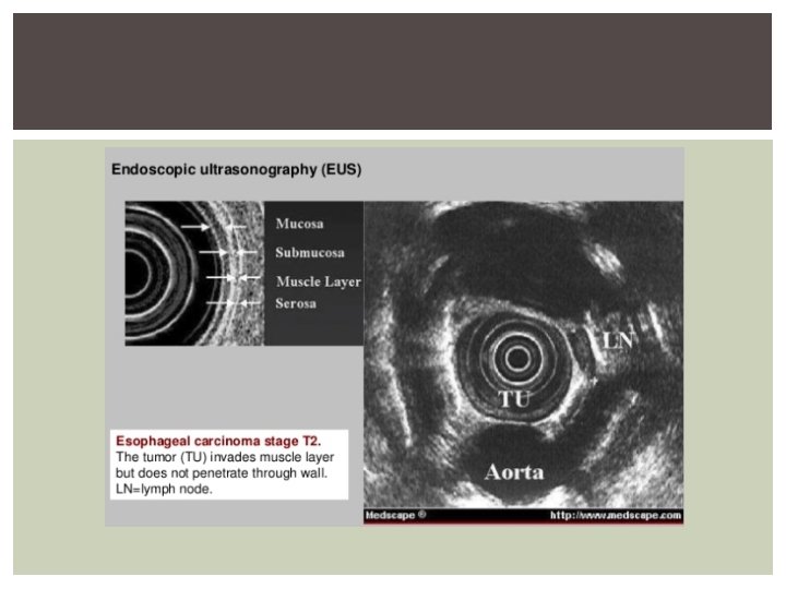
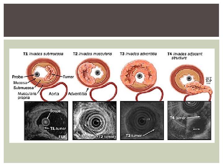
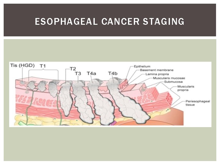
Endoscopic therapy for Barrett’s neoplasia has been developed on the evidence that HGD and T 1 a EAC is associated with a low rate of lymph node metastasis: endoscopic and surgical series indicate a 0– 10% risk in T 1 a cancer, while submucosal invasion carries a higher risk (up to 46%) ER should be considered therapy of choice for dysplasia associated with visible lesions and T 1 a adenocarcinoma (Recommendation grade B)
For patients at high surgical risk, endoscopic therapy can be offered as an alternative to surgery for treatment of good prognosis T 1 b adenocarcinomas (T 1 b sm 1, well differentiated and without lymph vascular invasion). For T 1 b adenocarcinomas with involvement of the second sub-mucosal layer or beyond (T 1 b sm 2–sm 3), endoscopic therapy should not be considered curative
In the presence of HGD or intramucosal cancer without visible lesions (flat HGD/intramucosal cancer), these should be managed with an endoscopic ablative technique. RFA currently has a better safety and side-effect profile and comparable efficacy. Eradication of residual Barrett’s esophagus after focal ER reduces the risk of metachronous neoplasia and is recommended.
In patients treated for HGD, endoscopic follow-up is recommended every 3 months for 1 year and yearly thereafter. This should include biopsies at the GOJ and within the previous extent of the Barrett’s epithelium.
- Slides: 54