Barium Studies for Oesophagus Anatomy of Oesophagus 25

Barium Studies for Oesophagus

Anatomy of Oesophagus • 25 cm in length • Extends from cervical region ( cricopharyngeus C 5 level ) to T 11.

Muscular layers • It has an inner circular and an outer longitudinal muscle coat. • These muscle layers comprise predominately striated muscle in the upper third of the oesophagus and predominantly smooth muscle in the lower two thirds, with the transition occurring at the level of the aortic knuckle.

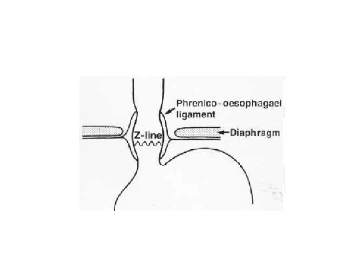
Mucosa • The mucosa of the oesophagus is stratified squamous epithelium. • In the region of the gastro-oesophageal junction it changes to columnar epithelium along an irregular horizontal line (Z line).

• Most of the impressions on the wall of the oesophagus are on the left side.
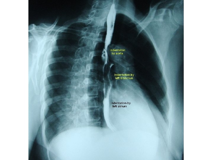

• The aortic arch indents the left wall of the oesophagus and immediately below this point the left main bronchus produces an indentation on the left anterolateral wall. • After passing through the diaphragmatic hiatus the oesophagus extends for approximately 2 cm before joining the fundus of the stomach.

Lymphatic drainage • Upper oesophagus is to cervical nodes, • Midoesophagus is to preaortic nodes and • Lower oesophagus drains to coeliac and left gastric nodes.

Parts of the oesophagus • • Oesophagus has got 4 parts. Cervical oesophagus extends from cricoid cartilage to sterno clavicular joint. Upper thoracic oesophagus extends from thoracic inlet to carina ( 8 cm). Lower thoracic oesophagus extends from carina to gastro oesophageal junction ( 8 cm). Gastro oesophageal junction.

1

• Endoscopic ultrasound of the oesophagus showing the layers of the bowel wall. • A = mucosa; • B = muscularis mucosa; • C = submucosa; • D = muscularis propria; • E = adventitia.

• What is the difference between serosa and adventitia?

• Serosa is the outer most covering which is lined by peritoneum. • When there is no peritoneum lining it is called adventitia.

• What does the oesophagus have?

• Oesophagus has serosa as well as adventitia. • In the thorax it has adventitia and in the abdomen when it crosses the diaphragm it has serosa.

CT • Normal wall thickness is 3 mm. • Cervical oesophagus is generally devoid of gas. • Mid and lower oesophagus may show some amount of gas. • The presence of an air-fluid level or a fluid-filled • lumen of more than I cm usually indicates the presence of functional or mechanical obstruction. • The gastro-oesophageal junction is a difficult area to evaluate with CT.

Barium studies • These are simple to perform, inexpensive and have high sensitivity. • Double-contrast studies may be difficult to achieve because of the transient nature of oesophageal dilatation during swallowing. • Good fluoroscopy is essential, aided by digital imaging for spot radiographs

• Begin the examination in erect position and turned obliquely to the left, so that the body of the oesophagus is thrown clear of the spine. • A barium suspension at 100 per cent w/v is ideal, as this will allow good mucosal coating and yet not be too dense. • Observe the initial bolus fluoroscopically, to ascertain if there is any obvious structural abnormality. • Then take spot radiographs of the upper mid and lower esophagus and OG junction.
 positon for")
• Then place the patient horizontally in prone oblique ( RAO) positon for assessing motility in the body of the oesophagus and to fully distend the gastro-oesophageal junction. • Full distension is required for the demonstration of both hiatal hernias and tumours.

• The examination is concluded by further spot radiographs of the stomach, remembering that problems as far away as the pylorus can present with dysphagia.

Additional examinations and/or modifications to barium swallow • If a lesion is suspected in the pharynx or pharyngo-oesophageal junction, take video or rapid digital images (4– 6 frames/s) of this area with the patient in the erect position. • Both anteroposterior (AP) and true lateral projections are utilized.

• If a motility disorder of the body of the oesophagus is strongly suspected, then it is best to avoid administering a spasmolytic agent before the prone swallow.

Suspected oesophageal rupture or tear. • A non-ionic water-soluble contrast agent is the best option; if this fails to show any obvious leak, it may be followed by thin barium is used. • If an ionic contrast agent such as Gastrografin (meglumine diatrizoate) is aspirated, it can cause severe pulmonary problems.

• What are the various oesophageal contractions?

• Primary wave • Secondary wave • Tertiary wave

• What is the primary stripping wave?

• Stripping wave is a muscular contraction that propels the bolus downwards and collapses the lumen of the oesophagus.

• Any residual barium will then be cleared by a secondary wave.

• Tertiary waves are sometimes seen, particularly in elderly patients, and these usually consist of non propulsive disorganized contractions that fail to advance the barium bolus.

• When assessing motility, only a single bolus of barium should be swallowed as repeated swallowing interferes with the assessment of normal motility.
 has largely replaced the barium swallow for the assessment")
• (UGIE or EGD) has largely replaced the barium swallow for the assessment of peptic ulcer disease and the assessment of haematemesis.
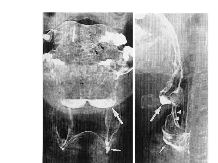

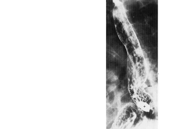
Normal Hypopharynx • • Large White arrows show valleculae. Small white arrow shows pyriform sinuses Arrow heads show ary epigllotic folds. Black arrow on AP view shows circumvallate papilla
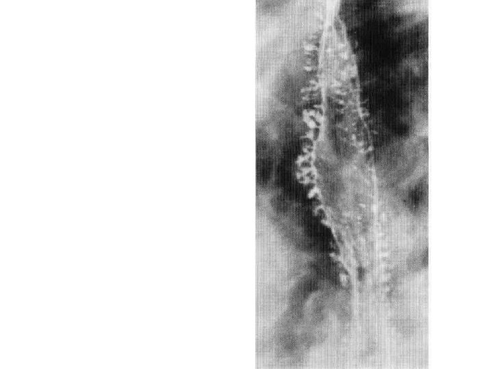
 is produced by failure of the")
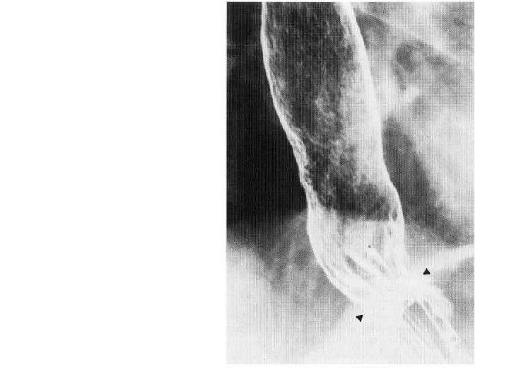
Cricopharyngeal muscle spasm • The posterior impression (arrow) is produced by failure of the cricopharyngeus muscle to relax.
.")
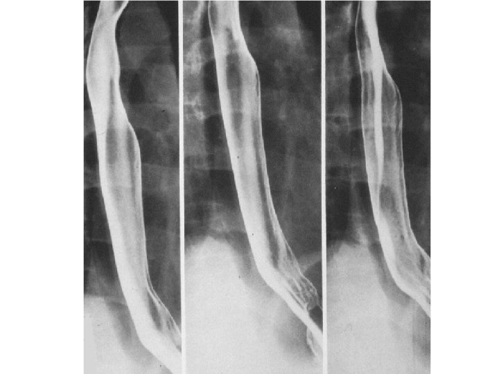
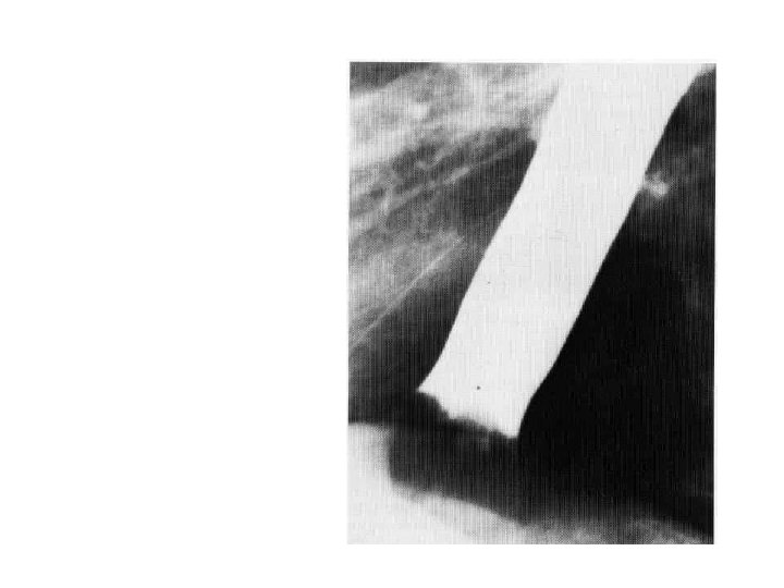
• Lateral cervical oesophageal diverticulum (Kill ian-lamieson diverticulum).

• Arrows show the indentation due to anterior venous plexus. • Arrow heads point to the impression due to the osteophytes.

• Arrow shows the web anteriorly • Posterior impression is due to the contracted cricopharyngeus.

• Why do you get the jet phenomenon?

9

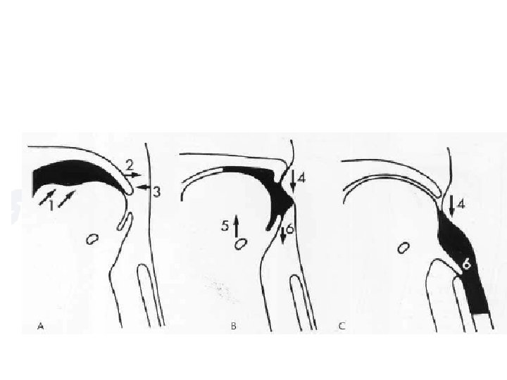
Motility Disorders • • • May be primary or secondary. Diffuse Esophageal Spasm. Nut cracker Oesophagus. Hypertrophic Oesophageal Sphincter Pres byoesophagus

• Secondary to a wide variety of diseases, including oesophagitis, diabetes, alcoholism, and collagen, endocrine and neuromuscular diseases. • Calcium channel blockers can be useful for treating primary motility disorders.

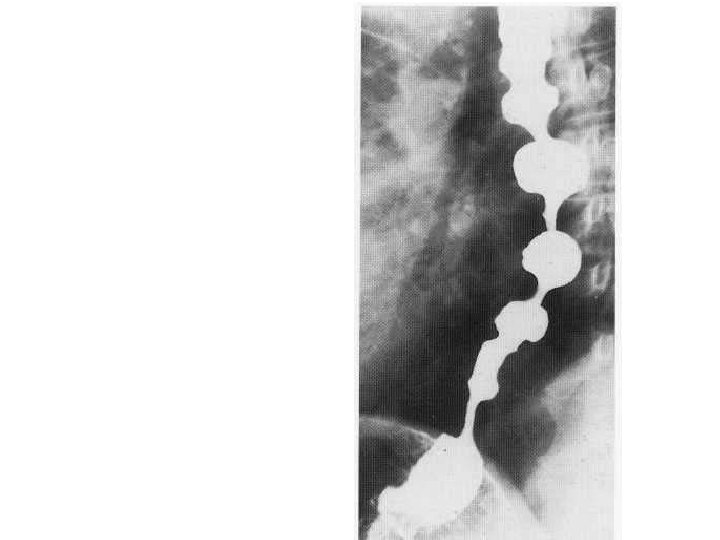
• Tertiary contractions seen as rippling of oesophageal wall.
.")
• A series of indentations resembling a corkscrew (hence the description 'corkscrew oesophagus').

• Which part of the oesophagus is affected in Myaesthenia Gravis ? And which in Slceroderma ? Why?

• Disorders involving striated muscle, such as motor neuron disease and myasthenia gravis, will only affect the pharynx and upper third of the oesophagus, whereas disorders of smooth muscle, such as scleroderma, affect the lower two-thirds.

• Striated muscle coats the pharynx and upper 4 cm of the oesophagus, while the lower half of the oesophagus has a smooth muscle coat; between the two, striated and smooth muscle intermingle. • The transition from smooth to striated muscle is at the level of the aortic knuckle.

• Name the components of Plummer Winson Syndrome.

Plummer-Vinson syndrome • • • Iron-deficiency anaemia, dysphagia, stomatitis, glossitis and Koilonychia. There may be an increased risk of developing pharyngeal and cervical oesophageal carcinomas.

Scleroderma • Incompetence of the gastro-oesophageal sphincter resulting in severe reflux oesophagitis with ulceration.

Pulsion diverticulam • Seen enface and in profile • Traction diverticulam are also seen in the thoracic region. • The neck of the diverticulam is a key feature in identifying both. • Wide neck is a feature of pulsion diverticulam.

• Epiphreinc Diverticulam
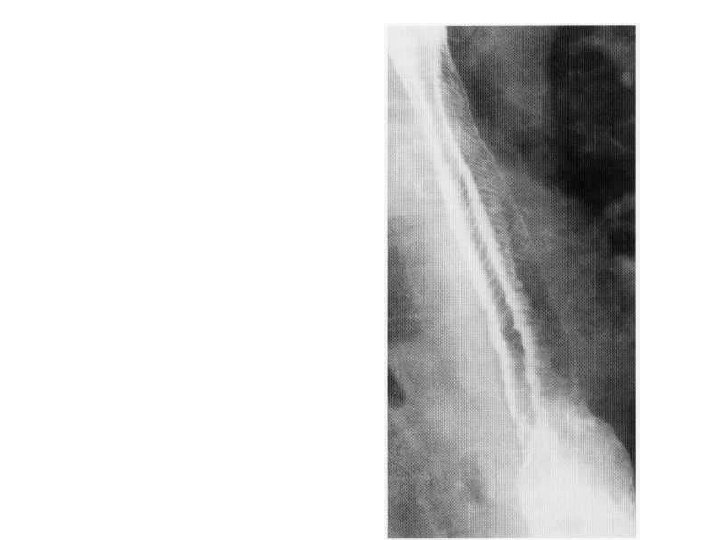
 marks the junction of squamous (oesophageal) and columnar")
• Z-line (between the arrows) marks the junction of squamous (oesophageal) and columnar (gastric) epithelium. • Gastric rugae are seen extending up to the line.
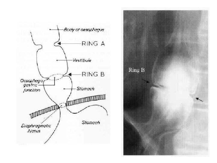

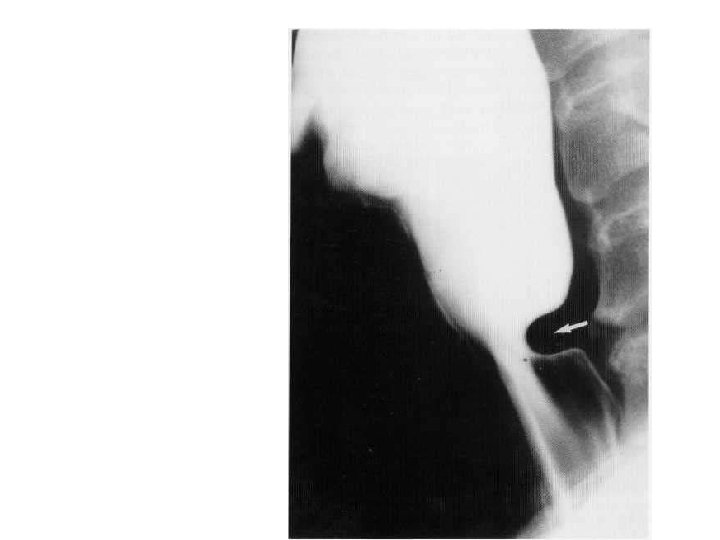
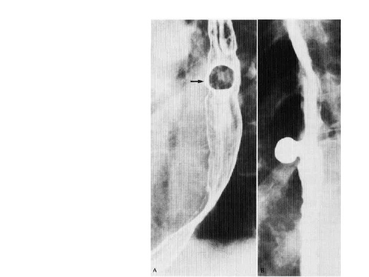
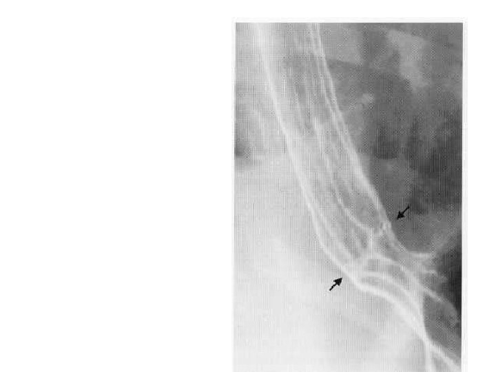
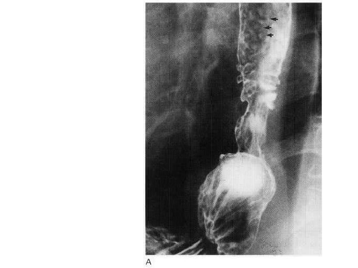
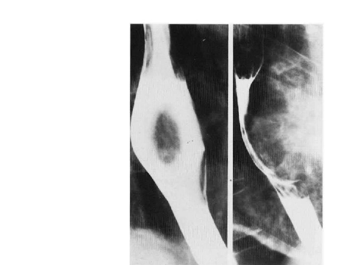
 Schatski's ring (between arrows) demonstrated by barium swallow. • (B)")
Schatski's ring • (A) Schatski's ring (between arrows) demonstrated by barium swallow. • (B) Bread soaked in barium has been swallowed and is lodged above the ring.

Schatzki ‘ Ring • It is defined as a pathological annular narrowing at the oesophago gastric junction causing dysphagia. • A Schatski or B ring is pathognomonic of a hiatal hernia because it marks the junction between squamous and columnar epithelium and represents the Z line. • These rings are very thin ( 2– 3 mm ) in thickness and, despite being mucosal, can be surprisingly symptomatic and may require dilatation.

• If the B ring is incomplete, part of it can sometimes be demonstrated as the incisural notch, which is inevitably on the greater curve aspect of the stomach.

• Before dysphagia occurs , the lumen of the oesophagus has generally narrowed to less than 13 mm diameter. • May be congenital, or inflammatory.

• Hiatus hernia with granular appearance of the oesophagus due to peptic oesophagitis.

• Assymetric stricture in the oesophagus with hiatus hernia.

Feline Oesophagus • Fine mucosal folds due to contraction of muscularis mucosae. • Generally a transient finding. • Will disappear in most cases after adequate distension. • Occasionally may be seen in oesophagiits. • Name given as a similar appearance is found in cats.

Barret’s Oesophagus • Hiatus hernia is seen. • Ulceration is seen. • Note the reticular pattern of gastric mucosa seen in the oesophago gastric junction.

Intramural pseudodiverticulosis • Multiple flask-shaped projections produced by barium entering dilated oesophageal glands.

Intramural pseudodiverticulosis • About 300 oesophageal glands may be seen to fill with Barium. • These glands show caudal angulation. • This condition is easily diagnosed on Barium studies rather than on endoscopy as the glands are very small and may not be seen on endoscopy. • Candida may be isolated from the glands but may be due to secondary infection.

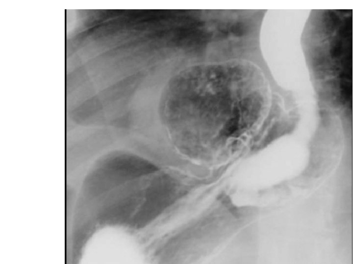
Leiomyoma • View showing features typical of an intramural or extrinsic lesion. • There is a broad-based filling defect bulging into, and widening the lumen of the oesophagus. • The lesion makes a wide angle with the wall of the oesophagus.

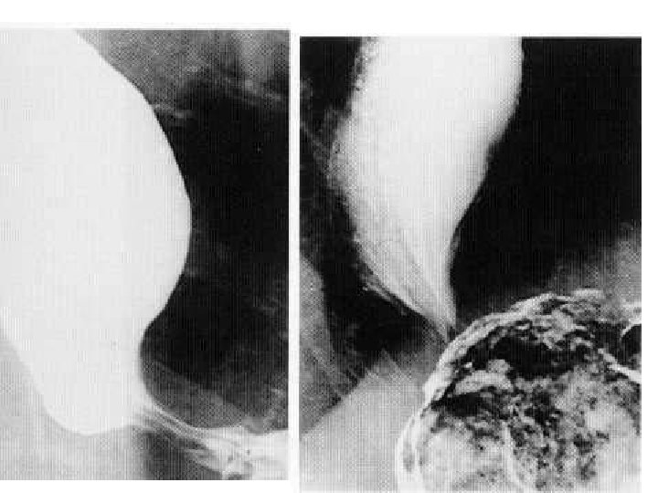
• Lower oesophageal obstruction produced by impaction of a large meat bolus
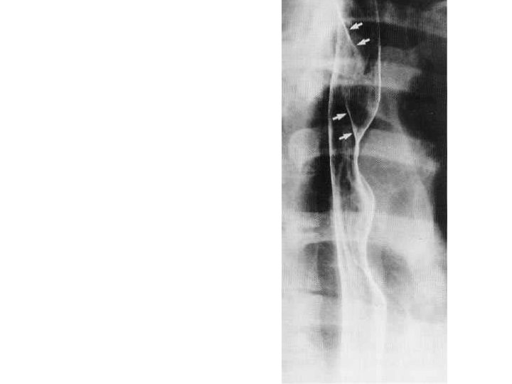
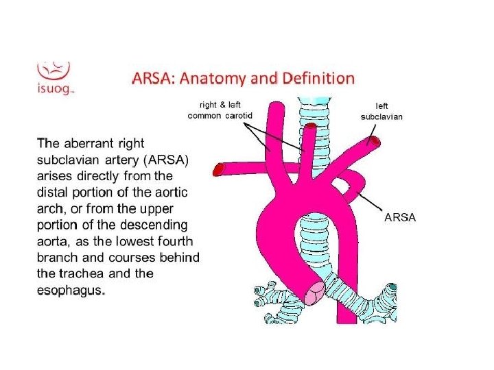

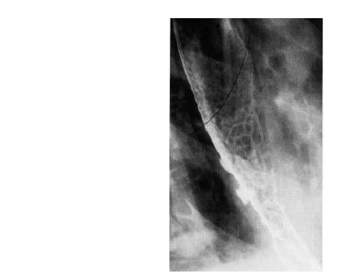
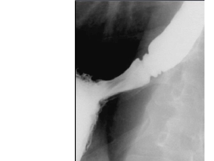
aberrant right subclavian artery • Aberrant right subclavian artery arises from the aortic arch distal to the origin of the left subclavian artery, and passes upwards and to the right behind the oesophagus. • This gives rise to a characteristic smooth, oblique indentation on the posterior wall of the barium-filled oesophagus on the oblique view.

• Characteristic radiological features of hiatus hernia and stricture in proximal oesophagus.

Barret’s Ulcer • Barret’s ulcer causing penetration of the posterior wall of the oesophagus.

Patient with sudden onset chest pain
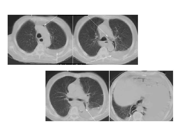

• Pneumomediastinum and pleural effusion

Boerhaave’s syndrome • Esophageal rupture is due to a sudden rise in intraluminal esophageal pressure produced during vomiting. • Neuromuscular in coordination causes failure of the cricopharyngeus muscle to relax. • The syndrome commonly is associated with overindulgence in food and/or alcohol.

Location of tear in Boerhaave syndrome • Left postero lateral wall of the lower third of the esophagus, 2 -3 cm proximal to the gastro esophageal junction.

GI Bleeding Definitions • Upper GI Bleeding = proximal to ligament of Treitz • Hematemesis = vomiting blood – This is diagnostic of upper GI bleeding • Melena = passage of tarry or maroon stool – Can be upper or lower (more commonly upper) • Hematochezia = Bright red blood per rectum – Usually characteristic of colonic hemorrhage

combined-type hiatus hernia. • Contrast study demonstrating a combinedtype hiatus hernia. • Note the rolling component with a large portion of stomach above the diaphragm, but in addition the gastro-oesophageal junction has also migrated cranially.

• Fixed sliding hiatal hernia together with several B or Schatski rings.

• • • Types of Hiatus hernia Sliding Para oesophageal Mixed Intrathoracic stomach

• Para oesophageal hernia is better diagnosed on Barium studies. • Hiatus hernia is better diagnosed on enodoscopy as the Z line of squamo columnar junction is better seen. • Z line is around 40 cm from incisor teeth.

• Why is hiatus hernia difficult to diagnose on Barium studies?

• It is difficult to diagnose Hiatus hernia on Barium studies because they do not normally define the mucosal junction and we have to utilize secondary features that indicate the position of the mucosal junction. • These include a Schatski or B ring, incisural notch and estimation of hiatal width.

• Ennumerate the techniques used for evaluating hiatus hernia. • What is the gold standard ?

• • Barium studies, Endoscopy with biopsy, p. H measurement and Radionuclide techniques.

• Barium studies and endoscopy assess the damage caused by gastro-oesophageal reflux disease. • Radionuclide studies and 24 hour p. H assess gastro-oesophageal reflux. • 24 -hour p. H measurement is the ‘gold standard’.
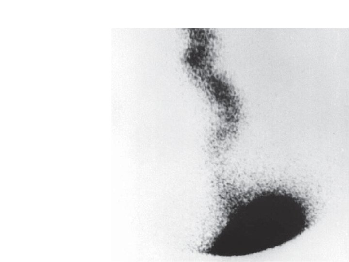

• A scintigraphic study demonstrating gross gastro-oesophageal reflux
. Note thin smooth wall and near-water-density contents. •")
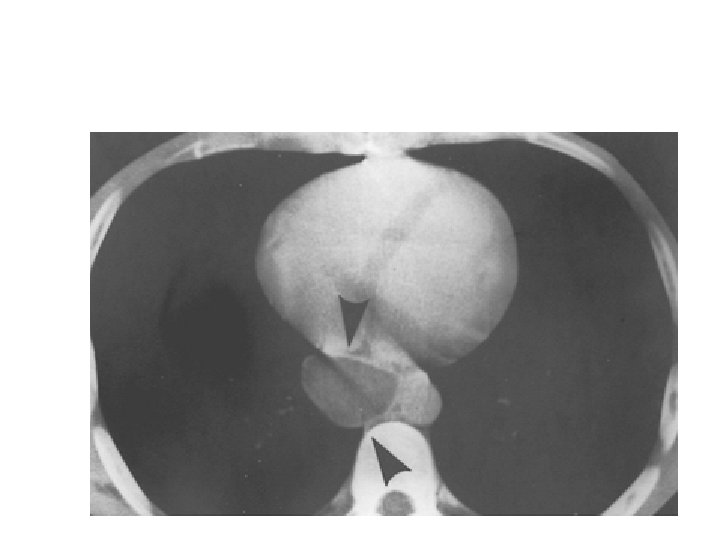
• Esophageal duplication cyst (arrowheads). Note thin smooth wall and near-water-density contents. • D/D esophageal retention cysts. ( They are smaller in size and may be multiple. They are rarer than duplication cysts. )
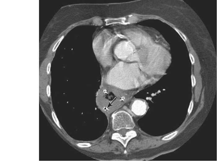

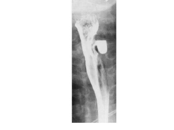
Achalasia • The esophagus is dilated and contains food and fluid and a coiled nasogastric tube (arrow) that could not be passed across the lower esophageal sphincter. • The wall thickening in achalasia is limited to the lower-esophageal sphincter and is generally less than 10 mm in thickness. • The remainder of the esophagus is dilated and often fluid-filled.
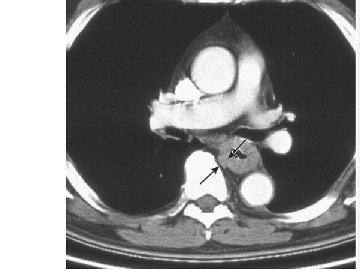
 of the entire thoracic")
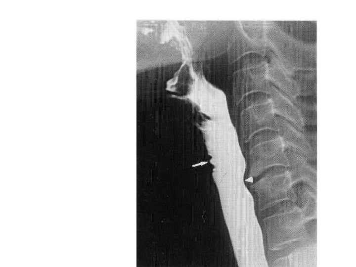
Diffuse esophageal spasm • There is symmetric mural thickening (arrows) of the entire thoracic esophagus. • The thickening is smooth, and there is no luminal dilatation.

What are uphill varices ? What are down varices?

Uphill varices • Result from reversal of venous flow in the coronary vein into the distal esophageal venous plexus. • These periesophageal veins drain into the systemic circulation via the azygous vein. • Most commonly seen in cirrhosis and portal hypertension. • Uphill varices can bleed or rupture, leading to massive GI hemorrhage

Downhill varices • Occur due to obstruction of the SVC. • This results in collateral drainage from the head, neck, and upper-extremity venous systems into the veins surrounding the midand upper-thoracic esophagus and into the azygous vein.
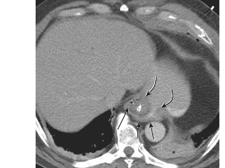
 is wrapped around the distal esophagus. • NG tube")
Fundoplication • The fundus (arrows) is wrapped around the distal esophagus. • NG tube courses through the esophagus. • Note that there is a sliding hiatal hernia, and the wrap lies above the esophageal hiatus (curved arrows).

• A Nissen fundoplication is a complete 360 ° wrap, and a Toupet fundoplication is partial 270 ° wrap. • On CT, a fundoplication has the appearance of a soft-tissue mass at the gastroesophageal junction. • CT does not provide functional information, it is ideal for assessing postoperative complications of leak and abscess.

• Fundoplication failure can be diagnosed on CT performed in the prone position after administering an effervescent agent. • Fundoplication dehiscence is diagnosed when there is lack of circumferential thickening around the distal esophagus and when there is gaseous distension of the lower esophageal sphincter

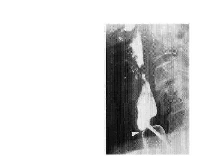
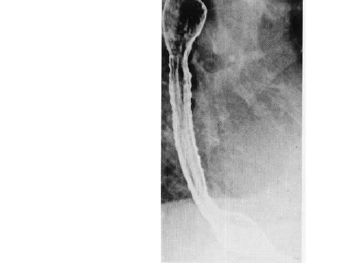
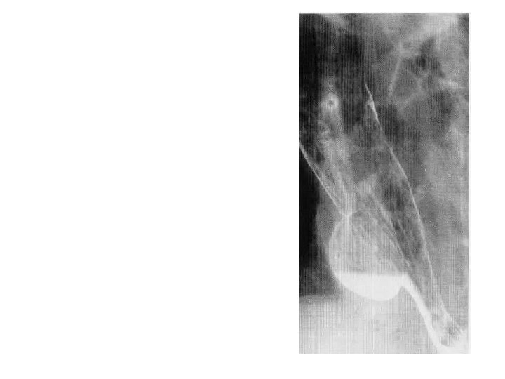
Achalasia • The oesophagus is distended. • Intact oesophageal folds pass through the tapered narrowing, which corresponds to the site of the lower oesophageal sphincter. • Fluoroscopy shows impaired motility. • Sufficient barium has entered the stomach to coat the fundus. This excludes an infiltrating gastric carcinoma.

• What is secondary achalasia ? • What is vigorous achalasia ?

• Achalasia developing rapidly, or after the age of 50 should arouse the suspicion of underlying neoplasm. • This is secondary or pseudo achalasia. • In early cases of primary achalasia the fluoroscopic examination might show tertiary contractions in the lower oesophagus. This is called vigorous achalasia.
- Slides: 130