BARIUM ENEMA Dr DEEPAK IT IS THE RADIOGRAPHIC

BARIUM ENEMA Dr. DEEPAK

�IT IS THE RADIOGRAPHIC STUDY OF THE LARGE BOWEL BY ADMINISTRATION OF THE CONTRAST MEDIUM THROUGH THE RECTUM. CONTRAST Barium sulphate. Particle size varies from 0. 6 -1. 4 to 4 -50 microns Particles are coated with various agents to achieve rapid flow , good mucosal adhesion , adequate radiographic density , an even coating which is plastic and does not crack and absence of artifact or foaming.
 PREPARATION Dietary")
�Additives Carboxy methyl cellulose Simethicone, Gum acaece, Pectin Dimethyl polysilicone(anti foaming agent) PREPARATION Dietary restriction Purgation Cleansing enema

�Success in colon cleaning is often a function of patient understanding and compliance with the preparation , as well as of the patient’s own baseline colonic motility. Baseline colonic motility low in diabetes, scleroderma, patients taking opiates or drugs with anticholinergic side effects. Before doing barium examn Appropriate clinical history Surgical history Medications Colonic disease Colonoscopy- biopsy

�DIET Low residue diet for two days prior to examination Iron containing medications should be stopped because they make stools adhere to mucosa LAXATIVES Castor oil Bisacodyl Magnesium citrate BOWEL WASH Previous night and two hours before the procedure

To come with empty stomach for investigation Preparation of the patient should not be done in 1. Diarrhoea 2. Total obstruction 3. Paralytic ileus 4. Children less than 8 years of age

�BARIUM ENEMA SINGLE CONTRAST STUDIES DOUBLE CONTRAST STUDIES Adv Better surface details Easy unravelling of the colon as it is possible to look through loops. Dis adv Fistulae and sinuses can be missed

Difficult in uncooperative patients Effacement of submucosal detail of the colon and overlooking of annular/polypoid lesion is possible. Difficult in uncooperative patients. If colon repeatedly gives contraction buscopan 1 ml iv can be given. For better retension of barium Make patient prone Distend the colon slowly Retension catheter In patients who have total obstruction , let patient evacuate part of the barium and then pump air. The froth goes through the obstruction and the proximal limit of the obstruction can be demonstrated.

�Indications of single contrast barium enema Uncoopertative, very debilitated or old patient Evaluation of acute obstruction or volvulus Reduction of intussusception Show configuration of colon Where gross pathology is to be excluded

�Indications of double contrast barium enema Preferred method for routine examination High risk patients-rectal bleeding , previous h/o carcinoma/polyp, colorectal ca/FAP Patients with severe diverticulosis Reduction of an intussusception.

�ABSOLUTE CONTRAINDICATIONS FOR BOTH DOUBLE AND SINGLE CONTRAST BARIUM ENEMA Toxic megacolon Pseudomembranous colitis If rectal biopsy has been done preferable to wait for 7 days Paralytic ileus Difficulty to pass tube in rectum-inflammed piles, growth

�RELATIVE CONTRAINDICATIONS Allergy to barium suspension Risk of perforation Peritonitis Following acute/fulminating ulcerative colitis
 �Hirschsprung's")
Barium enema Polyps �Diverticular disease �Ulcerative colitis �Colon cancer �Crohn's disease (regional enteritis) �Hirschsprung's disease �Intestinal obstruction �Intussusception (children) �Volvulus

Barium required �A routine double contrast enema requires 500 ml of barium sulphate suspension. � 1000 ml air or carbon dioxide. �The enema tube must be prepared. �If Gastrograffin is to be used then a 50 -50 mix of 400 ml (Gastrograffin and water)

Barium Enema

General views �Digital or fluoroscopic images �Hepatic flexure �Splenic flexure �Ascending colon �Descending colon �Transverse colon �Sigmoid colon �Cecum �rectum
 �Left")
General views �Non fluoroscopic images �Prone �Caudal angled sigmoid colon �Supine �Lateral (erect) �Left lateral decubitus �Right lateral Decubitus

Exposure �Exposure factors �For single contrast 100 -120 k. Vp �For double contrast and water soluble 80 -90 k. Vp is more suitable �Ensure you use the soft tissue filter for lateral decubitus images �All images are to taken on suspended respiration.

�Patient instruction �Patient to suspend respiration in full expiration �Image criteria �Entire opacified colon including flexures and rectum should be well demonstrated. �Abdomen should be seen with out rotation
 be")
Prone �Patient position prone �Prone � Have patient turn head to side (comfort) be careful not to allow rotation � Have patients arms up rested near head � In addition have the table in slight trendelenberg position � Demonstrate entire colon including the flexures and rectum. � Also the vertebral column centered so that ascending and descending colon are included.

Supine �Patient position supine �Supine � Place patient in supine position � Have arms extended down by side away from central ray. �Central ray � Perpendicular to film plane centered in midsagittal plane at level of iliac crests �Structures demonstrated � Demonstrate entire colon including the flexures and rectum. � Also the vertebral column centered so that ascending and descending colon are included.

Decubitus �Patient position Right lateral decubitus � Lay patient on right side with anterior or posterior aspect in contact with grid/film combination � Make sure patient does not fall from table. �Central ray � The x-ray beam should be in a horizontal direction and perpendicular to the film plane. � Centre the primary beam in the midsagittal plane �Structures demonstrated this view should demonstrate an AP or PA projection of the contrast filled colon � Area from splenic flexure to the rectum. � This view demonstrates the medial aspect of ascending colon �

�Patient position left lateral decubitus � Lay patient on left side with anterior or posterior aspect in contact with grid/film combination � Make sure patient does not fall from table. �Central ray � The x-ray beam should be in a horizontal direction and perpendicular to the film plane. � Centre the primary beam in the midsagittal plane �Structures demonstrated this view should demonstrate an AP or PA projection of the contrast filled colon � Area from hepatic flexure to the rectum. � This view best demonstrates the lateral side of ascending colon. �

Pathophysiology of Colorectal Cancer � Most colorectal cancers, regardless of etiology, arise from adenomatous polyps. Adenomatous polyps may be pedunculated or sessile. � Villous adenomas, most of which are sessile, become malignant more than three times as often as tubular adenomas. � Adenomatous polyps may be found in the colons of >30% of middle-aged or elderly folks; however, <1% or polyps ever become malignant. 1 cm polyp 3 cm carcinoma

Size Matters � The likelihood that any polypoid lesion in the large bowel contains invasive cancer is related to the size of the polyp � Adenomatous polyps are thought to require >5 years of growth before becoming clinically significant Polyp size Risk of malignancy <1. 5 cm <2% 1. 5 – 2. 5 cm 2 -10% >2. 5 cm >10%

Clinical Features �The majority of CRCs occur in the distal colon: � Distal colon (splenic flexure to rectum) ~ 60% � Proximal colon ~ 40% �Symptoms vary with anatomic location of the tumor � Right colonic lesion commonly ulcerate, leading to chronic, insidious blood loss without change in stool appearance � Transverse and descending colonic tumors often impede the passage of stool causing abdominal cramping, obstruction and even perforation � Rectosigmoid cancers are often associated with hematochezia, tenesmus, and narrowing of the stool caliber

Low/Average Risk Category • 70 -80% of all Colorectal Cancer • Age > 50 • No known risk factors This radiographic appearance is referred to as an “apple-core” lesion and is always highly suggestive of malignancy

Moderate Risk Category • 15 -20% of all Colorectal cancers • Single adenomatous polyp < 1. 5 cm Adenoma • Single adenomatous polyp > 1. 5 cm or multiple adenomatous polyps of any size • Personal history of CRC • CRC in a first degree relative < 60 years of age or CRC in 2 or more first degree relatives Single adenomatous colonic polyp

High Risk Category • 6 -10% of all Colorectal Cancers • Inflammatory bowel disease (Crohn’s and Ulcerative Colitis) • Familial Adenomatous Polyposis (FAP) • Hereditary non polyposis colorectal cancer (HNPCC) Colectomy resection specimen demonstrating classic familial adenomatous polyposis (FAP) with thousands of adenomatous polyps.

American Cancer Society Colorectal Cancer Screening Guidelines Beginning at age 50, both men and women at average risk should have 1 of the 5 of the screening options: 1. yearly stool blood test (FOBT) or fecal immunochemical test (FIT), or 2. flexible sigmoidoscopy every 5 years, or 3. yearly stool blood test plus flexible sigmoidoscopy every 5 years (Of these first 3 options, the ACS prefers the third option, that is, FOBT or FIT every year plus flexible sigmoidoscopy every 5 years. ) Or you may have: 4. double contrast barium enema every 5 years, or 5. colonoscopy every 10 years

Barium Enema Adenoma Carcinoma Sigmoid Rectum Double contrast barium enema of the colon Barium filled colon revealing a carcinoma
 Spot radiograph obtained with the patient")
Barium pool obscures polyp in splenic flexure. (a) Spot radiograph obtained with the patient in a right posterior oblique position shows the splenic flexure. The barium pool obscures the en face mucosal detail of the descending limb of the splenic flexure. The luminal contour is seen either as a continuous white line (black arrow) or as a smooth edge of the barium column (white arrow). (b) Spot radiograph obtained with the patient in an erect right posterior oblique position shows the splenic flexure. A 7 -mm polyp is manifested in the shape of a bowler hat. The brim of the hat (solid arrows) represents barium trapped between the base of the polyp and the adjacent normal mucosa. The dome of the hat (open arrow) represents the top of the polyp. The polyp is pointed inward, toward the longitudinal axis of the bowel.

Spot radiograph of the splenic flexure with the patient in an erect right posterior oblique position. Diverticula are filled with barium (short arrows) and coated with barium (long arrow).

Spot radiograph of the splenic flexure with the patient in a horizontal right posterior oblique position. The contour of the descending limb is sacculated. Subtle mucosal ulceration is manifested as shallow barium-filled ulcers surrounded by radiolucent halos (arrows). One week prior to this examination, this patient had acute rectal bleeding during an airplane flight. Endoscopic biopsy results revealed ischemic changes.

Spot radiograph of the rectum obtained with the patient in a left posterior oblique position shows a coarsely lobulated, barium-etched line (arrows) disrupting the normally smooth surface.

Overhead radiograph of the pelvis with the tube angled 30° caudad and the patient in a prone position shows the rolled edges (arrows) of a long, centrally ulcerated, plaquelike lesion, which in this position is seen in profile and is akin to the Carman meniscus sign. This is an adenocarcinoma at the rectosigmoid junction.

The mucosal surface en face. Close-up view from a spot radiograph of the sigmoid colon shows a 1. 9 -cm polypoid adenocarcinoma in a 68 -year-old man with right upper quadrant pain and subsequently proved liver metastases. The mass is manifested as a barium-etched hemispheric line (solid arrows) surrounding tiny radiolucent tumor nodules outlined by barium in the interstices of the tumor; representative nodules are identified by the open arrow. The normal mucosal surface is featureless and gray.
 shows")
Spot radiograph obtained with the patient in a left-side-down position (left lateral view) shows the rectum early in the examination. At the edge of the barium pool, there is a 7 -mm lobulated radiolucent filling defect (arrow). The enema tube tip obscures the distal rectum
 shows")
Spot radiograph obtained with the patient in a right-side-down position (right lateral view) shows the rectum after enema tube tip removal. The polyp is not depicted definitively. The distal rectum is no longer obscured by the enema tube tip. This polyp is a tubular adenoma.

Spot radiograph obtained with the patient in a right posterior oblique position shows a 3 -cm coarsely lobulated polypoid mass (arrows) on the anteromedial wall of the cecum and ascending colon, superior to and overlapping the ileocecal valve (arrowhead).

Close-up view from an overhead radiograph of the colon shows the edge of the ileocecal valve (arrow). The tumor is obscured by the barium pool. This is the best image of the cecum from of a series of overhead images, including the decubitus views.

Spot radiograph obtained with the patient in a prone position shows overlap of the sigmoid colonic loops.

Spot radiograph obtained with the patient in a prone position, with a compression balloon pushing on the anterior abdominal wall, shows separation of two of three sigmoid loops.

Spot radiograph obtained after enema tube tip removal with the patient in a supine position. The distal rectum is seen in air contrast. The most caudal loop (arrow) of sigmoid colon is filled with barium.

Spot radiograph obtained with the patient in a prone position, but the radiograph is printed in the same anatomic position as a to allow direct comparison of images. Barium in the distal rectum now obscures en face mucosal detail. The most caudal loop (arrow) of sigmoid colon is now seen with air contrast.

The granular mucosa typical of UC. Note the intact mucosal line.
 involving")
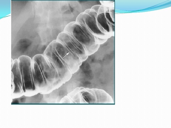
�“leadpipe” colon = rigidity with symmetric narrowing of lumen �“backwash ileitis” (5 -30%) involving 4 -25 cm of terminal ileum with patulous ileocecal valve + absent peristalsis + granularity Ø “burnt-out colon” ie. fairly distensible colon without haustral markings and without mucosal pattern. Ø hazy/fuzzy quality of bowel contour (excessive secretions) Ø Fine mucosal granularity ie. stippling of barium coat (from diffuse mucosal edema + � hyperemia + superficial erosions) Ø Spicules with serrated bowel margins (tiny superficial ulcers)
 protruding through the")
it an acute attack of UC with collar stud ulcers (arrow) protruding through the mucosal line (arrowhead).
")
Aphthoid ulcers (arrows)
 following an acute attack of UC. The mucosal surface and")
Filiform postinflammatory polyposis (arrow) following an acute attack of UC. The mucosal surface and haustration are normal as the colitis was inactive.
 in a patient")
Patulous, rigid ileocaecal valve with associated terminal ileal granularity ('back-wash ileitis') in a patient with ulcerative colitis

Innumerable aphthoid ulcers in Crohn's disease
 ie. discontinuous involvement with intervening normal areas Ø cobblestone appearance")
Ø skip lesions (90%) ie. discontinuous involvement with intervening normal areas Ø cobblestone appearance ie. serpiginous longitudinal and transverse ulcers separated by areas of edema Ø thick and blunted small bowel folds (inflammatory infiltration of lamina propria and submucosa) Ø straightening and rigidity of small bowel loops with luminal narrowing (spasm + submucosal edema) Ø Pseudopolyps ie. islands of hyperplastic mucosa between denuded mucosa Ø inflammatory polypoid masses Ø sessile/pedunculated/filiform postinflammatory polyps Ø diffuse mucosal granularity due to 0. 5 -1 -mm round lucencies Ø linear ulcers on mesenteric border Ø pseudodiverticula or pseudosacculations ie. bulging area of normal wall opposite affected scarred wall on antimesenteric side

Barium enema showing the typical pseudodiverticula found in crohn’s disease

Instant enema in Crohn's disease demonstrates extensive 'cobblestoning' due to linear ulceration and mucosal oedema. Note the rectum is relatively spared but contains aphthoid ulcers

Long stricture in Crohn's disease. A long segment of narrowing is seen in the ileum just proximal to the site of an ileocolic anastomosis in a patient who had undergone a previous resection for Crohn's disease.

Instant enema in a patient with ulcerative colitis reveals fine, continuous, symmetrical, left-sided ulceration.

Classical splenic flexure 'thumb-printing' diagnosing i schaemic colitis.
: Ø Single contrast - efface thumbprinting, but double contrast -sensitive!")
�BE (in 90% abnormal): Ø Single contrast - efface thumbprinting, but double contrast -sensitive! Ø thumbprinting (75%) due to submucosal hemorrhage and edema Ø transverse ridging ie. markedly enlarged mucosal folds (spasm) Ø serrated mucosa = inflammatory edema with superficial longitudinal/circumferential ulceration Ø deep penetrating ulcers (late)

Splenic flexure sacculation and stricturing as sequelae to ischaemic colitis.

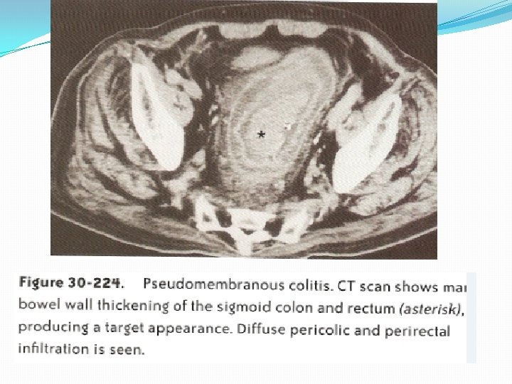
The barium column has a shaggy and irregular appearance because of the pseudomembrane and superficial necrosis with mucosa) ulceration. 48 -51 The pseudomembranes appear as multiple flat, raised lesions distributed circumferentially about the margin of the colon. 48 -52

Generally narrowed sigmoid and proximal rectum following radiotherapy.
.")
Radiation-induced colitis. A large, discrete penetrating ulcer is visible (arrow).

�AFTERCARE The patient should be told that bowel motion will be white for a few days after examination Laxatives should be used to avoid barium impaction in patients with patients. COMPLICATIONS Perforation-Intraperitoneal/extraperitoneal, barium granuloma Inspissation of barium Water intoxication and electrolyte imbalance Caustic colitis Transient bacteremia

Pharmacologic complications Buscopan Anaphylactic SPECIAL BARIUM ENEMA STUDIES Instant Barium enema To show extent and severity of known colitis No bowel preparation is required as residue does not accumulate in a segment of active colitis A plain radiograph is recommended to exclude toxic megacolon or perforation.

�SIGMOID FLUSH Used in patients with severe diverticular disease to improve visualisation of affected bowel. COLOSTOMY ENEMA For visualisation of bowel proximal to stoma A Non wash out bowel preparation is advised.

�REDUCTION OF AN INTUSSUCEPTION Air reduction is preferred More rapid reduction because low viscosity of air permits rapid filling of colon In the presence of a perforation air in peritoneal cavity is better than barium.

� Barium enema has low detection rate, limiting the use of this radiologic technique as a primary surveillance tool for colorectal cancer � Colonoscopy is currently the gold standard, but it is not ideal May not reach the cecum in all cases; the rate of complete examination ranges from 80 to 95 percent Expensive Risks � CT Colonography Increasing acceptability Detection rates similar to conventional colonoscopy Developing ways to digitally remove stool from the computerized image to avoid bowel prep.

The End of Barium Enema’s? � A study of 580 patients at increased risk of polyps (Winawer SJ, et al. N Engl J Med 2000; 342: 1766, 72) underwent double contrast barium enema and colonoscopy � Barium enema found abnormalities in only a third of patients who had one or more adenomas on colonoscopic examination (rate of detection, 39 percent; 95 percent confidence interval, 33 to 45 percent) � Barium enema detected only about half the adenomas larger than 1. 0 cm that were identified by colonoscopic examination (rate of detection, 46 percent; 95 percent confidence interval, 26 to 67 percent) � Barium enema was especially ineffective in identifying small polyps, detecting only 21 percent of the polyps identified by colonoscopic examination as being 0. 5 cm or smaller

Barium Enema Implications Contrast enemas remain a good test for: � Cases in which the colonoscope does not reach the cecum � Localization of colonic disease in preoperative patients � Assessment of LBO � Ileocecal intussusception reversal � Assessment of the status of a colon anastomosis After filling the colon with barium, the radiologist can view a clear silhouette of its shape and condition.

THANK YOU
- Slides: 73