BALLOON MITRAL VALVULOPLASTY Dr JANSHER SENIOR RESIDENT DEPARTMENT

BALLOON MITRAL VALVULOPLASTY Dr JANSHER SENIOR RESIDENT DEPARTMENT OF CARDIOLOGY CALICUT MEDICAL COLLEGE
 throughout the world is rheumatic")
Introduction • The leading cause of mitral stenosis (MS) throughout the world is rheumatic carditis • The rheumatic changes present in 99% of stenotic MV excised at the time of MVR • 25% --- isolated MS • 40%--combined MS + MR. • Multivalve involvement - 38% of patients with MS, with the aortic valve affected in approximately 35% • The tricuspid valve in approximately--- 6%. • Pulmonary valve- rare

EPIDEMIOLOGICAL ASPECTS WORLD WIDE BURDEN • At least 15· 6 million people have RHD, 0. 3 of about 0· 5 million individuals who acquire ARF every year go on to develop RHD • • 233 000 deaths annually are directly attributable to ARF or RHD. Carapetis JR. The current evidence for the burden of group A streptococcal diseases. WHO/FCH/CAH/05· 07

• The overall prevalence estimated to be about 1. 5 -2/1000 in all age groups, in India (total population about 1. 3 billion) suggests that there about 2. 0 to 2. 5 million patients of RHD in the country. R. Krishna Kumar & R. Tandon Rheumatic fever & rheumatic heart disease: The last 50 years Indian J Med Res 137, April 2013, pp 643 -658 review article

�In India, rheumatic fever is endemic and remains one of the major causes of cardiovascular disease, accounting for nearly 25 -45% of the acquired heart disease. �PRIMARY ATTACK RATE OF RF FOLLOWING STREPTOCOCCAL PHARYNGITIS ◦ EPIDEMICS: 3% ◦ SPORADIC: 0. 3%

FEATURES OF RHEUMATIC FEVER/RHD IN INDIA �High incidence of carditis �Erythema marginatum almost nonexistent �Chorea and subcutaneous nodules infrequent �Polyarthralgias more common than poly arthritis �High incidence of disease in young patients �Short interval from onset of ARF to RHD Rapid progression of symptoms Severe hemodynamic changes with PAH/CCF Past history of Rheumatic fever in < 50% High incidence of organic tricuspid valve disease

NATURAL HISTORY OF MS • In India, critical MS may be found in children as young as 6 to 12 years old. ( UP TO 20%) • In the asymptomatic or minimally symptomatic patient, survival is greater than 80% at 10 years, • 60% of patients having no progression of symptoms. • Once significant limiting symptoms occur, there is a 0% to 15% 10 -year survival rate • Once there is severe pulmonary hypertension, mean survival drops to less than 3 years.

• Natural history - pre surgical era indicate that symptomatic patients with MS have a poor outlook. • 5 -year survival rates 62% --- MS in NYHA III 15% -- class IV. • Data from un operated patients in the surgical era still reported a 5 -year survival rate of only 44% in patients with symptomatic MS who refused valvotomy

• 30 to 40% of patients with MS develop AF. • • AF occurs more commonly in older patients and is associated with a poorer prognosis, with a 10 -year survival rate of 25% compared with 46% in patients who remain in sinus rhythm. In more recent BMV studies, the prevalence of AF ranged from 4% in a series of 600 patients from India, with a mean age of 27 years, and 27% in a series of 4832 patients from China, with a mean age of 37 years.

�The mortality of untreated patients with MS 1. Progressive pulmonary and systemic congestion in 60% to 70%, 2. Systemic embolism in 20% to 30%, 3. Pulmonary embolism in 10%, 4. Infection in 1% to 5%. �Serial hemodynamic and Doppler-echo studies have reported annual loss of MV area ranging from 0. 09 to 0. 32 cm 2.
 • In developing countries, mitral stenosis is severe enough")
MS IN YOUNG( INDIAN SCENARIO) • In developing countries, mitral stenosis is severe enough to require commissurotomy before the age of 20 or even 15 years. • • • In 1408 patients with rheumatic heart disease seen at the G B Pant Hospital, New Delhi, between 1967 and-1973 713 (51 %) had mitral stenosis 140 patients below age 20 <10 10 -15 15 -20 4 (2. 8%) 55 (39. 4%) 81 (57. 8%)

• The interval between the initial episode of ARF and clinical evidence of MV obstruction is variable, ranging from a few years --- > 20 years. • In temperate zones, such as the US and Western Europe, patients who develop ARF have an asymptomatic period of approximately 15 to 20 yr. • In India, critical MS may be present in children as young as 6 to 12 years of age

• Rheumatic Carditis results in characteristic changes of the MV diagnostic features are thickening at the leaflet edges, fusion of the commissures, and chordal shortening and fusion • These anatomic changes lead to a typical functional appearance of the rheumatic MV. • In earlier stages of the disease, the relatively flexible leaflets snap open in diastole into a curved shape because of restriction of motion at the leaflet tips. • This diastolic doming is most evident in the motion of the AML and becomes less prominent as the leaflets become more fibrotic and calcified

• The symmetrical fusion of the commissures results in a small central oval orifice in diastole that on pathologic specimens is shaped like a fish mouth or buttonhole because the AML is not in the physiologic open position. • The most useful descriptor of the severity of obstruction is the degree of valve opening in diastole, or the MVA. • In normal adults, the cross-sectional area of the MV orifice is 4 to 6 cm 2

Severity of MS

Stages of Mitral Stenosis From Nishimura RA, Otto CM, Bonow RO, et al: 2014 AHA/ACCF guideline for the management of patients with valvular heart disease: A report of the ACC Foundation/AHA Task Force on Practice Guidelines. J Am Coll Cardiol 63: e 57, 2014.
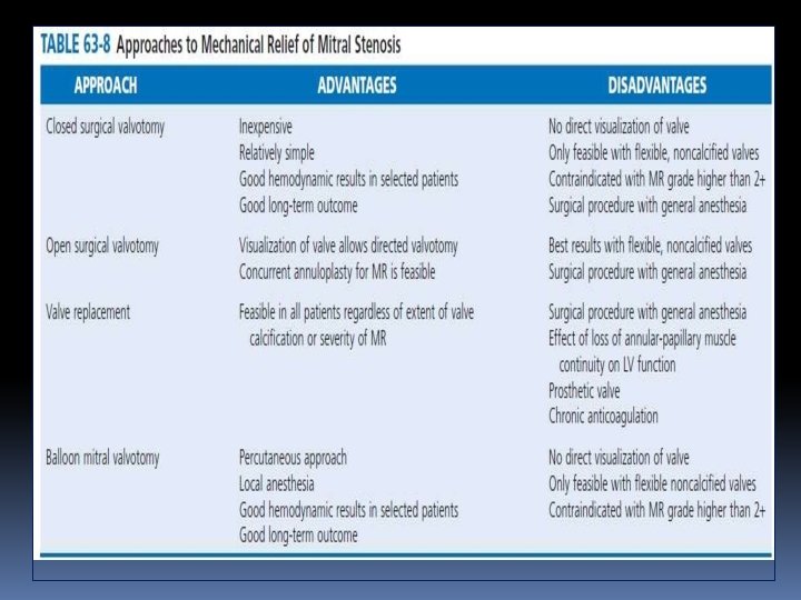

PBMC • Patients with mild to moderate MS who are asymptomatic frequently remain so for years • Severe or symptomatic MS, however, is associated with poor long-term outcomes if the stenosis is not relieved mechanically. • Percutaneous BMV is the procedure of choice for the treatment of MS. • Life saving emergency procedure in the patient with mitral stenosis and refractory pulmonary edema or cardiogenic shock • PMV is the remarkable landmark intervention in the field of interventional cardiology that leads to the great help in treatment of stenosed MV

Principle of the procedure PBMV TIPS AND TRICKS

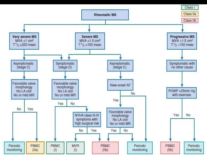
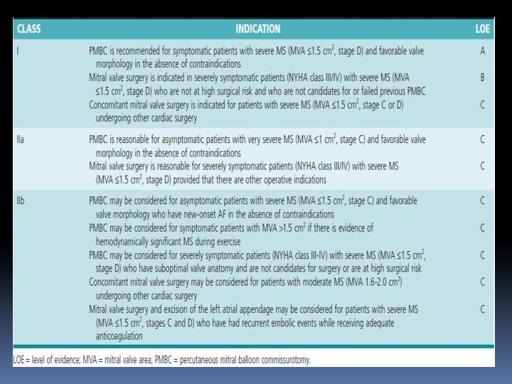
INDICATIONS OF PBMC • BMV is recommended for symptomatic patients with moderate to severe MS (i. e. , a MVA <1 cm 2/m 2 of BSA or <1. 5 cm 2 in normal-sized adults) • favorable valve morphology, • no or mild MR, • no evidence of left atrial thrombus

• Even mild symptoms, such as a subtle decrease in exercise tolerance, are an indication for intervention because the procedure relieves symptoms and improves long term outcome with a low procedural risk • BMV is a reasonable option for asymptomatic patients with very severe MS (<1 cm 2) with favorable anatomy or when obstruction has resulted in AF. • AF precipitates symptoms in most patients with significant MS.

• BMV also may be considered in symptomatic patients in whom surgery carries high risk for adverse events or outcomes, even when valve morphology is not ideal, including patients with restenosis after a previous BMV or previous commissurotomy who are unsuitable candidates for surgery because of very high risk. • Very old, frail patients; patients with associated severe IHD; patients in whom MS is complicated by pulmonary, renal, or neoplastic disease; women of childbearing age in whom MVR is undesirable; and pregnant women with MS

• BMV may be further considered for patients with mild MS in whom symptoms cannot be explained by other causes and who experience pulmonary hypertension (>25 mm Hg) with exercise
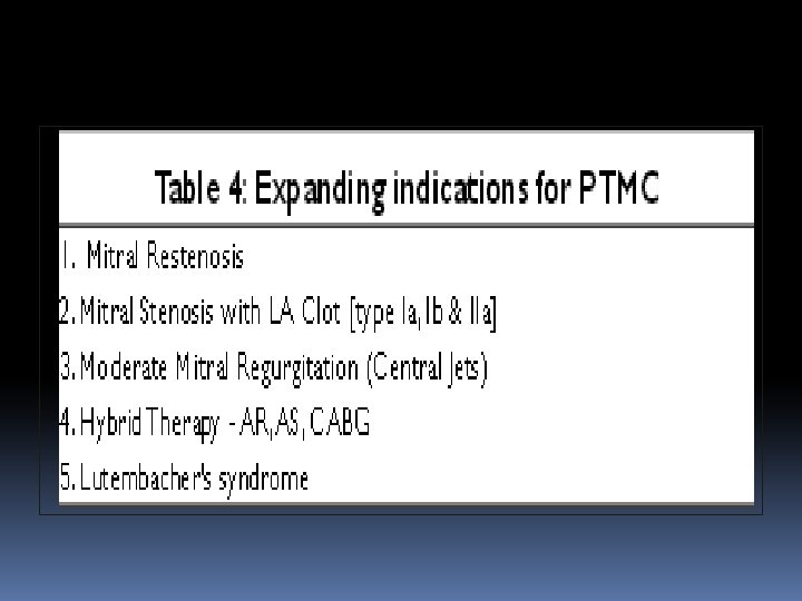

CONTRAINDICATIONS • The procedure can be performed at higher risk with thrombus localized to the LAA, thrombus within the LA itself is a contraindication to this procedure • Moderate or severe >2+ MR. • Mitral stenosis and aortic or tricuspid valve lesions that require cardiac surgery should be referred for surgery • Concomitant CAD can be treated with PCI in conjunction with valvuloplasty when the coronary anatomy is suitabl.

SUCCSES OF THE PROCEDURE • • Clinical profile of the patient Echocardiography Transseptal catheterization Balloon preparation Crossing of the MV Safe and effective balloon dilation Avoiding complications especially MR and cardiac tamponade.

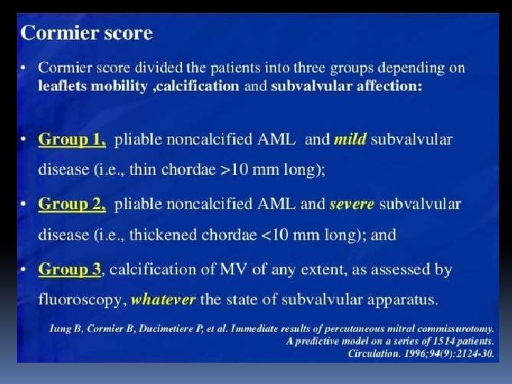
Clinical profile of patient • Usually the patients are in NYHA class II that can lie comfortably on cath table. • When a patient in NYHA class IV - can be undertaken for the procedure under anesthesia. • Younger patients with pliable noncalcific vaves with minimal subvalvular disease had better results.
 • Traditionally different grades of thickening, mobility, calcification and subvalvular disease")
Echocardiography (TTE &TEE) • Traditionally different grades of thickening, mobility, calcification and subvalvular disease of MV apparatus are assessed prior to PTMC. • A pliable valve responds to balloon dilation better than a non pliable valve. • However it is the calcification (that especially of commissures) out of these four characteristics that is of major concern during PTMC. • Even if the valve is heavily calcified with no calcification of commissures can be taken for PTMC with slight under dilation.

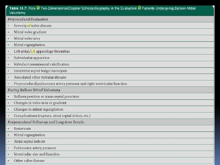
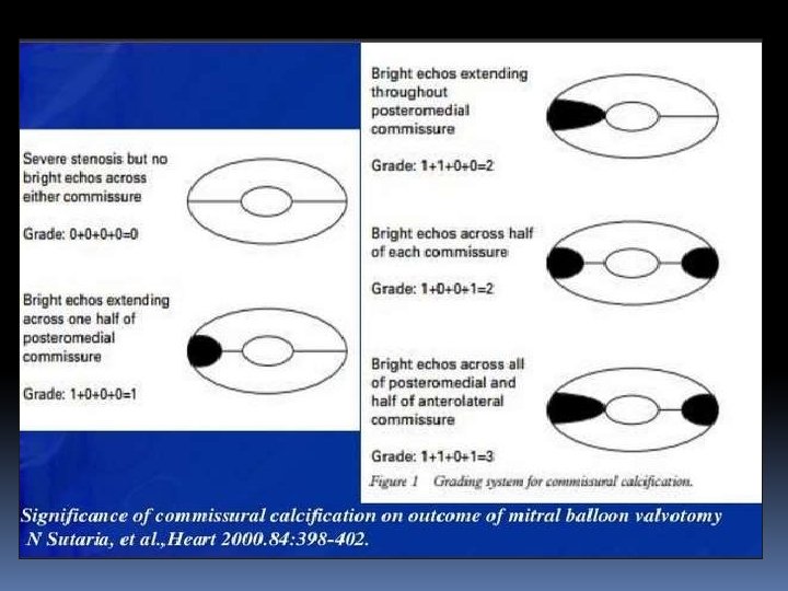
• High-quality TTE and TEE is an essential part of proper patient selection. • TEE prior to the planned procedure excludes the presence of LA thrombus and moderate or greater MR. • Dilating MV with commissural calcification may lead to leaflet tearing along non commissural lines and is associated with a higher incidence of procedure related MR

• Heavy calcification of the valve and/or bicommissural calcification are also associated with poorer acute and long term outcomes • Bicommisural symmetric fusion had a better success than assymetric.

• • A small LA size tells about less space available for manipulation of catheters, wires and balloons A very large RA size detected with TTE will be problematic during transeptal puncture.

• Aneurysm of IAS although rare should also be mentioned while doing TTE for a patient undergoing PTMC. • Similarly a thick IAS can identify prior to transseptal puncture that might require some extra effort during procedure.

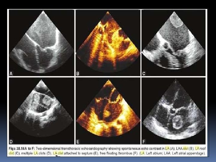
• A thrombus should be excluded in the cardiac chambers prior to PMV. • Most of the sites in LA can be effectively seen by TTE. • TEE is helpful when TTE is suboptimal. • The sites which should be specifically looked for thrombus are (a) LAA (b) at the junction of LAA and LA, (c) IAS, (d) layered thrombus in LA wall and (e) LA spontaneous echo contrast.

• One can also define the fresh or organized nature of the thrombus by its TTE features. • Degree of MR • To look for hemodynamically significant lesions of other valves. • This information is valuable during transseptal catheterization

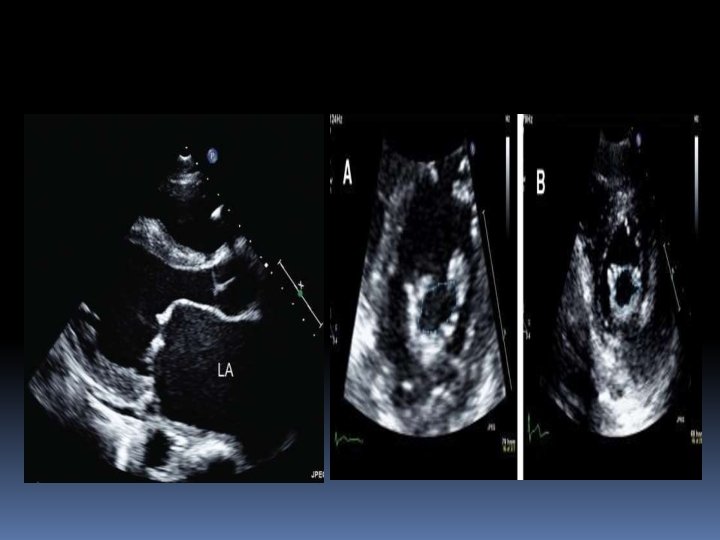
Manjunaths classification of LA Clot Catheterization and Cardiovascular In. Pt. Be. Mr. Vv. Te. Pn. I St. Aio. NDns. TR. I 7 CK 4 S: 653 -661(2009
(Boston or Abascal score)")
Echo Score (Wilkins Score)(Boston or Abascal score)
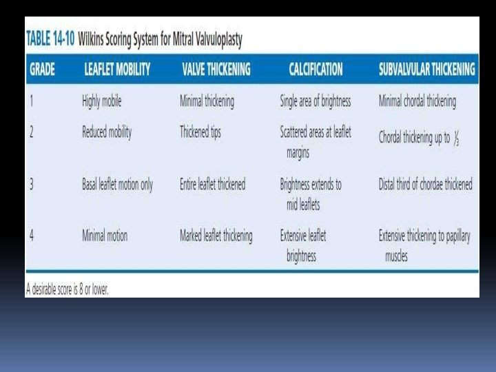

• A score of less than 8 gives better results and long term succes of the procedure than more than 8 • There is no absolute contraindication to PMV in patients with higher echo scores

LIMITATIONS OF WILKINS • Echocardiography limited in ability to differentiate nodular fibrosis from calcification • Assessment of commissural involvement is not included or underestimated. • Doesn’t account for uneven distribution of pathologic abnormalities. • Doesn’t account for relative contribution of each variable (no weighting of variables). • Frequent underestimation of subvalvular disease. • Doesn’t use results from TEE or 3 D echocardiography

• Chen et al is a modified Wilkins score parameter for subvalvular thickening according to the involved segment of chordal length • (1) if less than 1/3, (2) if more than 1/3, (3) if more than 2/3, and (4) if involved the whole chordal length with no separation

• Reid score includes leaflet motion, leaflet thickness, subvalvular disease, and commissural calcium • Leaflet motion was expressed as a slope by dividing the height (H) by the length (L) of doming of anterior leaflet. • • Leaflet thickness was expressed as the ratio between the thickness of the tip of MVand thickness of posterior wall of aortic root. The score was assigned as 0 for mild affection, 1 for moderate, and 2 for severe affection

Nobuyoshi score

MV score based on real-time 3 D echocardiography Normal=0, mild=1– 2, moderate= 3– 4, severe >5 b Normal=0, mild=1– 2, moderate= 3– 5, severe >6 (Modified from Anwar et al. [31 • • ]P; Bw. MVit. Th. PIp. SAe. Nr. Dm. Ti. Rs. Cs. IKio. Sn. )

• The individual RT 3 DE score points of leaflets and subvalvular apparatus RT 3 DE score were summed to calculate the total RT 3 DE score, ranging from 0 to 31 points. • Total score of mild MV involvement was defined as <8 points, moderate MV involvement 8– 13, and severe MV involvement >14.

APPROACH There are usually two approaches for the PBMC 1. Transvenous approach 2. Transarterial approach. • Percutaneous transvenous mitral valvuloplasty (PTMC) is the most common and time tested approach for this procedure.

TECHNIQUE S There are three main techniques for PTMC: 1. Single balloon technique 2. Double balloon technique 3. Inoue balloon technique
 • BMV was introduced in 1984 by the Japanese")
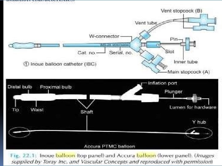
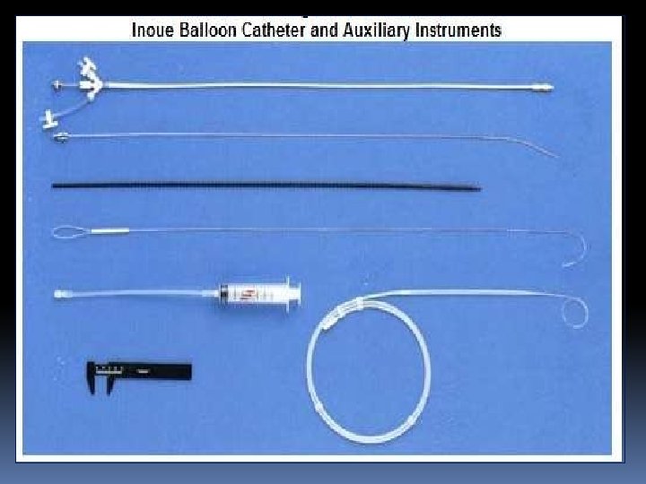
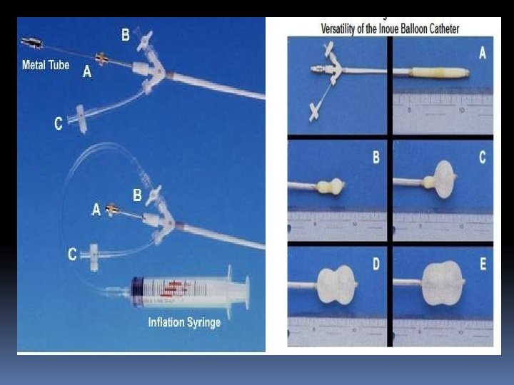
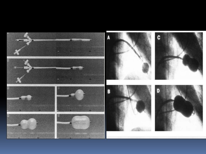
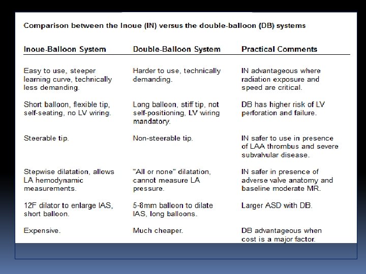
Inoue balloon technique (TV approach) • BMV was introduced in 1984 by the Japanese surgeon Kanjie Inoue, who developed the procedure as a logical extension of surgical closed commissurotomy. • The Inoue-Balloon Catheter is manufactured of polyvinyl chloride with a balloon attached to the distal end. • The balloon is two latex layers between which is polyester micromesh • Owing to the variable elasticity along its length, the balloon inflates in three distinct stages.

• The balloon section is stiffened and slenderized when stretched by the insertion of a metal tube. • The balloon size is pressure dependent and consists of 3 portions with slightly different compliance

• Inoue balloon technique is faster and less cumbersome and generally requires less fluoroscopy time. • Inoue balloon allows simple progressive upsizing of the balloon without withdrawing the balloon from the LA an important advantage if larger balloon sizes are needed. • The Inoue balloon system may, however, result in a slightly higher incidence of MR

Transeptal punture The FO is located superiorly and posteriorly to the ostium of the CS and well posterior of the TA and r. RAA. The fossa ovalis is posterior and caudal to the aortic root and anterior to the free wall of the right atrium The goal of trans-septal catheterization is to cross from the RA to the LA through the fossa ovalis

• Puncture of the fossa ovalis itself is quite safe, the danger lies in the possibility that the needle and catheter will puncture an adjacent structure (i. e. , the posterior wall of the RA, the CS, or the aortic root). • In aortic stenosis, the plane of the septum becomes more vertical and the fossa may be located slightly more anteriorly. • In mitral stenosis, the intra-atrial septum becomes flatter with a more horizontal orientation and the fossa tends to lie lower.

• Several algorithms using fluoroscopic landmarks determined by right and LA angiography, or the position of a pigtail catheter in posterior (noncoronary) aortic sinus of Valsalva, have been developed to aid localization of the best site for TSP. • Intraprocedural TTE , TEE or ICE may aid in identifying the optimal location for puncture of the IAS.

Septal puncture • First advance a flexible 0. 032 -inch, 145 -cm J guidewire into SVC with an end hole catheter. • For the femoral approach use a 70 -cm curved Brocken brough needle which tapers from 18 gauge to 21 gauge at the tip under continuous ECG and pressure monitoring. • Brockenbrough needle is put inside the Mullin’s sheath and dilator (usually 7 F size) and advanced over the wire into SVC.

• Either the Mullin’s dilators could be advanced over the PFO or the IAS can be punctured with Brockenbrough needle and then the Mullin’s dilator • In most cases PFO can be engaged with descending the atrial septum from the SVC to the TV level noting the aortic bulge and pulsations of the LA pressure on the tip of descending Mullin’s dilator with the “limbic” edge of PFO present just below the aortic bulge

• The catheter is advanced slightly to flex its tip against the limbus at the superior portion of the foramen ovale. • Once the operator is satisfied with this position, advance the Brockenbrough needle smartly so that its point emerges into LA. • Change in atrial pressure waveform and the ability to withdraw oxygenated blood from the needle, the demonstration of the typical fluoroscopic appearance of the LA during a contrast puff through the needle.

• Once the needle is in LA , the needle is removed and then a specially curved LA wire (a special solid-core coiled 0. 025 -inch guidewire) is introduced into the LA, and the Mullins sheath dilator system is removed. • Septal dilator is passed over the wire for adequate dilatation of the septal puncture site • Systemic anticoagulation after the septal puncture to prevent the formation of the thrmobi on the wires and cathters.
 and Bing stylet (left) can be used in conjunction")
The Brockenbrough needle (far left) and Bing stylet (left) can be used in conjunction with the traditional Brockenbrough catheter (center) and Mullins sheath/dilator system (right)

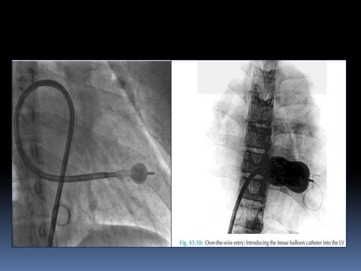
• The previously prepared, tested, and now slenderized Inoue balloon is then introduced over the guidewire into the LA. • After the slenderized balloon has been positioned within the LA, the stretching tube is removed, and a preshaped J • stylet is introduced into the Inoue balloon. • The distal portion of the balloon is inflated slightly to aid in crossing the valve and to prevent intra chordal passage

• AP , RAO 30° and lateral views are utilized with fluoroscopy for transseptal puncture. • Some anatomical changes in the position of PFO may occur due to hemodynamic consequences of mitral and aortic valve diseases.

• As viewed from the feet with the patient lying supine, the plane of the atrial septum runs from 1 o’clock to 7 o’clock. • The fossa ovalis is posterior and caudal to the aortic root, anterior to the free wall of the RA, superior and posteriorly to the ostium of the CS, and well posterior of the TA and the RAA. • It is approximately 2 cm in diameter and is bounded superiorly by the limbus

• In MV disease, rather than lying at the junction of lower and middle third of IAS the FO tends to lie lower, and in severe disease the limbic ledge is found in the lower third. • It means that there is inferior displacement of FO in presence of MS. • IAS becomes more horizontal and tends to bow into RA as LA pressure rises. • The septal bulge by displacing the FO and gutters by interfering the mobility of puncture set in RA present TIP. S difficulty in probing the. PBMFVO

• In this situation gentle anterior and posterior rotatory movements are given to the trans septal introducer set during its withdrawal from SVC to RA. • Another difficulty is the “jumping” of transseptal introducer set out of the gutter and onto the central septum. • A reinitiated descent from SVC in AP, RAO and lateral views is sometimes required to engage the FO.

• Reshaping the Brockenbrough needle’s curve at the last few centimeters is also sometimes helpful • Aortic valve diseases because of dilated and unfolded aorta displace the FO superiorly and anteriorly. • Hence IAS is more vertically oriented. • A peculiar problem in this condition is that the tip of the introducer set passing repeatedly from the aortic septal bulge to the lower septum, not engaging FO. • Here again reshaping the Brockenbrough needle by exaggerating the curve of the last few centimeters may help to engage the FO

• In case of massive RA enlargement, the transseptal introducer set may not descend against the aorta and then lower septum, but rather lie free within RA cavity. • To solve this problem a gentle curve is shaped approximately 10– 15 cm from the needle tip and in the same plane as the more distal curve.

• It should be remembered that inferior limit of IAS is best seen in AP view where the margins of LA “double contour” are seen and the lateral fluoroscopic view is the best for degree of posterior positioning of the catheters and angulation of the needle prior to its advancement

• A low puncture in the middle and posterior one-third of the IAS is desired for the double balloon mitral valvuloplasty. • A posterior but higher puncture is recommended for Inoue balloon technique.

So one should remember following things which require special attention while attempting septal puncture during PMV • Isolated MS • Mitral stenosis with AS and/or AR • Mitral stenosis with tricuspid stenosis (TS) and/or TR or huge RA dilation • Kyphoscoliosis • Interatrial septum aneurysm • Thick IAS (e. g. postoperated or diseased)

Following things either alone or in combination can help during difficult transseptal puncture • TTE/TEE • Biplane fluoroscopy • Pigtail catheters positioned in noncoronary aortic sinus • Septal injection of contrast • Single and/or biplane RA angiogram. • CT scan use has been described in rare cases of lipomatous hypertrophy of IAS.

• One important thing at this stage is complication in the form of cardiac perforation • Common sites of cardiac perforation during transseptal catheterization are: • Coronary sinus - the most common site of cardiac perforation. • Left atrium perforation: Sudden jerk and jump of the Brockenbrough needle after puncturing the IAS can lead to perforation of LA roof or posterior wall. • One has to be very careful especially in cases of thick septum and small LA as needle requires more than usual pressure at puncture site.

• Other rare sites of cardiac perforation while TSP are perforation of RA at the junction of SVC and perforation of right lateral aspect of root of aorta adjacent to SVC. • The common mechanism in RA and aortic root perforation is the superior slipping of the transseptal set along the IAS until it is held by crista terminalis. • At this point misinterpretation of resistance to further advancement of transseptal set being offered by FO leads to this complication. • A similar misunderstanding can occur with a thick IAS.

• Complications of trans-septal catheterization are generally infrequent (needle tip perforation <1%, tamponade <1%, and death <0. 5%) in experienced hands

BALLON SIZE • There are some guidelines for selecting optimal balloon size: • Effective Balloon Dilating Diameter (mm) = [(Height of patient in cm/10) + 10] • This is the most common and worldwide accepted formula for selecting a balloon size while performing PMV with Inoue balloon.

• BSA of the patient: A 26 -mm diameter balloons for patient of less than 1. 5 m² BSA, 28 mm diameter balloons for patient of 1. 5– 1. 7 m² BSA and 30 mm diameter balloons for patient of greater than 1. 7 m² BSA. • This can be useful in lean, thin and underweight patients.

CROSSING THE MITRAL VALVE • when a higher and posterior puncture is taken for the balloon can be passed across the MV orifice in a direct line from the atrial septum because of its specific curvature • During balloon catheter manipulation performed under a 30° RAO fluoroscopic view, the catheter in the LA should always be kept to the left of the pigtail catheter preplaced in the LV.
 or the angle between")
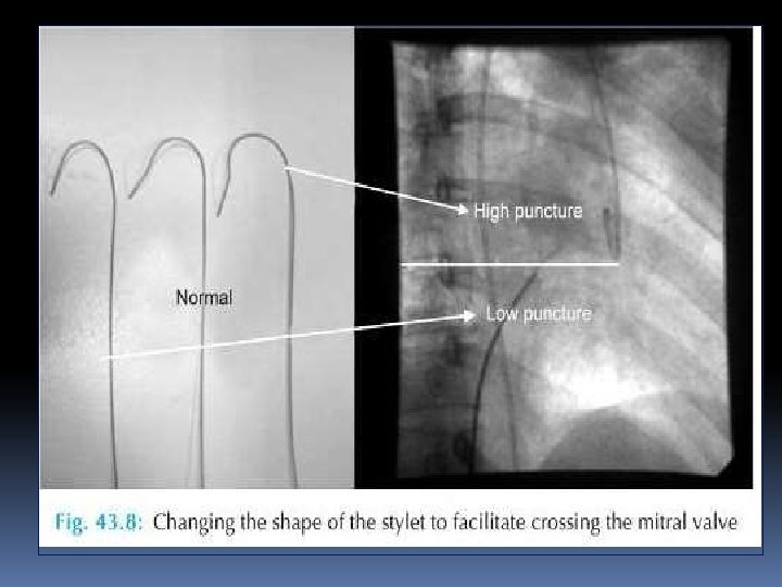
• When the site of TSP is superior (high) or the angle between the site of puncture and MV is very acute then • Clockwise rather than counterclockwise rotation of stylet so that balloon can be bounced of the posterior LA wall • 15– 20° angle in the stylet placed 10– 12 cm proximal to the distal tip.

When there is giant biatrial enlargement, a very large curve 20– 30 cm proximal to the distal tip of the stylet may help to give the catheter an arch appropriate to the large curvature of the path toward the mitral orifice

• If the site of puncture deviates rightwards or upwards, a large radius curve is made in the stylet with the apex of stylet curve placed on the puncture site in the septum, the ascending curve of the balloon catheter in RA and the descending curve in the LA. • This curve is larger than the curve placed in the LA alone with the conventional direct method.

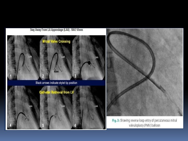
• When the puncture site is moved to the left, downwards or too close to the MV, the alternatives to cross the balloon are reverse loop method. • Another approach in this situation is first crossing the MV and placing the 0. 20 backup J-tipped wire in LV and then introduction of diagnostic JR coronary catheter over this wire. • Then positioning of the preshaped 0. 25 coil wire in LV and then introduction of Inoue balloon over the wire

C. N. Manjunath. DM 1, *, K. H. Srinivasa DM 1, C. B. Patil DM 1, H. V. Venkatesh DM 1, . S. Bhoopal MD 2 and. C. Dhanalakshmi RCDS 1 Article fir st published online: 6 DEC 1998 BALLOON MITRAL VALVULOPLASTY: OUR EXPERIENCE WITH A MODIFIED MITRAL VALVE IN DIFFICULT CASES TECHNIQUE OF CROSSING THE

• As the complexity of balloon crossing increases there is simultaneous increase in risk of development of MR, damage to the MV apparatus, cardiac perforation. • Sometimes withdrawing the steering stylet tip up to the septal puncture site inside the Inoue balloon which is minimally inflated at its distal portion can place the balloon toward MV.

• Re dilation of IAS with a 14 -F dilator or even with a 6– 10 mm peripheral arterial balloon may sometimes be necessary. • Applying negative pressure on the balloon can help to cross the MV and also placing the balloon at LV apex • Once the MV has been crossed, the free movements of the partially inflated distal balloon in the LV should be ascertained to prevent the disastrous consequences, i. e. rupture of chordae, papillary muscles or leaflets

• This is done by simultaneously pushing the catheter and pulling the stylet in opposite directions ("accordion" maneuver) to ensure that the partially inflated distal balloon slides freely along the orifice-apex axis.

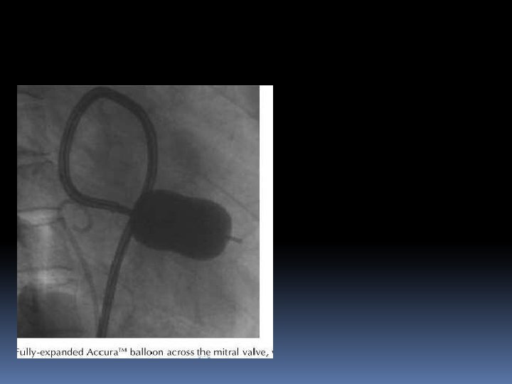
• After the balloon catheter is across the mitral orifice, the distal portion of the balloon is inflated more fully and the catheter is pulled back gently to confirm that the inflated distal portion of the balloon is secure across the valve. • As further volume is added to the balloon, the proximal end inflates to lock the valve between the proximal and distal balloon. • Inflation to precalibrated volume then dilates the valve orifice to the corresponding preset size

• It is then allowed to deflate passively before it is withdrawn into the LA. • Inflations in the low-pressure zone result in less mitral regurgitation than inflations in the high-pressure zone using smaller size ballon. EX…a 30 -mm balloon inflated to a maximum diameter of 28 mm will overall result in causing less MR than using a maximal nominal 28 -mm balloon inflated to 28 mm (in the high-pressure zone Goel PK, Garg N, Sinha N. Pressure zone used and the occurrence of mitral regurgitation in Inoue balloon mitral commissurotomy. Cathet Cardiovasc Diagn 1998; 43: 141– 146 Yamabe T. Nagata S. Ishikura F. Kimura K. Miyatake K. Influence of intraballoon pressure on development of severe mitral regurgitation after percutaneous transvenous mitral commissurotomy. Cathet Cardiovasc Diagn 1994; 31: 270– 276

• Stepwise Dilation • The nominal balloon diameter was decided according to the height of the patient (i. e. height (cm)/10 + 10 = balloon diameter). • The first inflation was performed to a balloon diameter of 2 mm less than nominal, followed by successive inflation by 0. 5 mm increment to the maximum nominal balloon diameter.

Low pressure zone* = balloon diameter < 2 mm of nominal balloon size. High pressure zone** = balloon diameter within 2 mm of nominal balloon size. SL = severe subvalvular lesions

• After each balloon inflation, the balloon catheter was withdrawn into the LA and the transmitral gradient was immediately reassessed. • An auscultation, examination of LA pressure waveform and on occasion color D examination and left ventriculography were repeated to evaluate any change in MR.

• If the transvalvular gradient persists and no increase in MR was observed, another balloon inflation was performed to a balloon diameter 0. 5– 2 mm larger than nominal. • This stepwise process was repeated till the mitral gradient was reduced as much as possible without a significant increase in MR

Fully inflated PMV balloon across MV in RAO view. Note the alignment of long axis of balloon catheter along the long axis of LV cavity
")
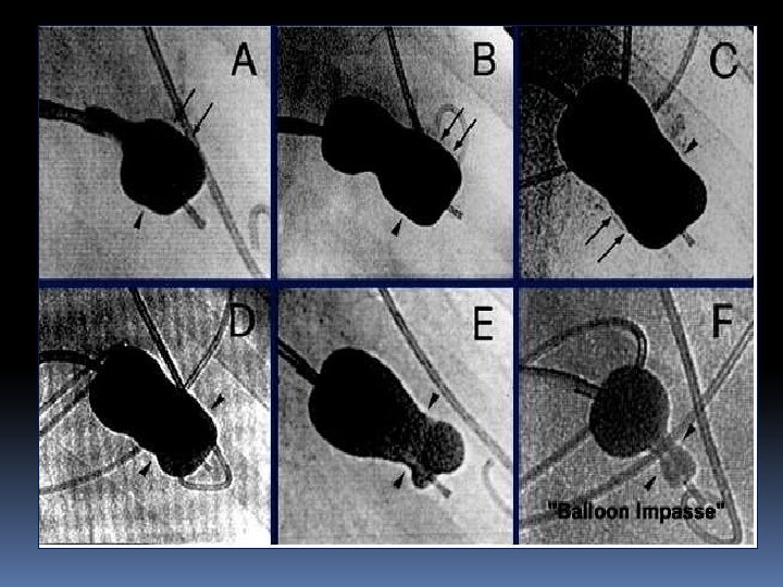
BALLON SUBVALVULAR TRAPPING • Gross indentation of the inflated distal balloon (balloon compression sign) • "Balloon impasse. " In cases of tight MS, valve crossing may be difficult even when the balloon is not inflated, the catheter is checked (or entrapped) at the mitral valve. This finding, which was termed "balloon impasse, " reflects resistance caused by severe obstructive subvalvular lesions This sign indicates the presence of the most severe subvalvular disease and signifies a extremely high-risk for creation of MR • Cogwheel resistance. Rarely, while withdrawing the partially inflated balloon to anchor it at the mitral valve, cogwheel resistance may be encountered. This suggests the presence of subvalvular disease.

SAFE AND EFFECTIVEBALLOON DILATION • The purpose of stepwise dilation is to prevent the onset of severe MR and to achieve the largest maximum mitral orifice possible. Termination of the procedure • More than or equal to 50% improvement in valve area • Final MVA greater than 1. 5 cm² or greater than 1 cm²/m² BSA • Complete opening of at least one commissure • Appearance or increment of regurgitation more than 1/4 grades.

• Following successful dilatation, the Inoue balloon is then reslenderized by first reintroducing the guidewire and then the stretching tube. • It is useful to leave the guidewire across the atrial septal puncture in the LA for 3 to 5 minutes after completion of the procedure, while monitoring the systemic arterial pressure

• If a wire is left in place at the end of the procedure and the BP drops precipitously after a couple of minutes, with the wire in place, a small balloon catheter can be passed back across the puncture site and inflated to stabilize the patient while pericardial centesis is performed and plans for further management are made
 Tearing")
COMPLICATIONS MR during PMV can occur because of following (from 2 to 9%) Tearing or stretching of commissures Failure of leaflet co-aptation Rupture of mitral leaflet Rupture of chordae Damage to papillary muscles. Same day surgical MVR 2 -3% •

• Skilled hands --the failure rate of the procedure should be <5%. • Failure usually results at septal puncture, positioning the balloon catheter successfully across the MV • Mortality 0 -3% • Hemopericardium 0. 5 -10% • Systemic embolization 0. 5 to 5%

PRECAUTIONS TO AVOID COMPLICATIONS • Careful clinical evaluation and echo prior to PMV (e. g. to rule out acute rheumatic activity, IE) • Appropriate size selection and preparation of balloon • Be sure that balloon catheter is free in LV cavity after MV has been closed. If balloon is having angulated course during its LV entry and appearing as entangled within chordae tendineae, balloon is withdrawn and then the balloon is passed again. • Recognizing “Impasse” sign indicative of severe subvalvular disease • Careful positioning of balloon which must move freely along the long axis of the LV cavity prior to inflation

• Procedure related thromboembolism (a missed intracardiac thrombus or vegetation or valve tissue or a newly formed thrombus/air) leading to stroke is another complication

PBMV During Pregnancy • Inoue balloon is preferred technique because of shorter procedure time and low radiation exposure • External shielding during procedure • Saving fluoroscopic images and avoiding high dose cineradiography • Reducing the frame rate of fluoroscopy (e. g. 15 frames/sec or lower). • Experienced operators are preferred • Keep the intensifier as close as possible to the patient • Avoid angulated projections—AP projection is preferred
")
• Procedure is best avoided until organogenesis is complete (5 months after conception) unless the patient is severely symptomatic and refractory to optimal medical therapy. • If emergency PMV becomes unavoidable during early months of pregnancy, then target of radiation exposure should be set as doses greater than 50 m. Gy are associated with congenital malformation and growth retardation

• Only at doses greater than 100 m. Gy should termination of pregnancy be considered on the basis of exposure. • We should strictly follow the as low as reasonable achievable principle so as to limit radiation exposure to mother and fetus.

DOUBLE BALLON TECHNIQUE • Used with two balloons advanced over separate guidewires from the femoral vein to the LA, across the MV into the LV. • The two balloons are then inflated simultaneously across the mitral valve. • • When properly performed, the DBT results in excellent improvement in MVA. • Multiple studies have shown no significant difference in hemodynamic results (MVG or MVA) post procedure between the doubleballoon technique and the Inoue balloon system

Mansfield balloon

• An adaptation of the double-balloon technique uses a monorail approach to deliver two balloons across the mitral valve over a single guidewire. • The first valvuloplasty balloon with a short monorail segment is passed over the wire across the MV, followed by a second conventional balloon that is then passed over the wire until it is parallel with the first balloon. • There are no substantial differences in the mechanism of delivery of force by two balloons using this approach compared with conventional double-wire, double-balloon technique • Bonhoeffer P, Piechaud JF, Sidi D, et al. Mitral dilatation with the Multi-Track system: an alternative approach. Cathet Card. Pi. Bo. Mv. Va. Tsc. IPSDANia. Dg. Tn. RCI 1 K 9 S 95; 36: 189– 193. .
 techniques,")
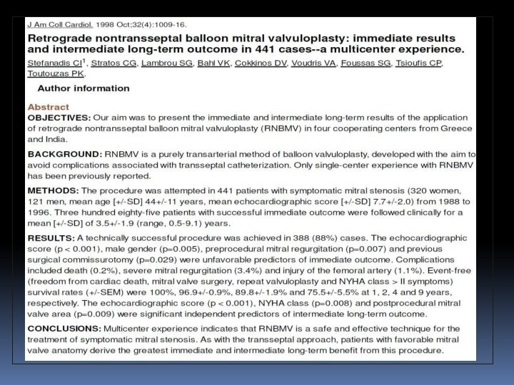
Retrograde transarterial techniques • Used alone or in combination with antegrade (transseptal puncture) techniques, have been used in some centers for single- and double-balloon PMV • Advantage of not requiring trans-septal puncture or using only minimal dilatation of the intra-atrial septum. • Disadvantages of these techniques include the opportunity for arterial injury because of the larger balloons used. • In addition, the procedures can be technically difficult and time consuming.

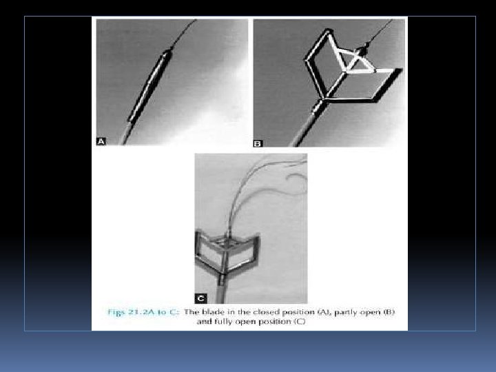
• A 19 F metallic commissurotome can be passed across the interatrial septum over a guidewire and used to accomplish mitral commissurotomy. • There has been some evidence that bicommissural splitting can be accomplished more frequently with the metal commissurotome. • Randomized comparisons of the Inoue balloon and metallic commissurotome have not demonstrated significant differences in long-term outcome. Zaki AM, Kasem HH, Bakhoum S, et al. Comparison of early results of percutaneous metallic mitral commissurotome with Inoue balloon technique in patients with high mitral echocardiographic scores. Catheter Cardiovasc Intervent 2002; 57: 312– 317 Cribier A, Rath PC, Letac B. Percutaneous mitral valvotomy with a metal dilatator [letter]. Lancet 1997; 349: 1967. .

Immediate Results • Immediate results of PMV are assessed by a combination of echo Doppler measurements and hemodynamics.

• Most investigators - valve area ↑ 1. 0 cm 2 2. 0 cm. • ↓ LA pressure and the transmitral pressure gradient, a reduction in PAP, and an increase in CO • ↓ in LA stiffness, resulting in an increase in LA pump function in patients in SR and an increase in LA reservoir function in those with AF.

• In some cases, a single commissure is split during one of the first balloon inflations. • This is often the result of asymmetric commissural fusion or calcification. • But splitting of a single commissure often makes it difficult to split the second commissure, since the inflated balloon will be displaced into the already opened side of the valve. • This typically results in an adequate rather than an excellent postprocedure valve area

Long-Term Hemodynamic Results • 1 YR after valvotomy, several parameters show continued improvement, including further reductions in PASP and increases in CO. • PVR declines and normalizes in many patients

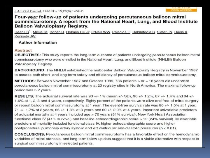
• The actuarial survival rate at one, two, three, and four years was 93, 90, 87, and 84 percent, respectively. • The event-free survival (freedom from death, mitral valve surgery, or repeat PMBV) at one, two, three, and four years was 80, 71, 66, and 60 percent

LONG-TERM RESULTS OF BMV FOR MS


Long term results up to 19 years of mitral balloon valvoplasty Asian Cardiovasc Thorac Ann, 17 (2009), pp. 627– 633

Multivariate predictions of mortality • • Echocardiographic score >8 Increasing age Prior surgical commissurotomy NYHA functional class IV Higher postprocedural PA pressure Preprocedural mitral regurgitation ≥ 2+ Postprocedural mitral regurgitation ≥ 3+

CONCLUSIONS • PBMV is a safe and effective procedure with >95% procedural success and excellent long term results. • Results depend on the experience of the operator and nature of the valve.

- Slides: 138