Bacterial Structure Function Genetics Prof Hanan Habib College

Bacterial Structure , Function & Genetics Prof. Hanan Habib College of Medicine , Department of Pathology , KSU

Objectives • Define the cellular organization of bacteria and recall the differences between Eukaryotes and Prokaryotes. • Recall major structures of bacteria and its function. • Describe the structure of cell wall of bacteria including the differences between Gram positive and Gram negative bacteria and main functions.

Objectives, cont. , • Describe the external and internal structures of bacteria and their functions. • Describe bacterial spores and its application in the practice of medicine. • Recall basic information about bacterial genetics and replication of bacteria.

Objectives, cont. • Describe plasmids , its origin , types and its importance in clinical practice. • Recall genetics variations, including ; mutation and mechanisms of gene transfer and its implication on bacterial resistance to antimicrobial agents.

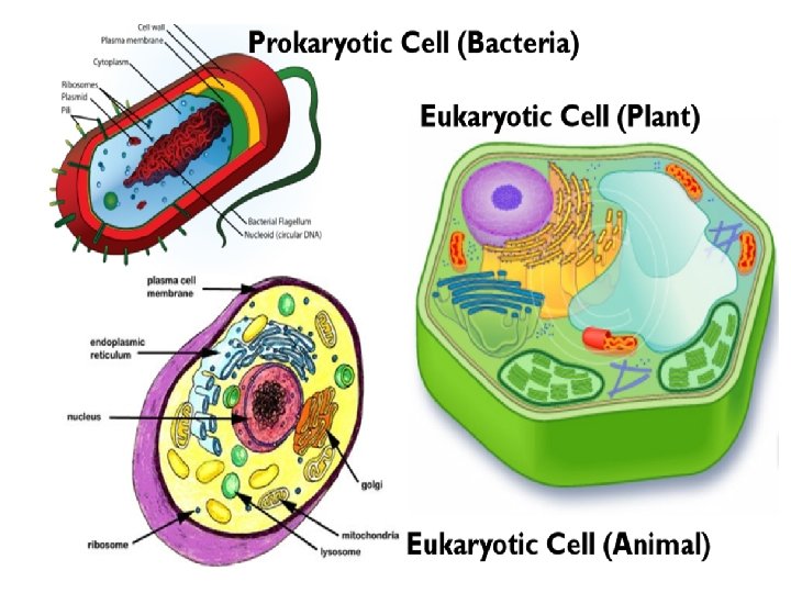
Definition Bacteria : a heterogenous group of unicellular organisms , about 1 -8 μm in diameter Bacteria is a Prokaryote (has a primative nucleus): - one chromosome - no nuclear membrane - no mitochondria - no sterols Bacteria contain Plasmids: an extra piece of DNA.

Shapes & Types of Bacteria • • • Spherical / Oval…………Cocci Rods……………Bacilli Very short Bacilli…………Coccobacilli Tapered end ……………Fusiform Club-shaped / Curved……Vibrio Helical / Spiral… …………Spirochaetes

Arrangements of Bacteria Arrangements among cocci : • Pairs…………. Diplococci • Chains………Streptococci • Clusters……. Staphylococci • In four………. Tetrad • Palisades…. . Corynebacterium


Structure of Bacteria

Cell Wall of Bacteria • Bacteria are cells with rigid cell wall surround cytoplasmic membrane and internal structures. Functions of cell wall: • Rigidity • Shapes bacteria • Protection • Porous / permeable to low molecular weight molecules • Cell division • Antigenic determinants

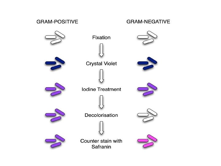
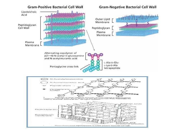
Cell Wall of Bacteria • Two groups of bacteria depending on reaction to GRAM stain : Gram positive: stain blue/purple. Gram negative: stain red. Note : Mycoplasma naturally have no cell wall. Chemical structure of bacterial cell wall: Peptidoglycan : Rigid part , mucopeptide composed of alternating strands of Nacetyl muramic acid and N- acetyle glucosamine linked with peptide sub units.


Cell Wall of Gram Positive Bacteria • Peptidoglycan is thick • Closely associated with cytoplasmic membrane. • Contain : Teichoic acid : anchors cell wall to cell membrane , epithelial cell adhesion. Antigens : - polysaccharides (Lancefield) - protein (Griffith)

Cell Wall of Gram Negative Bacteria • Thin Peptidoglycan • Has an outer membrane that contains : - specific proteins (porins) important in the transport of hydrophilic molecules - lipopolysaccharide (Endotoxin)


External Structures of Bacteria External protrude from the cell into the environment: • Flagella • Pili • Capsule

Flagella • • • Helical filaments Composed of protein FLAGELLIN. Found in Gram positive & Gram negative bacteria. Function : motility& chemotaxis Distribution: - Peritrichous - Monotrichous - Lophotrichous -Amphitricous

Pili Fine short filaments extruding from cytoplasmic membrane. Found on the surface of many Gram negative & Gram positive bacteria. Composed of protein Pilin. Two types: 1 - Common pili (fimbriae): covers the surface— responsible for: adhesion & colonization 2 - Sex pili : in some bacteria only, responsible for conjugation.

Capsules and Slime layer • These are the structures surrounding the outside of cell envelop. Can be seen by India ink or special stains • Usually consist of polysaccharide, however ; in some bacteria consist of polypeptide(protein). • They are not essential for cell viability, some strains within species produce capsule while others do not. Functions, include : • • Attachment Protection from phagocytic engulfment Resistant to dryness Reservoir for certain nutrient
 – Double layered structure composed of phospholipid & protein –")
Cytoplasmic Membrane (plasma membrane) – Double layered structure composed of phospholipid & protein – Act as semi- permeable membrane (passive diffusion) – Site of numerous enzymes involved in active transport of nutrients and various metabolic processes

Internal structures of bacteria Mesosomes : convolutes invaginations of cytoplasmic membrane. Function: 1. Involved in DNA segregation during cell division and respiratory activity 2. Contain receptors involved in chemotaxis 3. Permeability barrier (active transport of solutes).
 Ribosomes")
Core of Bacteria Core composed of : Cytoplasmic inclusions Nucleoid ( nuclear body) Ribosomes Cytoplasmic inclusions: Are nutritional storage granules , examples: - Volutin - Lipid - Starch / or Glycogen
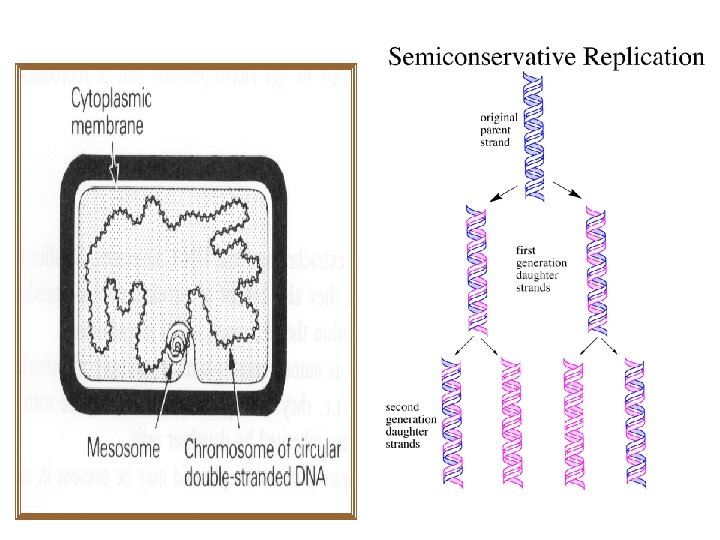
 • Circular single stranded chromosome (bacteria genome or DNA) •")
Nucleoid ( Nuclear Body) • Circular single stranded chromosome (bacteria genome or DNA) • No nuclear membrane • DNA undergoes semi-conservative replication , bidirectional from a fixed point.

Ribosomes of Bacteria • Distributed throughout the cytoplasm • Site of protein synthesis • Composed of RNA and protein

Bacterial Chromosomes • Haploid, circular molecule of double stranded. DNA attached to cell membrane. • Genetic code in Purine and Pyrimidine bases of nucleotides that makes DNA strand. • 3 bases comprise one code, each triplet codon codes for one amino acid. • Replication is semiconservative , takes place by binary fission.

Spores of Bacteria • Small , dense, metabolically inactive , nonreproductive structures produced by Bacillus & Clostridium • Enables the bacteria to survive adverse environmental conditions. • Contain high concentration of Calcium dipicolonate. • Resistant to heat, dissecation & disinfectants • Often remain associated with the cell wall

Spores of Bacteria-cont. • Spores are described as : 1 - Terminal spores 2 - Sub-terminal spores 3 - Central spores • Spores germinate when growth conditions become favorable to produce vegetative cells. • Application in medical practice : spore preparations used for checking the efficacy of Autoclaves, eg. Bacillus subtilis & Bacillus sterothermophilus.

Spores of Bacteria

BACTERIAL GENETICS

Bacterial Genetics: definitions • Genetics is the study of inheritance and variation. • Genetic information encoded in DNA. Function of genetic material: 1 - Replication of the genome 2 - Expression of DNA to m. RNA then to protein.

Definitions-cont. • Genotype: the complete set of genetic determinants of an organism. • Phenotype: expression of specific genetic material. • Wild type: reference (parent) strain • Mutant: progeny with mutation. Two types of DNA in bacteria • Chromosomal • Extra-chromosomal (Plasmid).

Plasmids • Extra chromosomal DNA composed of double stranded-DNA. • Found in most species of bacteria. • Origin? • Govern their own replication • Application : in genetic exchange, amplify genes • Transfer to other bacteria by conjugation

Plasmids

Types of Plasmids 1 - R-plasmids: genes code for antibiotic resistance particularly Gram negative bacteria. 2 -Col-plasmids: in Enterobacteria, codes for extracellular toxins. 3 - F-plasmids: (fertility) factor, transfer of chromosome during mating.

Genetic variation in bacteria Takes place by: 1 - Mutations 2 -Gene transfer
. • Chemical changes in")
Mutation • Inheritable changes in the structure of genes (DNA). • Chemical changes in one or more bases of DNA. Mutation /gene defect leads to alteration in: • Transcription, • Amino acid sequences, • Function eg. Bacteria become resistant to antibiotic.

Classification of Mutation Depends on biological sequencing: 1 - Resistance mutation: affects structure of cell protein. Main application in medical practice. Bacteria become resistant to antibiotics. 2 - Auxotrophic mutation: affects biosynthetic enzyme resulting in a nutritional requirement of mutant cell. 3 - Lethal mutation: leads to death of bacteria.

Mutation Causes Antimicrobial Resistance

Gene Transfer Among Bacteria Three mechanisms: 1 - Transformation 2 - Transduction 3 - Conjugation.
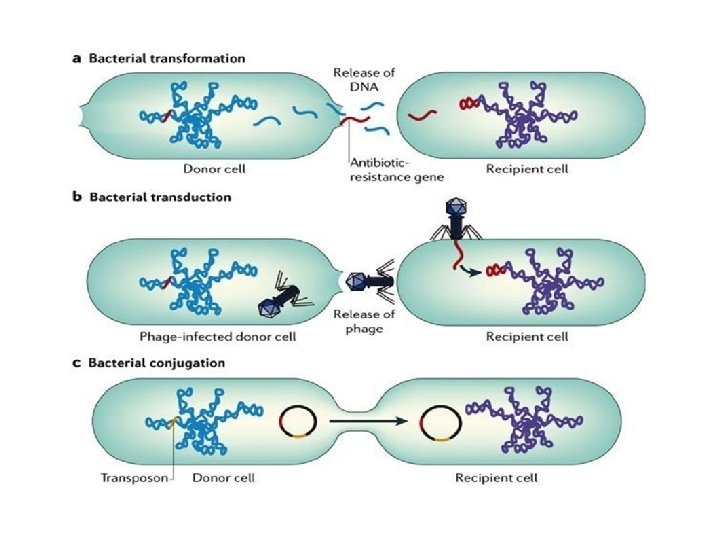

Transformation • A fragment of exogenous naked bacterial DNA are taken up and absorbed into recipient cells. • Common in Haemophilus influenzae & Streptococcus pneumoniae. • Bacteria become resistant to Ampicillin.

Transduction • Phage mediated transfer of genetic information from donor to recipient cells. Examples: • Beta – Lactamase production in Staphylococcus aureus : Bacteria becomes resistant to penicillin. • Toxin production by Corynebacterium diphtheriae.
 •")
Conjugation • Major way bacteria acquire additional genes. • Plasmid mediated( F factor) • Cell contact required and genes reside on plasmid resident within donor cells transfer to recipient cell (mating). • Conjugation is the common way of transfer of genes resistance to antibiotics among bacteria in hospitals.

Genetic Recombination After gene transfer, there are three possible fates: 1 -Exogenous DNA degraded by nuclease. 2 -Stabilized by circulization and become plasmid. 3 - Form a partially hybrid chromosome with segment derived from each source.

Reference Book Sherris Medical Microbiology, an Introduction to Infectious Diseases. Latest edition, Kenneth Ryan and George Ray. Publisher : Mc. Graw Hill.
- Slides: 49