BACTERIAL STAINING By Dr Hesnaa Saeed Al Mossawi

BACTERIAL STAINING By Dr. Hesnaa Saeed Al Mossawi

STAINING � � Stained preparations are needed in order to study their morphology and observe their cellular constituents Smears can be made from liquid or solid cultures or from the clinical specimen In Bacteriology, staining methods are divided into three categories Simple stains: This makes use of the direct staining method. Differential stains: This staining method divides bacteria into two groups Special stains: These are specialized staining methods to demonstrate certain bacterial components, e. g. spore.

Simple stain � � Direct Staining: This a simple one-step staining procedure in which the presence and morphology of bacteria are demonstrated Negative staining: This is when the organism remains unstained against a stained background. This is one of the few methods where acid stains such as nigrosin, are used

Gram stain Solutions Crystal Violet: 0. 5 to 1% in distilled water. Lugol's iodine: 10 g Iodine 20 g Potassium iodide 1000 ml Distilled water Decolouriser: Absolute ethyl alcohol or acetone and alcohol mixture (1: 1) Counterstain Aqueous solution of neutral red or safranin 0. 5%, or dilute carbol fuchsin. (1: 10 dilution of strong carbol fuchin in distilled water)

Procedure Make a smear, allow to dry and then fix with a gentle heat by passing the slide 2 or 3 times over a bunsen flame or placing the slide on a slide warmer. Stain with crystal violet for 1 minute. Wash with tap water. Apply Lugol's iodine and leave for 1 minute. Wash with tap water. Decolourise with acetone or alcohol until no more colour appears to ooze out of the smear (about 1 -2 seconds for acetone and 1 -2 minutes for alcohol and 10 seconds for acetone/alcohol mixture) � Wash immediately with tap water. � Counterstain with neutral red or safranin or dilute carbol fuchsin for 1 minute. � Wash with tap water. � Blot dry with a blotting or filter paper, and dry. �
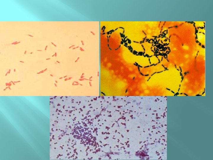

Gram stain � � is the most important staining procedure. Gram positive bacteria stain purple, whereas gramnegative bacteria stain pink. This difference is due to the ability of gram-positive bacteria to retain the crystal violet–iodine complex in the presence of a lipid solvent, usually acetone– alcohol. Gram negative bacteria, because they have an outer lipid-containing membrane and thin peptidoglycan, lose the purple dye when treated with acetone–alcohol. They become colorless and then stain pink when exposed to a red dye such as safranin
- Slides: 7