Bacterial morphology metabolism and growth Dr mer Kkbasmac

Bacterial morphology, metabolism and growth Dr Ömer Küçükbasmacı
")
Cell • Fundemental unit of living things (smallest bacterium-largest plants-animals)

Bacteria • The smallest cells • Visible only with the aid of a microscope • The smallest bacteria: Chlamydia and Rickettsia-0. 1 -0. 2 micrometer • Larger bacteria: many microns in length

A newly described species • Hundred of times larger than the average bacterial cell • Is visible to the naked eye Diversity!

Most bacterial cells • Approximately 1 micrometer in diameter • Visible by light microscope • Resolution: 0. 2 micrometer
 – Fluorescence – Phase")
Microscopes • Light: – Bright-field – Dark-field (Treponema pallidum. Syphilis_Frengi) – Fluorescence – Phase contrast (details of the living cell) • Electron

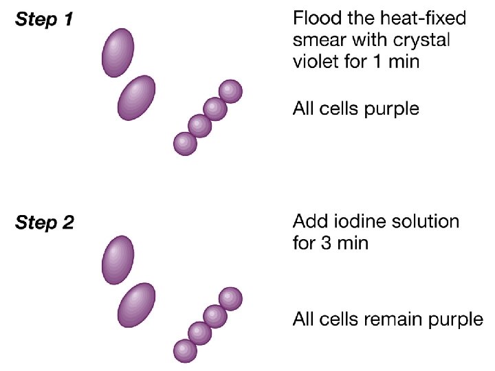
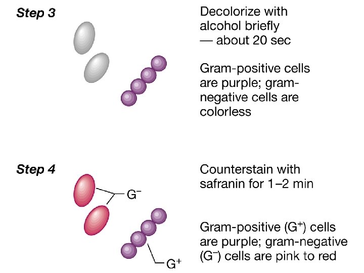
Staining • Simple • Differential: Gram and Acid-fast stain Gram-stain: cell wall Acid fast stain: Mycobacterium • Negative stain: Indian ink (capsule) • Special staining

Animal and plant cells • Much larger • Ranging from 7 micrometer (red blood cells) • To several feet (certain nerve cell)
 • Biochemical machinery (genetic information")
Each cell • Genetic basis for reproduction (DNA genome) • Biochemical machinery (genetic information is transcribed in m. RNA and m. RNA translated in proteins) • The machinery for energy production and biosynthesis • This is all packaged by a membrane.

Each cell • Replicates by cell division.
 • Prokaryotic (Greek for primitive nucleus)")
Cells • Eukaryotic (Greek for true nucleus) • Prokaryotic (Greek for primitive nucleus)
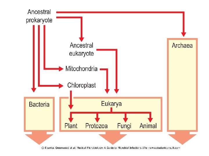

Eucaryotes • Animals • Plants • Fungi

Procaryotes • Bacteria • Blue-green algae

Major characteristics of Eucaryotic and prokaryotic cell Eucaryote >5 μm Prokaryote 0, 5 -3 μm • Size • Nuclear structure : Nucleus classic membrane no membrane Chromosomes strands of DNA single circular DNA diploid genome haploid genome

Major characteristics of Eucaryotic and prokaryotic cell Eucaryote Prokaryote • Cytoplasmic Structures Mitokondria + Golgi bodies + Endoplasmic reticulum + Ribosomes 80 S(60 S+40 S) Cytoplasmic membrane with sterols 70 S(50 S+30 S) no sterol

Major characteristics of Eucaryotic and prokaryotic cell Eucaryote • Cell wall -/composed of kitin Prokaryote complex structure (protein, lipits and peptidoglycans) • Reproduction sexual and asexual (binary fission) • Movement complex flagellum (If present) • Respiration via mitokondria simple flagellum (If present) via cytoplasmic membrane

Bacteria • Lack nucleus membrane and membrane bound organelles • A smaller ribosome • Peptidoglycan cell wall which protects it from environtment with low osmotic pressure, at temperature extremes (both hot and cold), dryness and with very dilute and diverse energy sources. • They have evolved their structures and functions to adopt these conditions.

Differences • Between Eukaryotes and prokaryotes
 -metabolic -antigenic and")
Differences between Prokaryotes • Bacteria differ: -morphology (size, shape, stainig characteristics) -metabolic -antigenic and -genetic characteristics

Size • They are diffucult to differentiate by size
 • Rod-shaped: bacillus (Escherichia) • Snakelike: spirillum (Treponema) •")
Shape • Spherical: coccus (Staphyloccus) • Rod-shaped: bacillus (Escherichia) • Snakelike: spirillum (Treponema) • Branched filamentous (Nocardia and Actinomyces) ( Clusters: diplococcus (Neisseria) chains (Streptococcus) grapelike (Staphylococcus) )
 Rod-shaped Spiral or spirillum Helix or spirochete")
Bacterial shape Sperical (coccus) Rod-shaped Spiral or spirillum Helix or spirochete

Bacterial arrangement Chains: streptococcus Clusters: staphylococcus Diplococcus Packets of eight: sarcina

Treponema by dark-field microscopy

Gram stain • Two major classes of bacteria are distinguished: • Gram-positive and • Gram-negative bacteria • Except: • Mycobacteria (waxy outer shell , distinguished by acid fast stain) • Mycoplasmas(no peptidoglycan)

Bacterial Ultrastructure • Internal structure • External structure • Gram-positive and gram-negative bacteria have -Similar internal structure -But different external structure

Cytoplasm • • • DNA chromosome m. RNA Ribosomes Proteins Metabolites

Bacterial chromosome • • • Unlike eukaryotes A single Double stranded circle Not in a membrane bound nucleus In a discrete area called nucleoid

Bacterial chromosome • Unlike eukaryotes • No histons

Plasmids • • • Smaller Circular Extrachromosomal DNAs Not usually essential for cellular survival Most commonly found in gram-negative bacteria • Often provide a selective advantage: resistance to antibiotics

Lack of a nuclear membrane • Simplifies the requirements and • Control mechanisms for the synthesis of proteins
ribosome • Bacterial 70 S chromosome")
Ribosomes • Unlike the eukaryotic 80 S(40 S+60 S)ribosome • Bacterial 70 S chromosome (30+50 S) • Proteins and RNA of the ribosome are significantly different • Major targets for antibacterial drugs

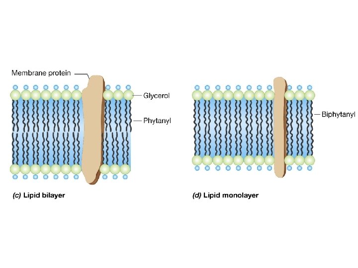
Cytoplasmic membrane • Lipid bilayer • Similar to eukaryotic membranes • But no sterols (cholesterol) Exception: Mycoplasmas

Cytoplasmic membrane • Responsible for many functions • Attributable to organelles in eukaryotes: -electron transport -energy production (mitokondria in eukaryotes)

Cytoplasmic membrane • Transport proteins: uptake of metabolites release of other substances • Ion pumps: to maintain a membrane potential • Enzymes

Mesosome • A coiled cytoplasmic membrane • An anchor to bind and pull apart daughter chromosomes during cell division.

Cell wall • Distinguishes gram-positive and gramnegative bacteria
 layer •")
The cytoplasmic membrane in most prokaryotes surrounded by • Rigid peptidoglycan (murein) layer • Except: Archaebacteria (pseudoglycan and pseudomurein) and mycoplasmas (no cell wall) • Peptidoglycan provides rigidity and determines the shape of a bacteria • Gram-negative bacteria. + outer membranes

Gram positive bacteria • Thick multilayered cell wall • Consisting mainly of peptidoglycan

Gram positive bacteria • Peptidoglycan • Sufficiently porous(allows diffusion of metabolites to the plasma membrane) • Essential for structure, replication, for survival
")
Peptidoglycan • • During infection İnterferes with phagocytosis Stimulates lymphoctes Pyrogenic activity (induces fever)

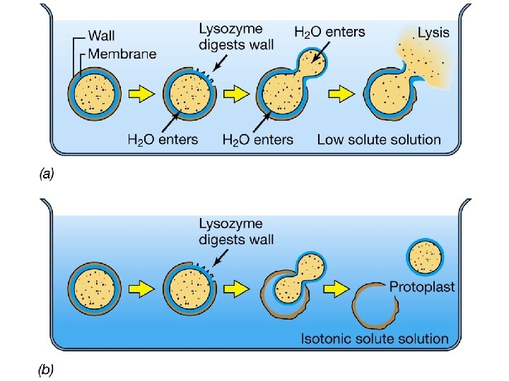
Peptidoglycan • Degraded by lysozyme • Enzyme in human tears, mucus (produced by bacteria and other organisms) • Degrades the glycan backbone of the peptidoglycan which protects it from osmotic pressure changes

Protoplast • Removal of cell wall with lysozyme • Lysis unless it is osmotically stabilized

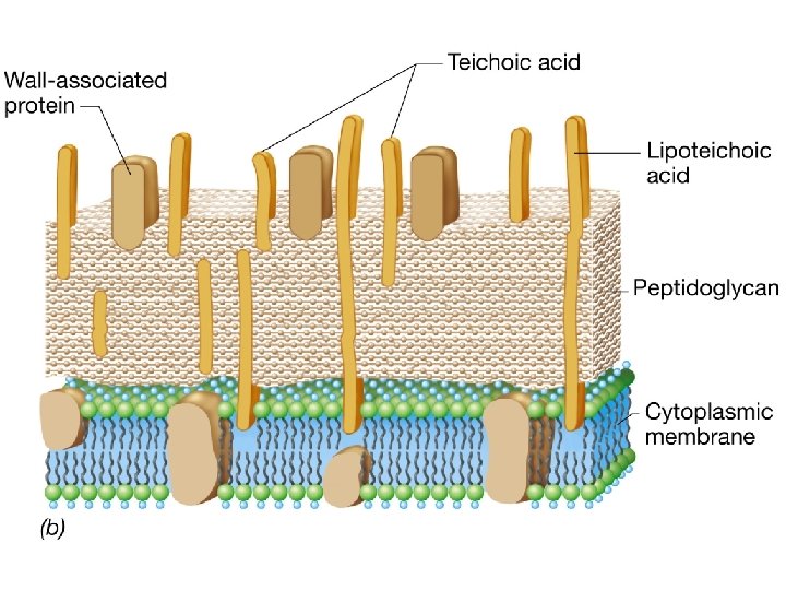
Gram-positive cell wall • • Peptidoglycan + Teicoic acid Lipoteichoic acid Complex polisaccarides (C polysaccharides) • M protein of streptococci • R protein of staphylococci

Gram-positive bacteria • Teicoic acid : covalently linked to peptidoglycan • Lipoteichoic acid : anchored in the cytoplasmic membrane • Common surface antigens • Distinguish bacterial serotypes • Promote attachment to other bacteria and to spesific receptors on mammalian cell surfaces (adherence)

Gram positive-bacteria • Teicoic acid: important virulance factors • Lipoteicoic acid are shed into media and host • Although weaker • Can initiate endotoxic-like activities.

Gram-negative bacteria • Cell wall is more complex • Both structurally and chemically

Gram-negative bacteria • Two layers external to the cytoplasmic membrane: • -Thin peptidoglycan layer • -Outer mebrane (unique to gram-negative bacteria) • No teicoic acid and lipoteicoic acid • +periplasmic space
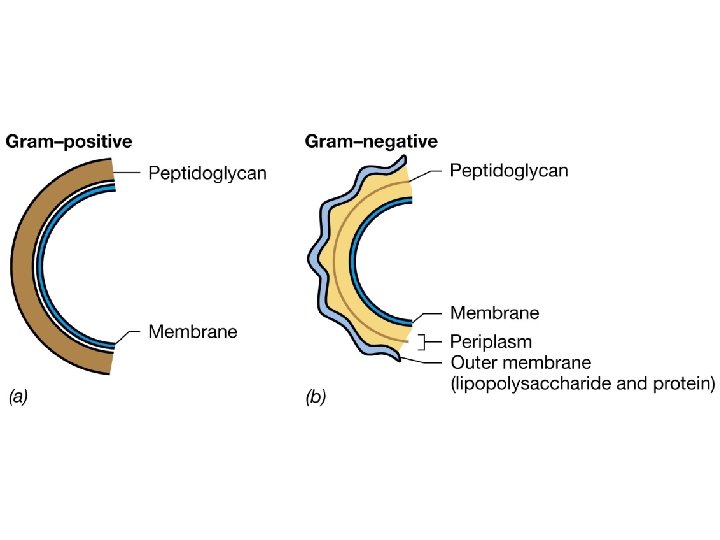
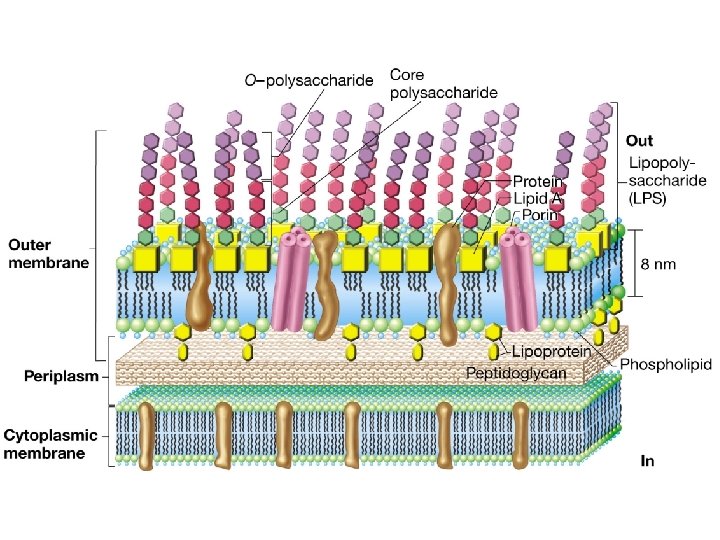

Periplasmic space • The area between the external surface of rhe cytoplasmic membrane and the internal surface of the outher membrane

Periplasmic space • A variety of hydrolytic enzymes • Breakdown of large macromolecules for metabolism • Lytic virulence factors ( collagenases, hyalurodidases, proteases, beta-lactamases) • Components of sugar transport system • Binding proteins for the uptake of different metabolites and of a chemotaxis system

Outer membranes • Unique to gram-negative-bacteria • Maintains the bacterial structure • Permeability barrier to large molecules and hydrophobic molecules • Provides protection from adverse environmental conditions (For Enterobactericeae from digestive system of the host)

Outer membranes • Asymetric bilayer • The inner leaflet: phospholipits normally found in bacterial membranes • Outer leaflet: amphipathic molecule( having both hydrophobic and hydrophilic ends) contains Lipopolysaccaride (LPS)

Outer membranes • LPS molecules are only found in the outer leaflet
 • Powerful stimulator of")
Lipopolysaccaride • Endotoxin (Lipid A+polysaccharide-O antigen, antigenic variety is great) • Powerful stimulator of immune responses • Causes fever and shock Shwartzman reaction (disseminated intravascular coagulation) follows the release of large amounts of endotoxin.

Outer membrane proteins • Porins: transmembrane proteins they form pores allow diffusion of hydrophilic molecules • Structural proteins and receptor molecules for bacteriophages

Disruption of the outher membrane • Weakens the bacteria • +lysozyme • Spheroplasts (osmotically sensitive) are formed.

External structures • • Capsule Pili Flagellum Proteins
 • Loose polysaccaride or proteinlayer • Slime")
Capsules • Some bacteria (gram-positive and gramnegative) • Loose polysaccaride or proteinlayer • Slime layer: loosely adherent and nonuniform in density and thickness • Glycocalyx: capsule and slime layer are also called.
")
Capsule • Polypeptide capsule: • Bacillus anthracis (polyglutamic acid)

Capsule • Hard to be seen by microscopy • Negative staining: Indian ink

Capsule • Unnecessary for growth • Very important for survival
 •")
Capsule • Poorly antigenic • Antiphagocytic and a major virulence factor (Streptococcus pneumoniae) • Barrier to toxic hydrophobic molecules such as detergents • Promote adherence (Streptococcus mutants: stick the tooth)

Quellung reaction • For vizualizing the capsule • Using antibodies • The capsule is swollen
 • • Motility Protein (flagellin) Antigenic and strain determinants Anchored in membranes")
Flagella (Kirpik) • • Motility Protein (flagellin) Antigenic and strain determinants Anchored in membranes through a hook and basal body • One or several

Flagella • • Monotrichous Polar: Pseudomonas aeruginosa Peritrichous : Escherichia coli Lophotrichous
 • Protein(pilin) • Different from flagella: smaller in diameter and not coiled")
Fimbriae (Pili) • Protein(pilin) • Different from flagella: smaller in diameter and not coiled in structure. • Promote adherence to other bacteria or to th host (adhesins, lectins, evasins, aggresins)
 • Fimbriae are important virulance factors as an adhesin in E. Coli")
Fimbriae (Pili) • Fimbriae are important virulance factors as an adhesin in E. Coli (urinary tract), Neisseria gonorrhoeae • The tips of fimbriae may contain proteinslectins that bind to spesific sugarsmannose
 • F pili (Sex) • Promote transfer of large segments of bacterial")
Fimbriae (Pili) • F pili (Sex) • Promote transfer of large segments of bacterial chromosome between bacteria • Encoded by a F plasmid.

Bacterial exceptions • • Mycobacteria Corynebacterium Nocardia Mycoplasmas
 Waxlikelipit coat of mycolic acid Cord factor Wax")
Mycobacteria • • Peptidoglycan (slightly different) Waxlikelipit coat of mycolic acid Cord factor Wax D Sulfolipids Acid-fast staining The coat responsible for virulence and antiphagocytic.

• Corynebacterium • Nocardia Produce mycolic acid lipids.

Mycoplasmas • No peptidoglycan cell wall

Structure of Bacterial Cell Wall • The components are large structures • They are made up of polymers of subunits • Synthesis of it occurs outside the bacteria
 • Linear polysaccaride chain: -repeating disaccarides: N-acetylglucosamine N-acetylmuramic acid • Cross-linked")
Peptidoglycan (Mucopeptide, Murein) • Linear polysaccaride chain: -repeating disaccarides: N-acetylglucosamine N-acetylmuramic acid • Cross-linked by peptides

Cell wall synthesis • Cross-linking reaction is catalyzed by: -membrane bound transpeptidases -DD-carboxypeptidasespenicillin-binding proteins (PBPs)
: -remove extra terminal D-alanines -These terminal D-alanines")
Cell wall synthesis • Penicillin-binding proteins (PBPs): -remove extra terminal D-alanines -These terminal D-alanines limit the extent of cross-linking -They are targets for penicillin and betalactam antibiotics

Cell wall • Peptidoglycan is constantly being synthesized and degraded. • Autolysins such as lysozyme are important for determining the shape of bacteria.

Cell wall • During starvation: -New peptidoglycan synthesis does not occur -Peptidoglycan is weakened -Gram-staining property changes (old cultures)

Biosynthesis of peptidoglycan • Unique to bacterial cells • İnhibited with no adverse effect of human cells • An important target for antibiotics (selective toxicity)

Lipopolysaccaride • Lipid A • Core polysaccaride • O antigen

Lipoppolysaccaride • Lipid A: basic component essential for bacterial viability endotoxin activity • O antigen: antigenic part (serotypes)
 Glycogen Polyphosphate")
Inclusion granules • • Storage of energy Poly-beta-hydroxybutyric acid (PHB) Glycogen Polyphosphate

Inclusion granules • Polymetaphosphate: Corynebacterium -Babes-Ernst bodies

Spores • Resistant to harsh conditions • (loss of nutritional requirement, dessication, intense heat, radiation and attack by most enzymes and chemical agents)

Spores • Exist for centuries • Diffucult to decontaminate with standart disinfectants

Spores formers: • Some gram-positive • Never gram-negative

Spore formers • Bacillus • Clostridium

Kinds of spores • Endospore: Bacillus subtilis • Terminal endospore: Clostridium tetani ‘drumstick’ • Subterminal: Clostridium botulinum
")
Under harsh conditions • Vegetative state is converted to dormant state (spore)

Localisation of the spore within a cell • Characteristic of bacteria • Can assist in identification of the bacterium.

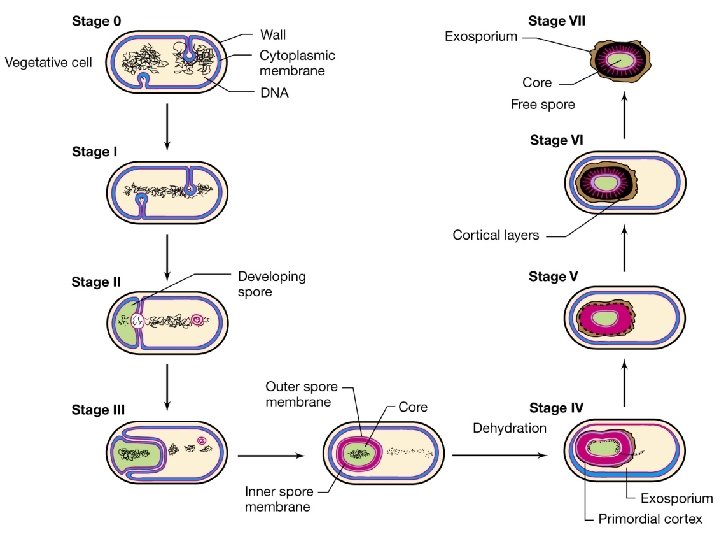
Spore • Dehydrated • Multishelled structure • A complete copy of chromosome • Minimum concentration of proteins and Ribosomes + High concentration of calcium bound dipicolinic acid

Spore • Outside the core: -inner membrane -spore wall -cortex -outher membrane -keratin-like protein coat -exosporium

Sporulation • 6 -8 hours
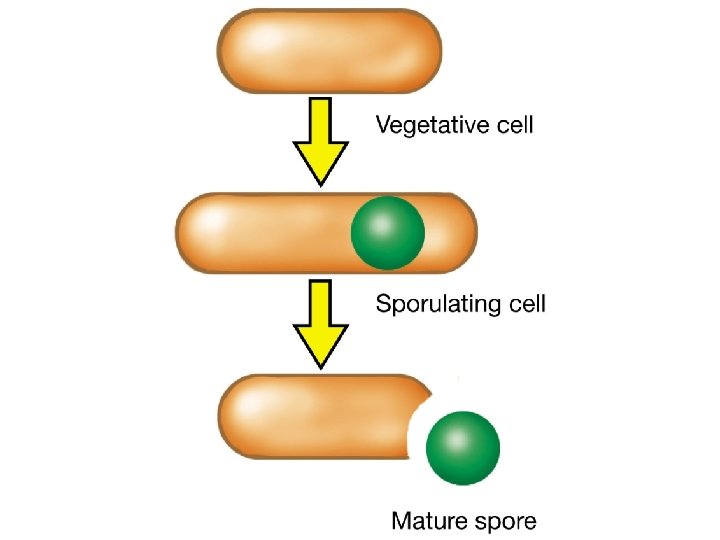

Germination • Spor__vegetative state: disruption of the outher coat by mechanical stress, p. H, heat or another stressor It takes about 90 minutes

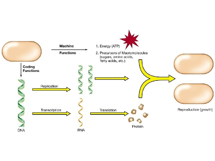
Bacterial metabolism • Bacterial growth: -a source of energy -raw materials *To build the proteins, structures and membranes *That make up the structure and biochemical machines of the cell

Bacterial metabolism • Bacteria should obtain or synthesize: -aminoacids -carbohydrates -lipids as building blocks of the cell

The minimum requirement for growth • • • Carbon Nitrogen Energy source Water Various ions

• Chemotrophs: derive energy from inorganic material • Chemoorganotrophs: Most bacterial

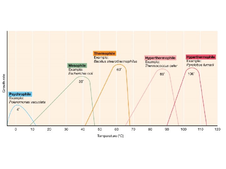
Heat • Cardinal temperatures: -minimum temperature -optimum temperature -maximum temperature

Temperature • • Psychrophiles Mesophiles Thermophiles Hyperthermophiles
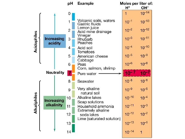
 • Alkaliphiles: Above p. H")
ph • Acidophiles: Below p. H 5 (many fungi) • Alkaliphiles: Above p. H 9 (Vibrio) • Neutral p. H: most pathogens

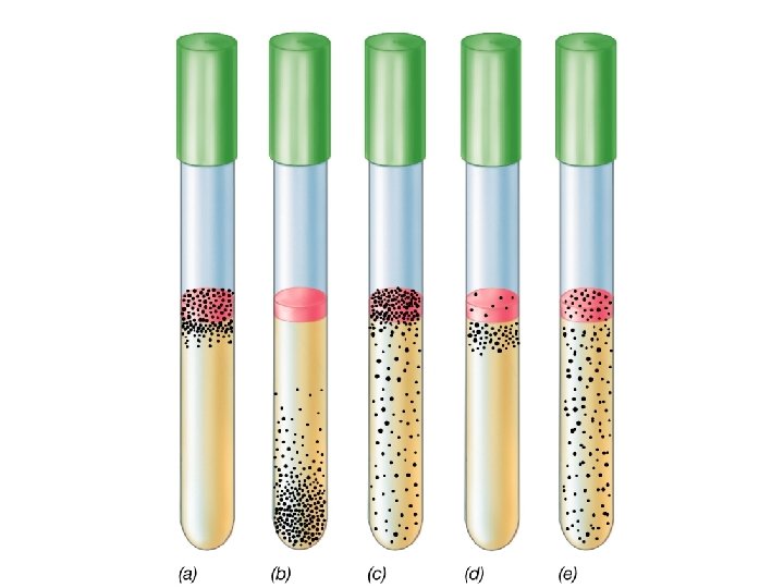
Oxygen requirement • Obligate anaerobes : Clostridium perfringens • Obligate aerobes • Facultative anaerobes • Microaerophilic
")
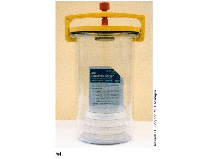
Incubation for growth • Aerobic • Anaerobic • Capneic (%5 Carbon dioxide)

Methabolism • Catabolism: substrate breakdown and conversation into usable energy • Anabolism: synthesis of cellular constituents (cell wall, proteins, fatty acids, nucleic acids

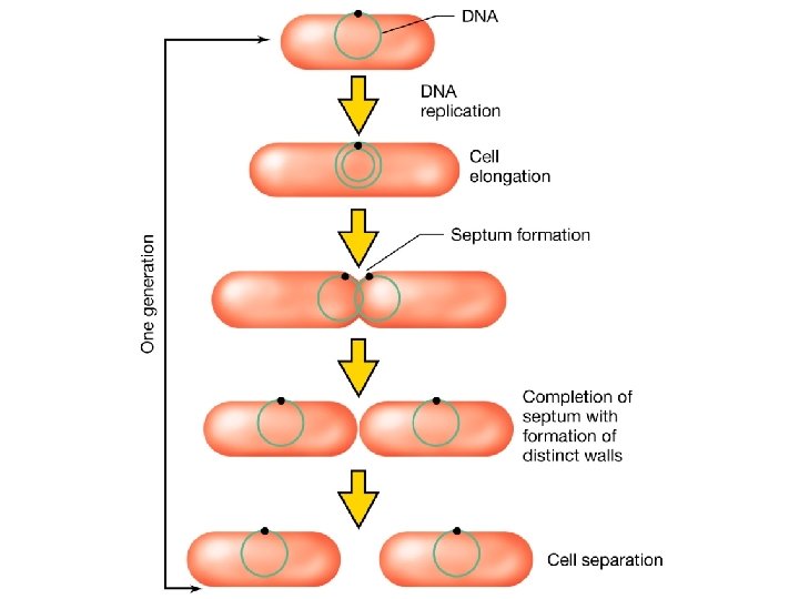
Bacterial growth • Bacterial replication • Two equivalent daughter cells • Binary fission (Escherichia coli: 20 minutes Mycobacterium much slower: 12 -24 h)

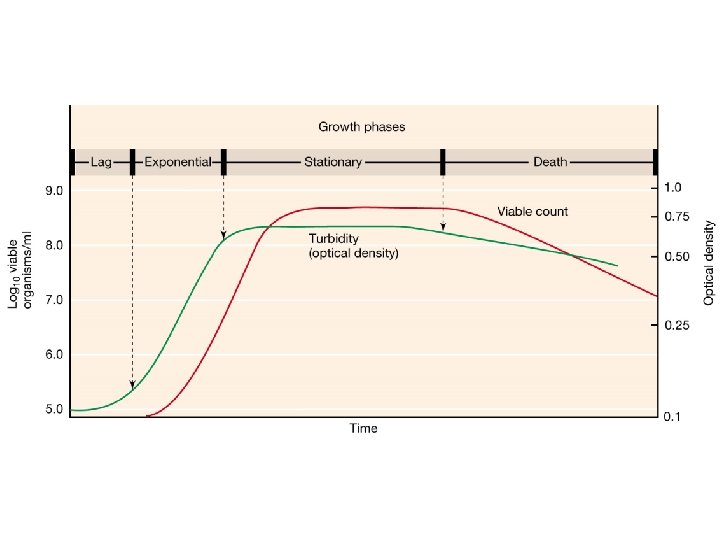
Bacterial growth curve • • Lag phase Exponential phase Stationary phase Death phase
- Slides: 123