Bacterial Meningitis Dr Hamed Al Zoubi School of

Bacterial Meningitis Dr Hamed Al Zoubi School of Medicine / University of Mu’tah.

Bacterial Meningitis • Meningeal symptoms: Ø Headache, vomiting, photophobia, fever and neck stiffness • Cerebral involvement: Ø Confusion, altered LOC, seizures. . • CSF analysis
: • Unwell, irritability and excessive crying, failure")
Bacterial Meningitis In infants (Usually nonspecific symptoms): • Unwell, irritability and excessive crying, failure to thrive or feed, vomiting and fever. • Respiratory distress, apnea, cyanosis

Clinical picture / signs in adults • Kernig’s sign: Passive knee extension on flexed hip in supine patient elicits neck pain and hamstring resistance. • Brudzinski’s sign: Passive neck flexion is accompanied by involuntary flexion of both hips.

Kernig’s sign

Brudzinski’s sign

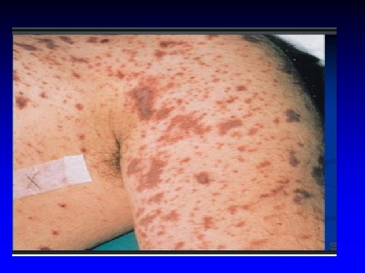
Clinical picture / signs • Papilloedema: presents in only about 30 -50% of meningitis patients with increased ICP; it takes at least several hours to develop. • Ptechial rash on skin: non blanching rash which are seen classically with N meningitidis; however, these also can sometimes occur with other bacterial and viral infections.

Papilloedema

Clinical picture / signs • Signs of a focus of infection e. g chest, heart or skin and soft tissue infection. • Also in Infants (< 1 year) Bulging fontanelle Hypotonia Paradoxic irritability (ie, quiet when stationary, cries when held)

Bacterial Meningitis • The causative bacteria and outcome depend on different factors such as: Ø Age Ø Immune status Ø Recent head trauma or neurosurgery Ø Bacterial virulence: Ig. A protease, capsules, fimbria. .
 • Gm - rods")
Bacterial meningitis / causal organisms – Neonates (< 1 Month) • Gm - rods (E. coli, Klebsiella species (sp)) 50 -60% • Grp B Streptococcus (G + cocci) 20 -40% • Listeria monocytogenes (G + Rods) 2 -10% Streptococcus pneumonia 0 -5% • Haemophilus influenza (G - coccobacilli) 0 -3%
 • Haemophilus")
Bacterial meningitis / causal organisms – Children (1 month to 15 years) • Haemophilus influenzae 40 -60% – Declining dramatically in many geographic regions • Neisseria meningitidis (G- diplococci) 25 -40% (Epidemic cases, late complement deficiency increase susceptibility) • Streptococcus pneumonia 10 -20%
 • S. pneumo 30")
Bacterial meningitis / causal organisms – Adults (> 15 years) • S. pneumo 30 -50% • N. Meningitidis 10 -35% • Gm (-) Bacilli 1 -10% – Elderly • S. aureus 5 -15% • H. influenzae 1 -3%

Bacterial meningitis / causal organisms – >50 years add Listeria, E. coli, Pseudomonas – In Ventriculoperitoneal shunts, post trauma and neurosurgery: usually S. aureus, diphtheroid sp and Pseudomonas – In immunocompromised patient, any organism has the potential to be involved
")
Meningitis / Pathogenesis: • Balance between host (Ig. A antibodies and epithelial cells status) and organism factors (Ig. A protease, capsule and normal flora status) • Organism spread via different route: 1. Blood (haematogenous): • From nasopharynx or a septic emboli e. g Lung abscess ad infective endocarditis • The commonest route 2. Direct spread: • Sinusitis, otitis media , trauma and skull opened fractures • Ventriculoperitoneal shunts and post neurosurgery 3. Vertical and Horizontal routes
 > cross the blood")
Meningitis / pathogenesis Ø Bacteria in Blood (bacteraemia or sepsis) > cross the blood brain barrier> CSF > Increased cytokines production > Ø increased BBB permeability > albumin into CSF plus Ø neutrophils and bacterial toxins > vasogenic and cytotoxic Ø oedema > increased intracranial pressure > brain oedema (increased ICP) and cell death with serious Central nervous system sequale such as:

Meningitis / pathogenesis 1. damaging cranial nerves (eg, cranial nerve VIII, with resultant hearing loss) 2. Brain ischemia > decreased level of consciousness 3. Severe Handicap – Hemiplegia – Blindness – Deafness – Severe Learning Difficulty – Severe behavioural disturbances – Severe Epilepsy. Ø Without treatment > death
, Liver and")
Diagnosis 1. History and examination. 2. Laboratory: • Full blood count (FBC), Liver and Kidney function tests • Coagulation profile and serum glucose • Blood culture and throat swabs 3. Radiology: • Chest X rays. • Computed topography scan (CT scan): Ø Infection, masses and abscess > increased intracranial pressure (ICP).

Diagnosis Ø Obtain a CT scan in: • • History of CNS disease New onset seizures Papilledema Altered level of consciousness or Focal neurologic signs • Lumbar puncture (LP) – Defer LP until CT scan or MRI obtained if any of above present – If suspect meningitis and awaiting neuroimaging • Obtain BC’s and start empiric Abx

LP tubes • Tube # 1 Protein & Glucose • Tube # 2 bacterial Gram stain & Culture (acid-fast bacillus (AFB) stain and tuberculosis (TB) cultures, India ink stain and fungal cultures, and cryptococcal antigen, if indicated). • Tube # 3 Cell count & differential • Tube # 4 Store ( PCR, viral studies if available, or for repeat cell count if needed)

CSF findings Normal Cells 0 -5 WBC/mm 3 ~25 cells <6 months old Bacterial Viral TB >1000/mm 3 <1000/mm 3 25 -500/mm 3 Polymorphs 0 predominate early +/- increased Lymphocytes 100% late predominate increased Glucose 40 -80 mg/dl decreased normal decreased 66% < 40% Normal < 30% Protein 15 -40 mg/dl increased +/-increased Culture negative positive negative +TB Gram stain negative positive negative Positive AFB

Treatment General: § Stabilize the patient (antipyretics, fluid, anti seizures if there. . . ) • Specific: 1. Antibiotics Empiric treatment consists of bactericidal antibiotics that have good CSF penetration, such as a third-generation cephalosporin (eg cefotaxime, ceftriaxone, ceftazidime) , Ampicillin, vancomycin Change according to clinical response and CSF results. Duration 10 -14 days, 3 weeks in Listeria. • Patient must be isolated for the first 24 hours after starting treatment.
: Controversial ? . Preferable in paediatric patients. Must be given")
Treatment 2. Steroids (dexamethasone): Controversial ? . Preferable in paediatric patients. Must be given before antibiotics or shortly after ! Why? . 3. If TB meningitis > anti TB medications

Antibiotics • The right antibiotic choice depends on many factors such as, age, likely organism and immune status and local susceptibility patterns of bacteria; However, a general good protocol is to give: Ø In < 1 month old Cefotaxime + Amoxicillin Ø 1 month Cefotaxime or Ceftriaxone + Ampicillin > In VP shunted patients and post neurosurgery meningitis use add vancomycin and ceftazidime > Always follow the CSF culture and sensitivity results and adjust the antibiotic treatment if needed.
 N meningitidis: • Indicated")
Prophylaxis Ø Prophylaxis for close contacts of patients with (suspected) N meningitidis: • Indicated for those at increased risk, such as those who were in close contact with patient for at least 4 hours during the week before onset (eg, house mates, daycare center, cell mates) Or • were exposed to patient's nasopharyngeal secretions (eg, kissing, mouth-to-mouth resuscitation, intubation, nasotracheal suctioning). Ø Give Rifampicin 4 doses over 2 days (Ciprofloxacin or Ceftriaxone single dose is an alternative) Ø In pregnancy: Ceftriaxone.
 of patients with")
Prophylaxis Ø Prophylaxis for household or daycare contacts (>4 hrs contact) of patients with (suspected) H influenzae type b: Give Rifampicin twice daily for 2 days as before. Ø Vaccination: Available for: Meningococcus types A, C, Y and W 135 but not for type B. H. Influenza serotype B. Streptococcus pneumonia: For 23 serotypes or 17 serotypes.

Take home messages Ø Acute bacterial meningitis is a bacterial emergency Ø Diagnosis and initiation of treatment relies heavily on clinical picture aided by lab. Tests Ø Start antibiotics as quick as possible Ø Avoid lumbar puncture in cases of increased ICP > brain herniation Ø Think of other causes also such as viral and fungal cause Ø Do not hesitate to ask your seniors

The end
- Slides: 29