
Bacterial Diseases by Dr Mouchira Mohamed Mohi Eldin
Bacterial Diseases by Dr. Mouchira Mohamed Mohi Eldin Pathology and Clinical Pathology Department, Faculty of Veterinary Medicine, south Valley University, Qena

Infections due to Corynebacterium Ovine Caseous lymphadenitis (Pseudo-tuberculosis in sheep and goats) (Cheesy gland)
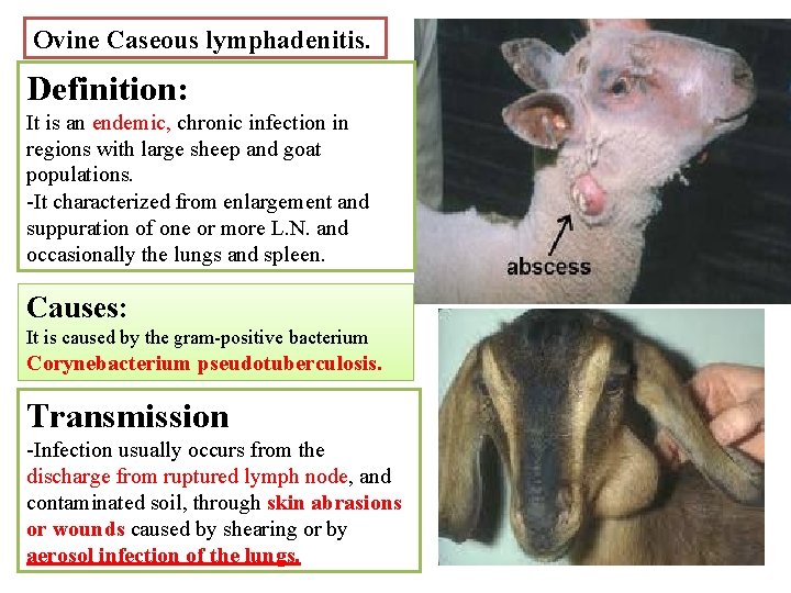
Ovine Caseous lymphadenitis. Definition: It is an endemic, chronic infection in regions with large sheep and goat populations. -It characterized from enlargement and suppuration of one or more L. N. and occasionally the lungs and spleen. Causes: It is caused by the gram-positive bacterium Corynebacterium pseudotuberculosis. Transmission -Infection usually occurs from the discharge from ruptured lymph node, and contaminated soil, through skin abrasions or wounds caused by shearing or by aerosol infection of the lungs.
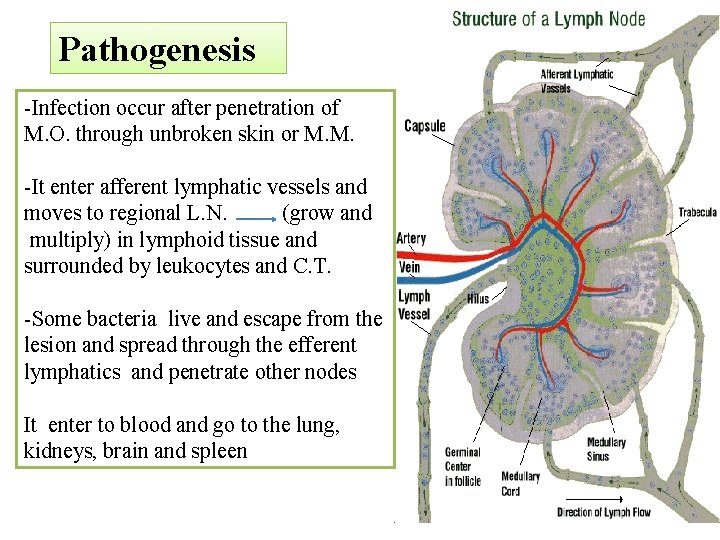
Pathogenesis -Infection occur after penetration of M. O. through unbroken skin or M. M. -It enter afferent lymphatic vessels and moves to regional L. N. (grow and multiply) in lymphoid tissue and surrounded by leukocytes and C. T. -Some bacteria live and escape from the lesion and spread through the efferent lymphatics and penetrate other nodes It enter to blood and go to the lung, kidneys, brain and spleen
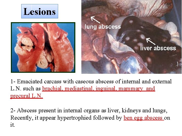
Lesions 1 - Emaciated carcass with caseous abscess of internal and external L. N. such as brachial, mediastinal, inguinal, mammary and precural L. N. 2 - Abscess present in internal organs as liver, kidneys and lungs, Recently, it appear hypertrophied followed by ben egg abscess on it.
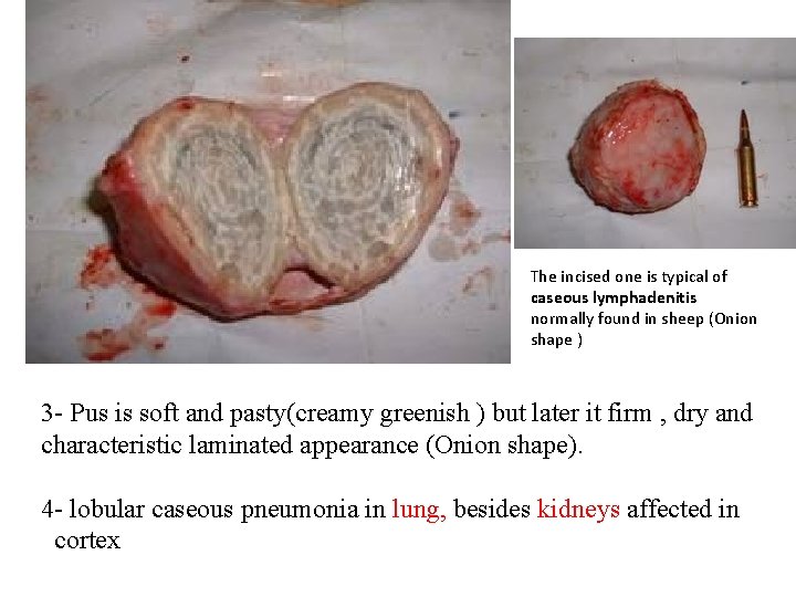
The incised one is typical of caseous lymphadenitis normally found in sheep (Onion shape ) 3 - Pus is soft and pasty(creamy greenish ) but later it firm , dry and characteristic laminated appearance (Onion shape). 4 - lobular caseous pneumonia in lung, besides kidneys affected in cortex
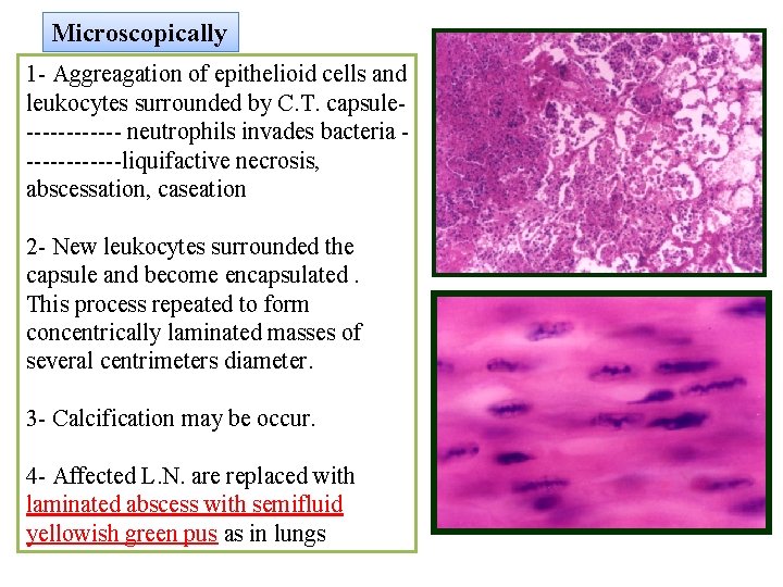
Microscopically 1 - Aggreagation of epithelioid cells and leukocytes surrounded by C. T. capsule------ neutrophils invades bacteria ------liquifactive necrosis, abscessation, caseation 2 - New leukocytes surrounded the capsule and become encapsulated. This process repeated to form concentrically laminated masses of several centrimeters diameter. 3 - Calcification may be occur. 4 - Affected L. N. are replaced with laminated abscess with semifluid yellowish green pus as in lungs
Infections due to Bordetella 1 - Colibacillosis or white scour disease
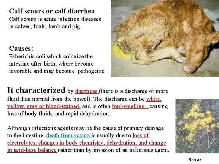
Calf scours or calf diarrhea Calf scours is acute infection diseases in calves, foals, lamb and pig. Causes: Esherichia coli which colonize the intestine after birth, where become favorable and may become pathogenic. It characterized by diarrheas (there is a discharge of more fluid than normal from the bowel), The discharge can be white, yellow, grey or blood-stained, and is often foul-smelling , causing loss of body fluids and rapid dehydration. Although infectious agents may be the cause of primary damage to the intestine, death from scours is usually due to loss of electrolytes, changes in body chemistry, dehydration, and change in acid-base balance rather than by invasion of an infectious agent. Scour
2 - Actinobacillosis A- Actinobacillus equali Joint-ill in foals B- Actinobacillus lignieresi (Wooden tongue)
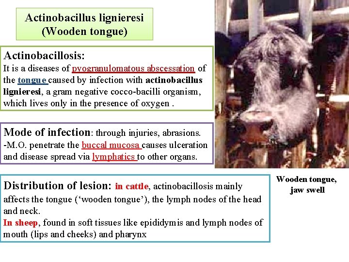
Actinobacillus lignieresi (Wooden tongue) Actinobacillosis: It is a diseases of pyogranulomatous abscessation of the tongue caused by infection with actinobacillus lignieresi, a gram negative cocco-bacilli organism, which lives only in the presence of oxygen. Mode of infection: through injuries, abrasions. -M. O. penetrate the buccal mucosa causes ulceration and disease spread via lymphatics to other organs. Distribution of lesion: in cattle, actinobacillosis mainly affects the tongue (‘wooden tongue’), the lymph nodes of the head and neck. In sheep, found in soft tissues like epididymis and lymph nodes of mouth (lips and cheeks) and pharynx Wooden tongue, jaw swell
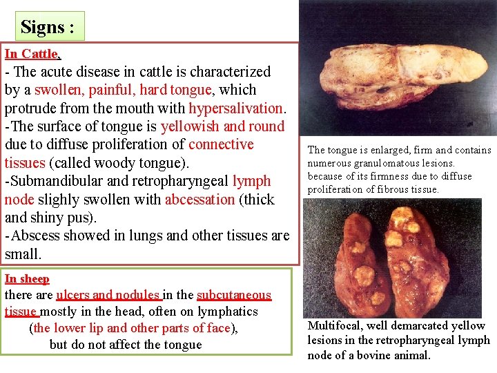
Signs : In Cattle, - The acute disease in cattle is characterized by a swollen, painful, hard tongue, which protrude from the mouth with hypersalivation. -The surface of tongue is yellowish and round due to diffuse proliferation of connective tissues (called woody tongue). -Submandibular and retropharyngeal lymph node slighly swollen with abcessation (thick and shiny pus). -Abscess showed in lungs and other tissues are small. The tongue is enlarged, firm and contains numerous granulomatous lesions. because of its firmness due to diffuse proliferation of fibrous tissue. In sheep there are ulcers and nodules in the subcutaneous tissue mostly in the head, often on lymphatics (the lower lip and other parts of face), but do not affect the tongue Multifocal, well demarcated yellow lesions in the retropharyngeal lymph node of a bovine animal.
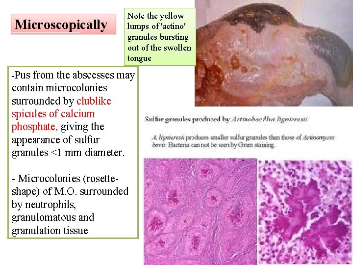
Microscopically Note the yellow lumps of 'actino' granules bursting out of the swollen tongue -Pus from the abscesses may contain microcolonies surrounded by clublike spicules of calcium phosphate, giving the appearance of sulfur granules <1 mm diameter. - Microcolonies (rosetteshape) of M. O. surrounded by neutrophils, granulomatous and granulation tissue
Diseases caused by higher bacteria Actinomycosis (Lumpy jaw)
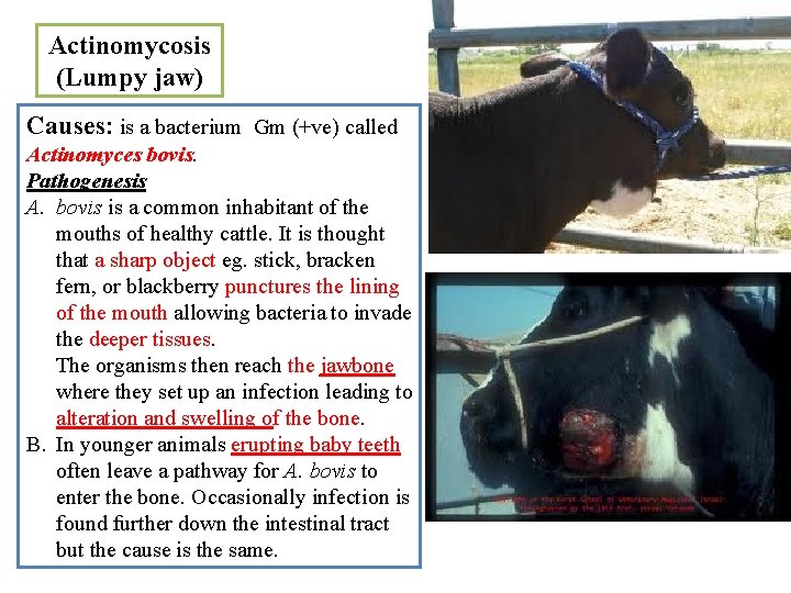
Actinomycosis (Lumpy jaw) Causes: is a bacterium Gm (+ve) called Actinomyces bovis. Pathogenesis A. bovis is a common inhabitant of the mouths of healthy cattle. It is thought that a sharp object eg. stick, bracken fern, or blackberry punctures the lining of the mouth allowing bacteria to invade the deeper tissues. The organisms then reach the jawbone where they set up an infection leading to alteration and swelling of the bone. B. In younger animals erupting baby teeth often leave a pathway for A. bovis to enter the bone. Occasionally infection is found further down the intestinal tract but the cause is the same.
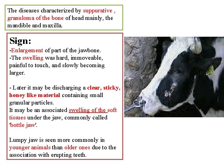
The diseases characterized by suppurative , granuloma of the bone of head mainly, the mandible and maxilla. Sign: -Enlargement of part of the jawbone. -The swelling was hard, immoveable, painful to touch, and slowly becoming larger. - Later it may be discharging a clear, sticky, honey like material containing small granular particles. It may be an associated swelling of the soft tissues under the jaw, commonly called 'bottle jaw'. Lumpy jaw is seen more commonly in younger animals than older ones due to the association with erupting teeth.
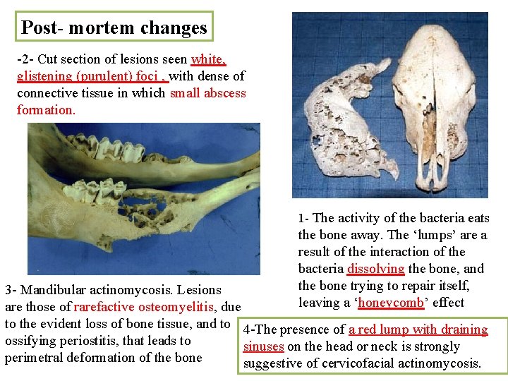
Post- mortem changes -2 - Cut section of lesions seen white, glistening (purulent) foci , with dense of connective tissue in which small abscess formation. 1 - The activity of the bacteria eats the bone away. The ‘lumps’ are a result of the interaction of the bacteria dissolving the bone, and the bone trying to repair itself, leaving a ‘honeycomb’ effect 3 - Mandibular actinomycosis. Lesions are those of rarefactive osteomyelitis, due to the evident loss of bone tissue, and to 4 -The presence of a red lump with draining ossifying periostitis, that leads to sinuses on the head or neck is strongly perimetral deformation of the bone suggestive of cervicofacial actinomycosis.
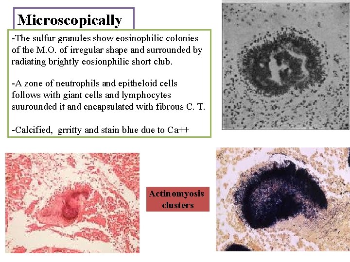
Microscopically -The sulfur granules show eosinophilic colonies of the M. O. of irregular shape and surrounded by radiating brightly eosionphilic short club. -A zone of neutrophils and epitheloid cells follows with giant cells and lymphocytes suurounded it and encapsulated with fibrous C. T. -Calcified, grritty and stain blue due to Ca++ Actinomyosis clusters
Tuberculosis
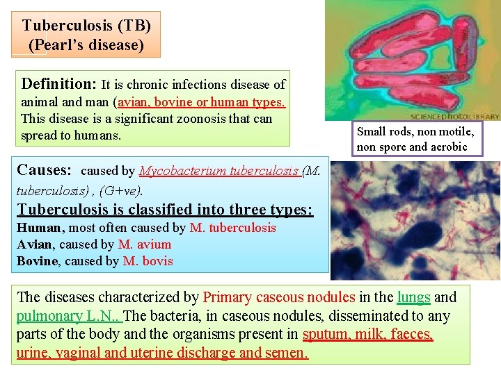
Tuberculosis (TB) (Pearl’s disease) Definition: It is chronic infections disease of animal and man (avian, bovine or human types. This disease is a significant zoonosis that can spread to humans. Small rods, non motile, non spore and aerobic Causes: caused by Mycobacterium tuberculosis (M. tuberculosis) , (G+ve). Tuberculosis is classified into three types: Human, most often caused by M. tuberculosis Avian, caused by M. avium Bovine, caused by M. bovis The diseases characterized by Primary caseous nodules in the lungs and pulmonary L. N. . The bacteria, in caseous nodules, disseminated to any parts of the body and the organisms present in sputum, milk, faeces, urine, vaginal and uterine discharge and semen.
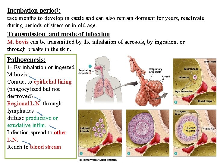
Incubation period: take months to develop in cattle and can also remain dormant for years, reactivate during periods of stress or in old age. Transmission and mode of infection M. bovis can be transmitted by the inhalation of aerosols, by ingestion, or through breaks in the skin. Pathogenesis: 1 - By inhalation or ingested M. bovis Contact to epithelial lining (phagocytized but not destroyed) Regional L. N. through lymphatics diffuse productive or exudative inflm. Infection spread to other L. N. Reach to blood stream
2 - Serous surface as pleura rich in lymphatics , so, spread of M. O. is extensive…. . Pearl-like nodules (Pearl-disease) attach to one another to form grape-like lesion (grapes disease). 3 - Organs like lungs are showed with bloodborne bacteria, resulting in miliary T. B. (early generalization) acute form mostly in lungs, liver, kidneys and spleen), eventually generalized tuberculosis chronic form. -The disease kill the animal with complete localization and destruction of L. N. (underlying toxemia) 4 - Local manifestation depend on the route of invasion: - by inhalation…………. . route via lungs and bronchopulmonary L. N…. blood. - by ingestion…………… route via mesenteric L. N…. intestinal wall…. and to liver via portal system. Organisms from nodes may reach to thoracic cavity, enter blood…. arrest in lungs (caseous nodules)…bacteria enter pulmonary vein , with subsequent general disseminstion. 5 - In most instances, a secondary reinfection does not occur due to development of tuberculin hypersensitivity and cellular immunity. The presence of antigen concentrations at the initial site of infection brings about necrosis and eventually fibrosis and calcification of the tissues, which arrests the infection and renders the disease inactive. -If, the infection is not controlled, the symptoms of progressive primary tuberculosis is develops
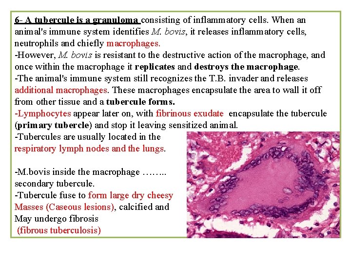
6 - A tubercule is a granuloma consisting of inflammatory cells. When an animal's immune system identifies M. bovis, it releases inflammatory cells, neutrophils and chiefly macrophages. -However, M. bovis is resistant to the destructive action of the macrophage, and once within the macrophage it replicates and destroys the macrophage. -The animal's immune system still recognizes the T. B. invader and releases additional macrophages. These macrophages encapsulate the area to wall it off from other tissue and a tubercule forms. -Lymphocytes appear later on, with fibrinous exudate encapsulate the tubercule (primary tubercle) and stop it leaving sensitized animal. -Tubercules are usually located in the respiratory lymph nodes and the lungs. -M. bovis inside the macrophage ……. . secondary tubercule. -Tubercule fuse to form large dry cheesy Masses (Caseous lesions), calcified and May undergo fibrosis (fibrous tuberculosis)
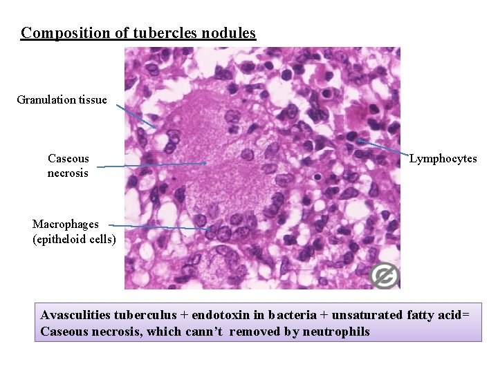
Composition of tubercles nodules Granulation tissue Caseous necrosis Lymphocytes Macrophages (epitheloid cells) Avasculities tuberculus + endotoxin in bacteria + unsaturated fatty acid= Caseous necrosis, which cann’t removed by neutrophils
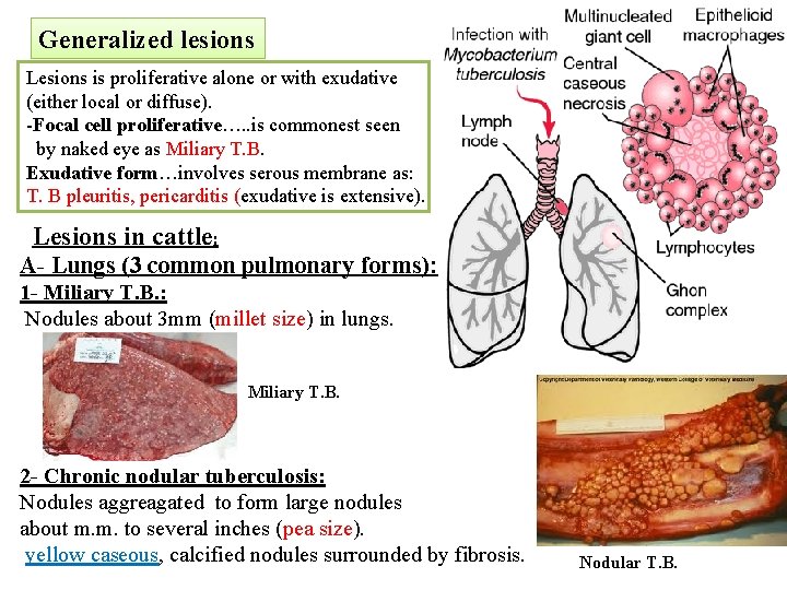
Generalized lesions Lesions is proliferative alone or with exudative (either local or diffuse). -Focal cell proliferative…. . is commonest seen by naked eye as Miliary T. B. Exudative form…involves serous membrane as: T. B pleuritis, pericarditis (exudative is extensive). Lesions in cattle: A- Lungs (3 common pulmonary forms): 1 - Miliary T. B. : Nodules about 3 mm (millet size) in lungs. Miliary T. B. 2 - Chronic nodular tuberculosis: Nodules aggreagated to form large nodules about m. m. to several inches (pea size). yellow caseous, calcified nodules surrounded by fibrosis. Nodular T. B.
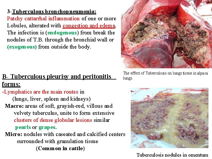
3 -Tuberculous bronchopneumonia: Patchy cattarrhal inflammation of one or more Lobules, alterated with congestion and edema. The infection is (endogenous) from break the nodules of T. B. through the bronchial wall or (exogenous) from outside the body. B- Tuberculous pleurisy and peritonitis forms: The effect of Tuberculosis on lungs tissue in alpaca lungs. -Lymphatics are the main routes in (lungs, liver, spleen and kidneys) Macro: areas of soft, grayish-red, villous and velvety tuberculus, unite to form extensive clusters of dense globular lesions similar pearls or grapes. Micro: nodules with caseated and calcified centers surrounded with granulation tissue (Common in cattle) Tuberculosis nodules in omentum
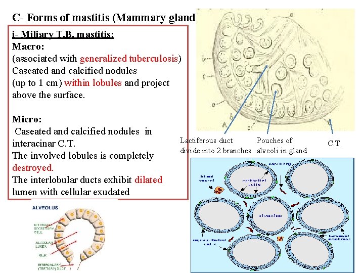
C- Forms of mastitis (Mammary gland) i- Miliary T. B. mastitis: Macro: (associated with generalized tuberculosis) Caseated and calcified nodules (up to 1 cm) within lobules and project above the surface. Micro: Caseated and calcified nodules in interacinar C. T. The involved lobules is completely destroyed. The interlobular ducts exhibit dilated lumen with cellular exudated Lactiferous duct Pouches of divide into 2 branches alveoli in gland C. T.
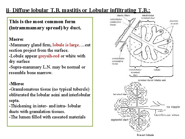
ii- Diffuse lobular T. B. mastitis or Lobular infiltrating T. B. : This is the most common form (intrammamary spread) by duct. Macro: -Mammary gland firm, lobule is large…. cut section project from the surface. -Lobule appear greysih-red or white with dry surface -Supra-mammary L. N. may be normal or resemble bone marrow. -Micro: -Granulomatous tissue (no typical tubercle) obliterated the lobular acini and interlobular septa. -Thickening in inter- and intra- lobular ducts with granulation tissues. -The lumen filled with caseated materials
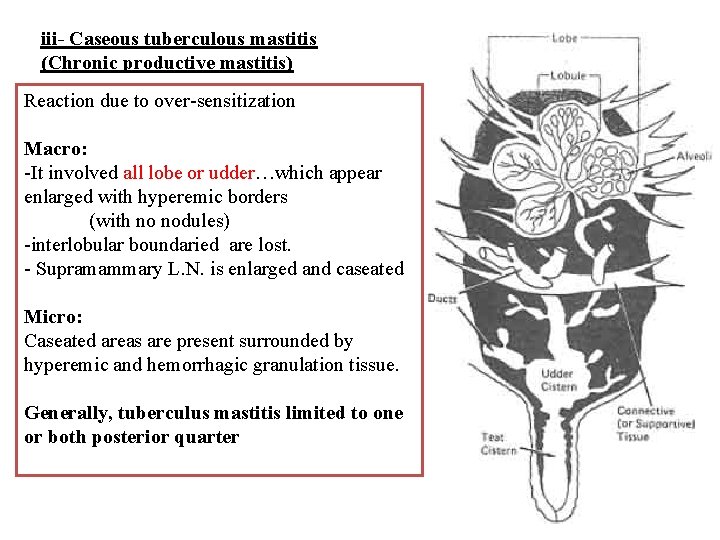
iii- Caseous tuberculous mastitis (Chronic productive mastitis) Reaction due to over-sensitization Macro: -It involved all lobe or udder…which appear enlarged with hyperemic borders (with no nodules) -interlobular boundaried are lost. - Supramammary L. N. is enlarged and caseated Micro: Caseated areas are present surrounded by hyperemic and hemorrhagic granulation tissue. Generally, tuberculus mastitis limited to one or both posterior quarter
Alimentary canal: I secondary except in calves with congenital T. B. -- primary lesions in ileum or colon particulary in peyer’s patches or solitary follicles Lesions: Nodules and ulcers in mesentric L. N. (3 -5 mm in diameter), tonsils, esophagus, stomach, ileum and cecum of old cattle. - In liver caseated and calcificated foci in sheep (cirrhotic). C. N. S: Lesions: in calves and young cattle Large dry caseous nodule present on cerebrum and cerebellum. Tubercles may show along the meninges T. B. in horse: Caused by bovine type…. . -Primary lesions…. . retropharngeal and mesentric L. N. (blood & lymph spread the infection). . infected L. N. enlarged & caseated. -Splenic form showed tumor-like sweling (caseated, suppurative without calcification) confused with lymphosarcoma. -Secondary lesions. . pulmonary lesions a- miliary T. B. : Small nodules along the lung. . coalase to appear as sarcoma b- chronic nodular T. B. : Few encapsulated with caseated (10 mm indiameter).
Johne’s disease (Chronic Pseudotuberculosis enteritis) (Paratuberculosis)
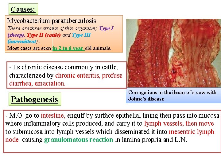
Causes: Mycobacterium paratuberculosis There are three strains of this organism; Type I (sheep), Type II (cattle) and Type III (intermittent). Most cases are seen in 2 to 6 year old animals. - Its chronic disease commonly in cattle, characterized by chronic enteritis, profuse diarrhea, emaciation. Pathogenesis Corrugations in the ileum of a cow with Johne's disease - M. O. go to intestine, engulf by surface epithelial lining then pass into mucosa where inflammatory cells produced, and carry it to lymph vessels, then move to submucosa into lymph vessels which disseminated it into mesentric lymph node causing granulomatous reaction in lamina propria and L. N.
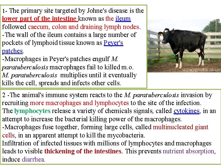
1 - The primary site targeted by Johne's disease is the lower part of the intestine known as the ileum followed caecum, colon and draining lymph nodes. -The wall of the ileum contains a large number of pockets of lymphoid tissue known as Peyer's patches. -Macrophages in Peyer's patches engulf M. paratuberculosis macrophages fail to killed m. o. M. paratuberculosis multiplies until it eventually kills the cell, spreads and infects other cells. 2 -The animal's immune system reacts to the M. paratuberculosis invasion by recruiting more macrophages and lymphocytes to the site of the infection. The lymphocytes release a variety of chemicals signals, called cytokines, in an attempt to increase the bacterial killing power of the macrophages. -Macrophages fuse together, forming large cells, called multinucleated giant cells, in an apparent attempt to kill the mycobacteria. Infiltration of infected tissues with millions of lymphocytes and macrophages leads to visible thickening of the intestines. This prevents nutrient absorption, induce diarrhea.
Signs: In cattle, the main signs of paratuberculosis are diarrhea and wasting. -The initial signs are loss of body weight, decreased milk production, or roughening of the hair coat. -The diarrhea is usually thick, without blood, mucus, or epithelial debris, and may be intermittent. Several weeks after the onset of diarrhea, a soft swelling may occur under the jaw. Known as "bottle jaw" or intermandibular edema. Animals die due to, emaciated and dehydration and severe cachexia lesions -Ileum showed mild, velvety thickening of the mucosa to severe thickening of the bowel with transverse corrugation of the mucosal surface. 2 - Pure granulomatous enteritis and mesentric lymphadenitis without necrosis, ulceration and calcification in small and large intestine. - Necrotic foci in the intestinal mucosa may be found in goats, sheep.
Microscopically: -The presence of aggregations of large macrophages (epithelioid cells), in the intestinal mucosa and submucosa, lymphatics and in the cortex of mesenteric lymph nodes, accompanied by lymphocytes, eosinophils and occasional neutrophils. - Multinucleate giant cells are seen in the intestinal mucosa and cortex of the mesenteric lymph nodes of cattle, deer and small ruminants. -The diffuse infiltration in the intestinal mucosa is associated with atrophy of villi and decrease in the number of crypts. -Focal areas of caseation and calcification have been observed in the bowel and mesenteric lymph nodes of sheep and goats. - Extensive fibrosis and necrosis in the mesenteric lymph nodes.
- Slides: 36