
Bacterial Classification The second lecture Dr Sahar Mahdi

Bacterial Classification (The second lecture) Dr. Sahar Mahdi
Classification Systems The classification of bacteria serves a variety of different functions. Because of this variety, bacteria may be grouped using many different typing schemes. The critical feature for all these classification systems is an organism identified by one individual is recognized as the same organism by another individual. At present the typing schemes used by clinicians and clinical microbiologists rely on phenotypic typing schemes. These schemes utilize the bacterial morphology and staining properties of the organism, as well as O 2 growth requirements of the species combined with a variety of biochemical tests. Scientists interested in the evolution of microorganisms are more interested in taxonomic techniques that allow for the comparison of highly conserved genes among different species. As a result of these comparisons a phylogenetic tree can be developed that displays the degree of relatedness of different organisms.
Phenotypic classification systems Gram stain and bacterial morphology: This method was Discovered by. Gram, H. C in 1884 it remains an important and useful technique to this day. It allows a large proportion of clinically important bacteria to be classified as either Gram positive or negative based on their morphology and differential staining properties. Slides are sequentially stained with crystal violet, iodine, then destained with alcohol and counter-stained with safranin. Gram positive bacteria stain blue-purple and Gram negative bacteria stain red. The difference between the two groups is believed to be due to a much larger peptidoglycan (cell wall) in Gram positives.
As a result the iodine and crystal violet precipitate in the cell wall and are not eluted by alcohol in contrast with the Gram negatives where the crystal violet is readily eluted from the bacteria. As a result bacteria can be distinguished based on their morphology and staining properties. Some bacteria such as mycobacteria (the cause of tuberculosis) are not stained due to the large lipid content of the peptidoglycan. Alternative staining techniques (acid fast stain) are therefore used for stained bacteria
Growth Requirements: Microorganisms can be grouped on the basis of their need for oxygen to grow. Facultative anaerobic bacteria can grow in high oxygen or low oxygen content. In contrast, strictly anaerobic bacteria grow only in conditions where there is minimal or no oxygen present in the environment. Bacteria such as bacteroides found in the large bowel are examples of anaerobes. Strict aerobes only grow in the presence of significant quantities of oxygen. Pseudomonas aeruginosa, an opportunistic pathogen, is an example of a strict aerobe. Microaerophilic bacteria grow under conditions of reduced oxygen and sometimes also require increased levels of carbon dioxide. Neisseria species (e. g. , the cause of gonorrhea) are examples of microaerophilic bacteria.
Biochemical reactions: Clinical microbiology laboratories typically will identify a pathogen in a clinical sample, purify the microorganism by plating a single colony of the microorganism on a separate plate, and then perform a series of biochemical studies that will identify the bacterial species. Serologic systems: Selected antisera can be used to classify different bacterial species. This may be based on either carbohydrate or protein antigens from the bacterial cell wall or the capsular polysaccharide. (Group A streptococcal M proteins or O and H polysaccharide antigens of Salmonella)
Environmental Reservoirs: When considering likely pathogens it is also important to know which of the different species are found in different locations. Environmental reservoirs are generally divided into those that are endogenous (i. e. , on or within the human or animal body) and exogenous (somewhere in the environment). Requirement for oxygen like aerobic bacteria as Bacilli and Anaerobic bacteria, like Clostridium, also Facultative anaerobe like Staphylococci , In addition sometime bacteria were Lactose fermenters as E. coli, Klebsiella and Non lactose fermenters as Salmonella, Shigella
Genotypic systems: Universal Phylogenetic Tree( )ﺍﻟﻨﺸﻮﺀ ﻭﺍﻟﺘﻄﻮﺭ : depend of ribosomal RNA sequences. These sequences are highly conserved and undergo change at a slow, gradual and consistent rate. They are therefore useful for making comparisons among the different living organisms. Molecular subtyping: Sometimes it is necessary to determine whether strains from the same species are the same or different. For example, if there is an outbreak of infections that appear due to the same bacterial species, the hospital epidemiologist will want to know if all of the infections are due to the same strain. Clues can be obtained by examining the biochemical studies or the antibiotic susceptibility profile, but a more reliable method is by molecular analysis.
A microaerophile is a microorganism that requires oxygen to survive, but requires environments containing lower levels of oxygen than are present in the atmosphere (i. e. <21% O 2; typically 2 -10% O 2). Many microaerophiles are also capnophiles, ( requiring high concentration of carbon dioxide )e. g. 10% CO 2 in the case of Campylobacter spp. )
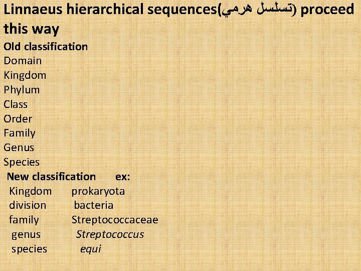
Linnaeus hierarchical sequences( )ﺗﺴﻠﺴﻞ ﻫﺮﻣﻲ proceed this way Old classification Domain Kingdom Phylum Class Order Family Genus Species New classification ex: Kingdom prokaryota division bacteria family Streptococcaceae genus Streptococcus species equi
Scientific nomenclature Binomial nomenclature A binomen or a scientific name; more informally it is also called a Latin name. The first part of the name identifies the genus to which the species belongs; the second part identifies the species within the genus. For example, humans belong to the genus Homo and within this genus to the species Homo sapiens. The binomial names of species are usually typeset in italics; for example, Staphylococcus aureus. Generally, the binomial should be printed in a font style different from that used in the normal text; for example; Streptococcus . When handwritten, each part of a binomial name should be underlined; for example, Staphylococcus aureus. The first part of the binomial, the genus name, is always written with capital letter, the second name (species ) is written with small letter. ex: Pasteulla maltucida
. The binomial name should generally be written in full. The exception to this is when several species from the same genus are being listed or discussed in the same paper or report, or the same species is mentioned repeatedly; in which case the genus is written in full when it is first used, but may then be abbreviated to an initial (and a period/full stop). For example, a list of members of the genus Canis might be written as "Canis lupus, C. aureus, C. simensis". In rare cases, this abbreviated form has spread to more general use; for example, the bacterium Escherichia coli is often referred to as just E. coli, The abbreviation "sp. " is used when the actual specific name cannot or need not be specified. The abbreviation "spp. " (plural) indicates "several species". These abbreviations are not italicized (or underlined).
Most genus and species name are chosen to tell us something about the organism its: ▀ Appearance ex: Streptococcus spp. ▀ Source ex : Canis Spp. ▀ Characteristic property ex: Staphylococcus aureus ▀ The scientist associated with it ex : Shigella For example : Escherishia coli is named for microbiologist Escherich and its habitat is the colon Shigella is named for Japanes microbiologist Shiga Staphylococcus aureus is named for the appearance and characteristics feature (staphyl means cluster) and (aureus means golden). Also the species is used for : ▀ The infection like Bacillus anthracis & Mycobacterium tuberculosis ▀ The host ex: Streptococcus eque & Brucella ovis ▀ the locality like the name of town or country ex. Salmonella Dublin & S. London.
The characters used to classify bacteria: Bergeys manual of determinative Bacteriology ► The characters used to classify bacteria fall into two groups A- Traditional 1 -morphology 2 -cultural characteristics 3 - gram staining reaction 4 - spore forming 5 - flagellation 6 - biochemical tests 7 - antigenic properties 8 - pathogenisity & toxin production
B- Genomic 1 - present G+C (Guanine- Cytosine). 2 - Sequences bases in DNA. 3 - DNA Hybridization.
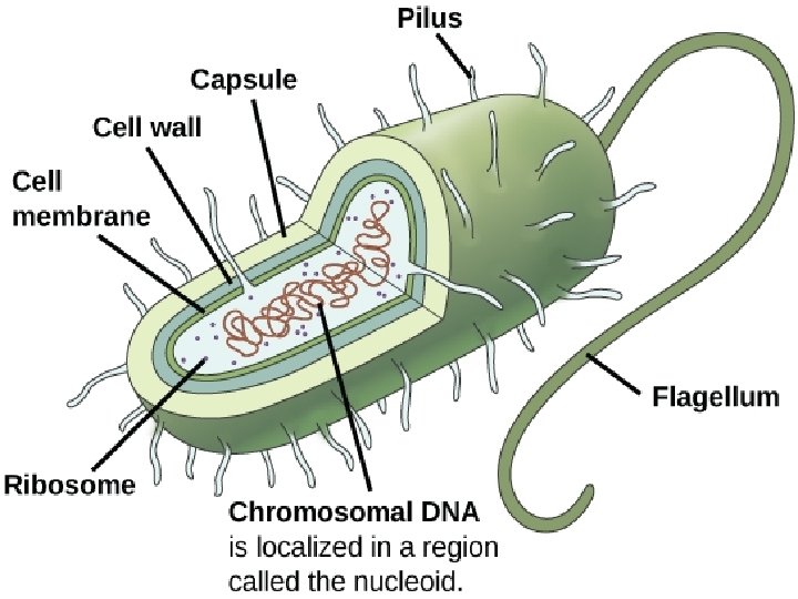
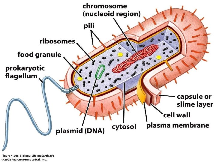
Basic Structures of Prokaryotic Cells Prokaryotes are unicellular organisms that lack organelles or other internal membrane-bound structures. Therefore, they do not have a nucleus, but, instead, generally have a single chromosome: a piece of circular, double-stranded DNA located in an area of the cell called the nucleoid. Most prokaryotes have a cell wall outside the plasma membrane
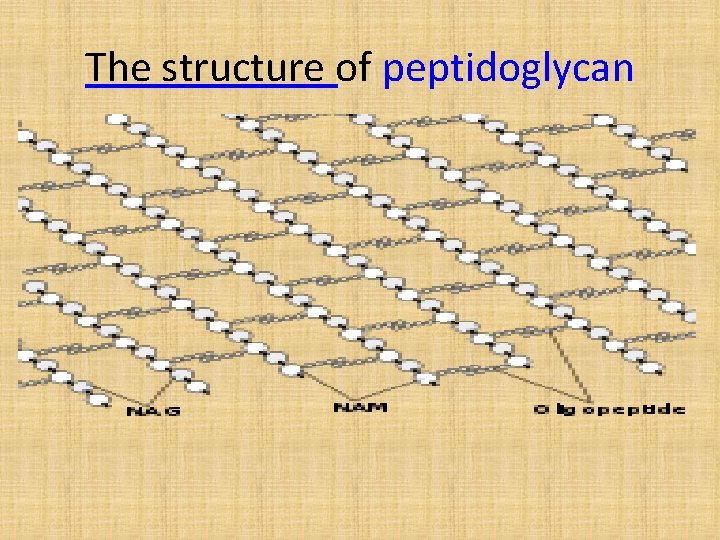
The structure of peptidoglycan
cell wall: A cell wall is a structural layer surrounding cell membrane. It can be tough, flexible, and sometimes rigid. It provides the cell with both structural support and protection, and also acts as a filtering mechanism. They may give cells rigidity and strength, offering protection against mechanical stress. Outer covering of most cells that protects the bacterial cell and gives it shape ► the primary function of the cell wall is to protect the cell from internal turgor pressure( )ﺿﻐﻂ ﺍﻻﻣﺘﻼﺀ caused by the much higher concentrations of proteins and other molecules inside the cell compared to its external environment.
►composed of peptidoglycan which is located immediately outside of the cytoplasmic membrane. Peptidoglycan is made up of a polysaccharide backbone consisting of alternating N-Acetylmuramic acid (NAM) and N- acetylglucosamine (NAG) residues in equal amounts. ►There are two main types of bacterial cell walls, gram-positive bacteria and gram-negative bacteria, which are differentiated by their Gram staining characteristics. For both these types of bacteria, particles of approximately 2 nm can pass through the peptidoglycan. If the bacterial cell wall is entirely removed, it is called a protoplast while if it's partially removed, it is called a spheroplast
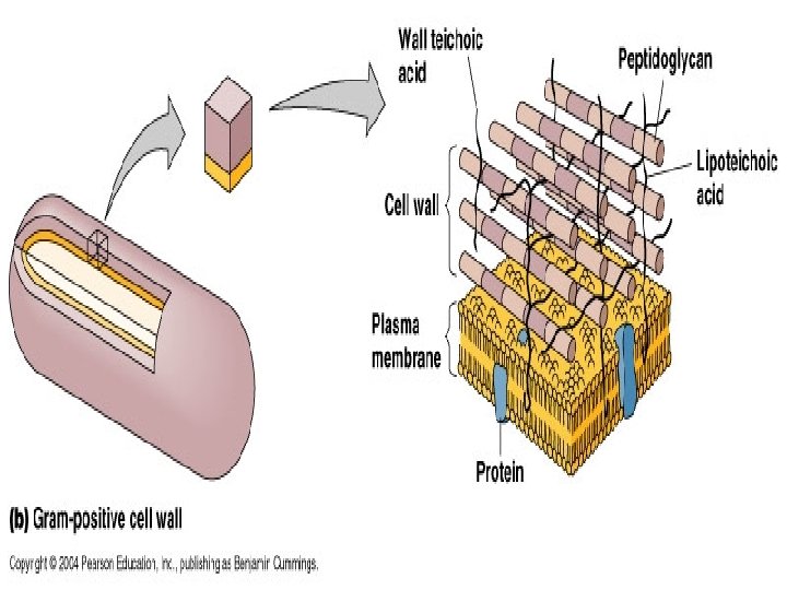
Gram positive bacterial cell wall The gram-positive cell wall Gram-positive cell walls are thick and the peptidoglycan ( also known as murein) layer constitutes almost 95% of the cell wall in some gram-positive bacteria and as little as 5 -10% of the cell wall in gram-negative bacteria. ◙The gram-positive bacteria take up the crystal violet dye and are stained purple. The matrix substances in the walls of gram -positive bacteria may be polysaccharides or teichoic acids. There are two main types of teichoic acid: ribitol teichoic acids and glycerol teichoic acids. However, the exact function of teichoic acid is debated and not fully understood. A major component of the gram-positive cell wall is lipoteichoic acid. One of its purposes is providing an antigenic function. The lipid element is to be found in the membrane where its adhesive properties assist in its anchoring to the membrane.
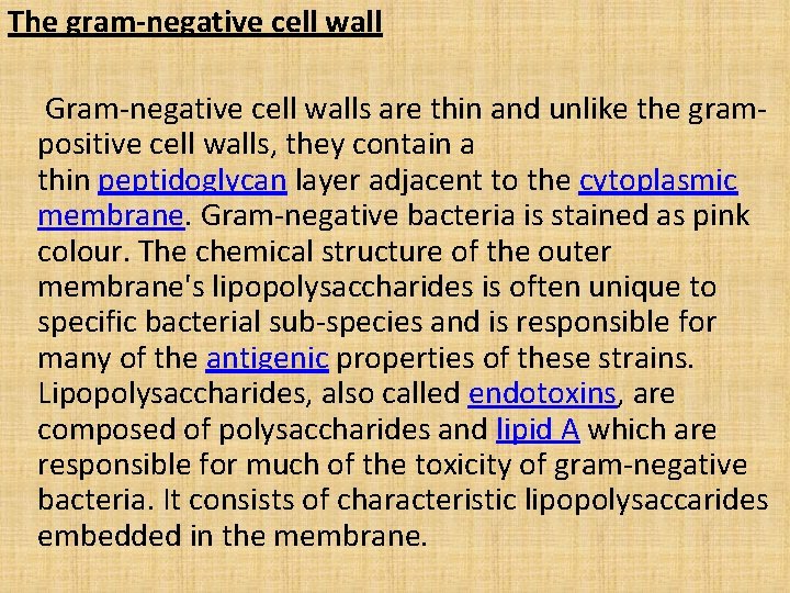
The gram-negative cell wall Gram-negative cell walls are thin and unlike the grampositive cell walls, they contain a thin peptidoglycan layer adjacent to the cytoplasmic membrane. Gram-negative bacteria is stained as pink colour. The chemical structure of the outer membrane's lipopolysaccharides is often unique to specific bacterial sub-species and is responsible for many of the antigenic properties of these strains. Lipopolysaccharides, also called endotoxins, are composed of polysaccharides and lipid A which are responsible for much of the toxicity of gram-negative bacteria. It consists of characteristic lipopolysaccarides embedded in the membrane.
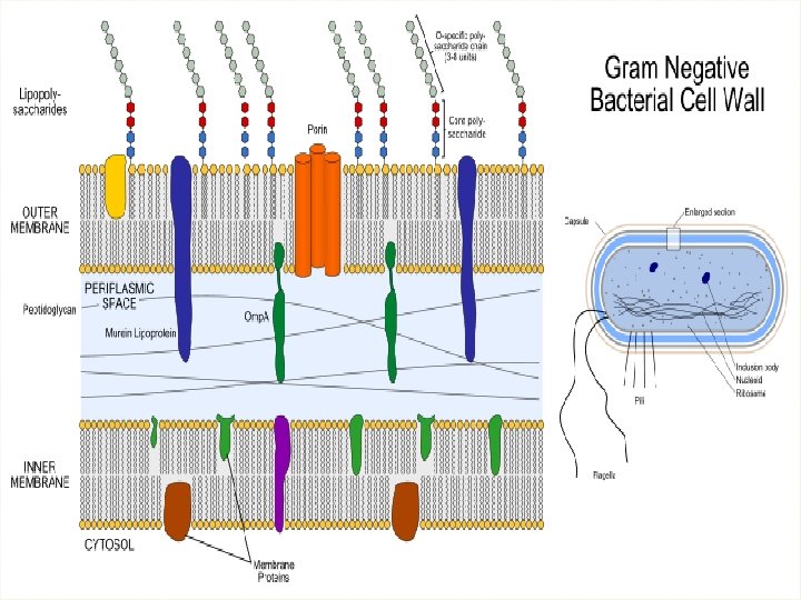
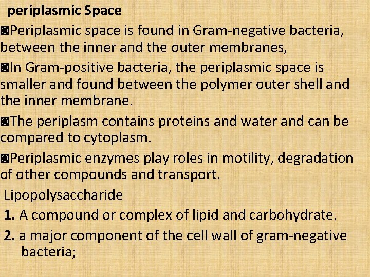
periplasmic Space ◙Periplasmic space is found in Gram-negative bacteria, between the inner and the outer membranes, ◙In Gram-positive bacteria, the periplasmic space is smaller and found between the polymer outer shell and the inner membrane. ◙The periplasm contains proteins and water and can be compared to cytoplasm. ◙Periplasmic enzymes play roles in motility, degradation of other compounds and transport. Lipopolysaccharide 1. A compound or complex of lipid and carbohydrate. 2. a major component of the cell wall of gram-negative bacteria;
3. The lipopolysaccharide (endotoxin) released from t he cell walls of gram -negative organisms that produces septic shock. 4 - lipid protein known as lipid A ◙ dead cells release lipid A when cell wall disintegrates ◙ may trigger fever vasodilation , inflammation shock, and blood clotting ◙ can be released when antimicrobial drugs kill bacteria
Wall-less forms of Bacteria When bacteria are treated with 1 - enzymes that are lytic for the cell wall e. g. lysozyme or 2 - antibiotics that interfere with biosynthesis of peptidoglycan, wall-less bacteria are often produced Usually these treatments generate non-viable organisms. 3 - Wall-less bacteria that can not replicate are referred to as sphero-plasts (when an outer membrane is present) or protoplasts(if an outer membrane is not present). 4 - L-form bacteria, also known as L-phase bacteria, Lphase variants, and cell wall-deficient (CWD) bacteria, are strains of bacteria that lack cell walls.
Components external to the cell wall Glycocalyx ( sugar coat) sticky , gelatinous polymer outside the cell wall composed of polysaccharide, polypeptide or both if attached to cell wall , considered a capsule contributes to bacterial virulence important component of biofilms help attach to various surfaces, protects, facilitates communication (extracellular polymeric substance EPS) If unorganized and loosely attached, considered a slime layer
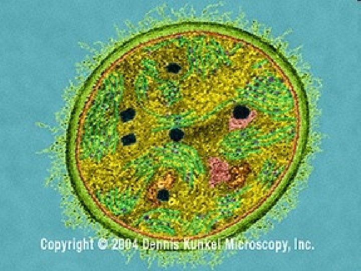
Capsule layer of highly organized material external to composed of polysaccharide (usually ) or poly peptide (sometime e. g. Bacillus has D- glutamic acid ) functional: ◙ protects it from dehydration ◙ protects against attack by phagocytic cells, ◙ increases its resistance to the immune responses. ◙ contributes to virulence of certain pathogens ( Streptococcus pneumonia , Nisseria meningitides, Klebsiella pneumonia …. etc… ◙ part of glycocalyx.
Slime layer ◙ part of glycocalyx ◙ less well organized than capsules ◙ aid in adherence to surfaces (biofilms ) ◙ Not as tight bound to cell as capsules ◙ protects against dehydration and loss of nutrients.
S layer ◙ part of glycocalyx ◙ highly structured layer made up of protein or glycoprotein (crystalline) ◙ Adheres to outer membrane(OM) in gram negative bacteria ◙ Adheres to peptidoglycan (PTG) in gram positive bacteria Slime layer and S layer Provide protection from * digestion by enzyme *ingestion by bacteria * ion and p. H changes * osmotic tress *phagocytosis by leucocytes (virulence factor) *Aide in adherence to surfaces (virulence factor) *protects from complement attack (virulence factor)
- Slides: 34