Bacterial Cell Structure Function Dr QuratUlAin Department Of

Bacterial Cell Structure & Function Dr. Qurat-Ul-Ain Department Of Microbiology KEMU, Lahore

Two Basic Types of Cells _____________________

Size of Living Things 1 m = 100 cm = 1, 000 mm = 1, 000 µm = 1, 000, 000 nm 1 mm = 1000 µm = 1000000 nm 1 µm = 1000 nm

Size of Bacteria Average bacteria 0. 5 - 2. 0 um in diam. ◦ RBC is 7. 5 um in diam. Surface Area ~12 um^2 Volume is ~4 um Surface Area to Volume is 3: 1 Typical Eukaryote Cell SA/Vol is 0. 3: 1 Food enters through SA, quickly reaches all parts of bacteria Eukaroytes need structures & organelles

Shapes of Bacteria Coccus ◦ Chain = Streptoccus ◦ Cluster = Staphylococcus Bacillus ◦ Chain = Streptobacillus Coccobacillus Vibrio = curved Spirillum Spirochete

Bacterial Structures � Flagella � Pili � Capsule � Plasma Membrane � Cytoplasm � Cell Wall � Lipopolysaccharid es � Teichoic Acids � Inclusions � Spores

Prokaryotes Cytoplasm: Also known as proto-plasm. Gel-like matrix of water, enzymes, nutrients, wastes, (organic n inorganic solutes) and gases and contains cell structures like numerous ribosomes and polysomes. No ER n memb. bound organelles. Shows signs of internal mobility like cytoplasmic streaming , amoeboid movement and formation and disappearance of vacoules. Location of growth, metabolism, and replication. Granules or inclusions: Ø Bacteria’s way of storing nutrients. Ø Staining of some granules aids in identification.

Prokaryotes Ribosomes: Small electron dense particles Involved in prt. synthesis 70 S(30 S + 50 S) Different from host cell ribosomes in SR. Streptomycin interferes with bacterial metabolism sparing the host cell ribosomes. 3 types of RNAs: Ribosomal, transfer , m. RNA Found within cytoplasm or attached to plasma membrane.

Plasma Membrane Ø Ø Ø Ø Ø Separates the cell from its environment. Limits the protoplast Thin n elastic , can be only seen with electron microscope With the exception of mycoplasma , bacterial cytoplasmic memb. lacks sterol. Phospholipid molecules oriented so that hydrophilic, water-loving heads directed outward and hydrophobic , water-hating tails directed inward. Proteins embedded in two layers of lipids (lipid bilayayer) FUNCTIONS: Semipermeable membrane Housing enzymes for cell wall, outer membrane synthesis, assembly n secretion of extractoplasmic n extracellular substances Generation of ATP Cell motility Mediation of chromosomal segragation during replication
 Unique to bacteria Sugars; NAG &")
Cell Wall Peptido-glycan Polymer (amino acids + sugars) Unique to bacteria Sugars; NAG & NAM ◦ N-acetylglucosamine ◦ N-acetymuramic acid D form of Amino acids used not L form ◦ Hard to break down D form Amino acids cross link NAG & NAM
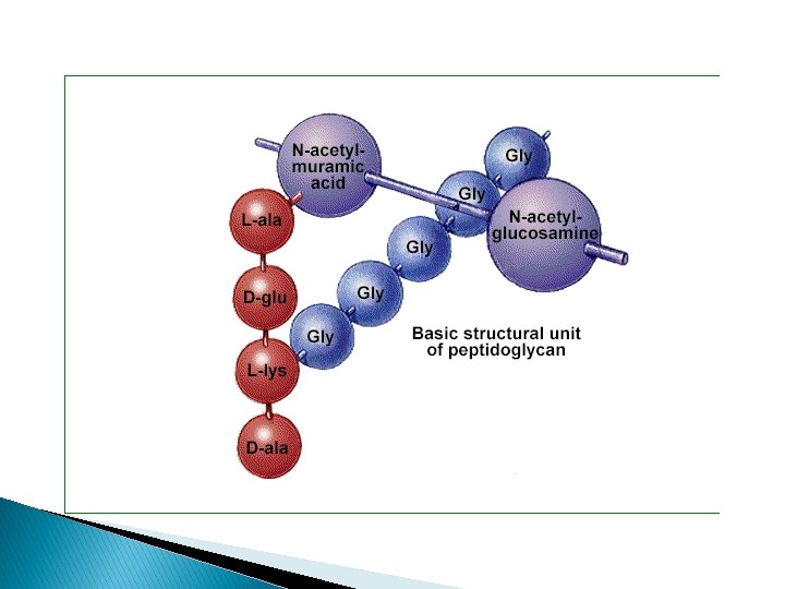

Prokaryotes – Cell Wall Ø Peptidoglycan is a huge polymer of interlocking chains of identical peptidoglycan monomers. Ø Provides rigid support while freely permeable to solutes. Ø Backbone of peptidoglycan molecule composed of two derivatives of glucose: - N-acetylglucosamine (NAG) - N-acetlymuramic acid (NAM) Ø NAG / NAM strands are connected by interpeptide bridges.

Prokaryotes - Cell Wall From the peptidoglycan inwards all bacteria are very similar. Going further out, the bacterial world divides into two major classes (plus a couple of odd types). These are: Gram Positive Negative Gram

Prokaryotes - Cell Wall Gram-Positive & Gram-Negative

Q: Why are these differences in cell wall structure so important?

Teichoic Acids Gram + only Glycerol, Phosphates, & Ribitol Attachment for Phages Participate in MG supply to the cell Antigenic determinant
 Endotoxin or Pyrogen ◦ Fever causing ◦ Toxin nomenclature Endo- part of")
Lipopolysaccharide (LPS) Endotoxin or Pyrogen ◦ Fever causing ◦ Toxin nomenclature Endo- part of bacteria Exo- excreted into environment Structure ◦ Lipid A ◦ Polysaccharide O Antigen of E. coli, Salmonella G- bacteria only ◦ Alcohol/Acetone removes primary stain durind gram’s staining.

Chapter 4
 Appearance of Colonies ◦ Mucoid = Smooth (lots of LPS or")
LPS (cont’d. ) Appearance of Colonies ◦ Mucoid = Smooth (lots of LPS or capsule) ◦ Dry = Rough (little LPS or capsule) O Antigen of Salmonella and E. coli ◦ 2, 000 different O Ags of Salmonella ◦ 100’s different O Ags of E. coli O 157 O Ags differ in Sugars, not Lipid A
 ◦ Osmotic Shock important DNA is circular, Haploid ◦")
Cytoplasm 80% Water {20% Salts-Proteins) ◦ Osmotic Shock important DNA is circular, Haploid ◦ Advantages of 1 N DNA over 2 N DNA ◦ More efficient; grows quicker ◦ Mutations allow adaptation to environment quicker Plasmids; extra circular DNA ◦ Antibiotic Resistance No organelles (Mitochondria, Golgi, etc. )

Prokaryotes - Glycocalyx Some bacteria have an additional layer outside of the cell wall called the glycocalyx. This additional layer can come in one of two forms: 1 - Glycoproteins loosely associated with the cell wall. - Slime layer causes bacteria to adhere to solid surfaces and helps prevent the cell from drying out. - Streptococcus The slime layer of Gram+ Streptococcus mutans allows it to accumulate on tooth enamel (yuck mouth and one of the causes of cavities). Other bacteria in the mouth become trapped in the slime and form a biofilm & eventually a buildup of plaque.

Prokaryotes - Glycocalyx 2. Polysaccharides to the cell wall. Ø Ø Ø firmly attached Capsules adhere to solid surfaces and to nutrients in the environment. Adhesive power of capsules is a major factor in the initiation of some bacterial diseases. Capsule also protect bacteria from being phagocitized by cells of the hosts immune system.

Prokaryotes – Surface Appendages Ø Ø Some prokaryotes have distinct appendages that allow them to move about or adhere to solid surfaces. Consist of delicate strands of proteins. Flagella: Long, thin extensions that allow some bacteria to move about freely in aqueous environments. Endoflagella: Wind around bacteria, causing movement in waves.

Flagella Motility - movement Swarming occurs with some bacteria ◦ Spread across Petri Dish ◦ Proteus species most evident Arrangement basis for classification ◦ ◦ Monotrichous; 1 flagella Lophotrichous; tuft at one end Amphitrichous; both ends Peritrichous; all around bacteria

Pili Short protein appendages ◦ smaller than flagella Adhere bacteria to surfaces ◦ E. coli has numerous types K 88, K 99, F 41, etc. ◦ Antibodies to it will block adherence. F-pilus; used in conjugation ◦ Exchange of genetic information

Endospores Resistant structure ◦ Heat, irradiation, cold ◦ Boiling >1 hr still viable ◦ Takes time and energy to destroy spores Location important in classification ◦ Central, Subterminal, Terminal Bacillus stearothermophilus -spores ◦ Used for quality control of heat sterilization equipment Bacillus anthracis - spores ◦ Used in biological warfare

Spore Formation This is what happens …………. . Cell

Spore forms in cell

Cell disintegrates

Spore is released

Spore starts to germinate

Spore continues to germinate
…………….")
Now see as, in suitable conditions, the cell begins to divide (binary fission)…………….


Prokaryotes – Arrangements of Cells Bacteria sometimes occur in groups, rather than singly. _____ divide along a single axis, seen in pairs or chains. _____ divide on one or more planes, producing cells in: - pairs (diplococci) - chains (streptococci) - packets (sarcinae) - clusters (staphylococci). Size, shape and arrangement of cells often first clues in identification of a bacterium. Many “look-alikes”, so shape and arrangement not enough for id of genus and species.
- Slides: 35