BACTERIA CLS 212 Medical Microbiology Prokaryotes Prokaryotic cells

BACTERIA CLS 212: Medical Microbiology

Prokaryotes • Prokaryotic cells possess simpler structures than eukaryotic cells , since they do not have a nucleus or a lot of cytoplasmic organelles. • There are two major types of prokaryotes: 1. Bacteria. 2. Archaea (also called archaebacteria) are often found in extreme environments, and while they are clearly prokaryotic, they have evolved separately from bacteria.
, Bacterium (singular). • Bacteria are unicellular microscopic prokaryotes.")
Introduction • Bacteriology • Bacteria (plural), Bacterium (singular). • Bacteria are unicellular microscopic prokaryotes. • Bacteria are vital in recycling nutrients such as the fixation of nitrogen from the atmosphere and decomposition of dead organic materials but also can cause disease to our body.

Classification of Bacteria • Bacteria can be classified on the basis of cell structure, cellular metabolism or on differences in cell components such as DNA, fatty acids, pigments, and antigens. • The most common method to classify pathogenic bacteria is on the basis of Gram Staining and Shape. Bacteria Gram Positive Gram +ve Cocci Gram +ve Bacilli Gram Negative Gram -ve Cocci Gram -ve Bacilli
 maintains international rules for the")
Taxonomy • The International Committee on Systematic Bacteriology (ICSB) maintains international rules for the naming of bacteria and taxonomic categories and for the ranking of them in the International Code of Nomenclature of Bacteria. Kingdom Phylum Class Order Family Genus Species Bacteria Proteobacteria Gamma Proteobacteria Enterobacteriales Enterobacteriaceae Escherichia coli e. g. Escherichia coli

Structure of Bacteria

1 -Cell Envelope The cell envelope is made up of two to three layers: A. the interior cytoplasmic membrane B. the cell wall C. in some species of bacteria an outer capsule.
 A Layer of phospholipids and proteins (lipid bilayer),")
A-Cytoplasmic Membrane ( cellular membrane ) A Layer of phospholipids and proteins (lipid bilayer), encloses the interior of the bacterium, regulating the flow of materials in and out of the cell.

B-Cell Wall • Each bacterium is enclosed by a rigid cell wall composed of peptidoglycan, a protein-sugar (polysaccharide) molecule. • Peptidoglycan is responsible for the rigidity of the bacterial cell wall and for the determination of cell shape. • Several antibiotics (Penicillins and Cephalosporins) stop bacterial infections by interfering with cell wall synthesis, while having no effects on human cells.
 and N-acetylmuramic acid (NAM),")
The “glycan” part consist of ulternating units of Nacetylglucosamine (NAG) and N-acetylmuramic acid (NAM), which is located immediately outside of the cytoplasmic membrane. The “peptido” part consist of a short string of amino acids. It cross-links the adjacent polysaccharide strands at the NAM subunit.

Functions of the Cell Wall: 1. The rigid cell wall gives the bacterium its shape and surrounds the cytoplasmic membrane, protecting it from the environment. 2. The strength of the wall is responsible for keeping the cell from bursting when there are large differences in osmotic pressure between the cytoplasm and the environment. 3. It also helps to anchor appendages like the pili and flagella, which originate in the cytoplasmic membrane and protrude through the wall to the outside.

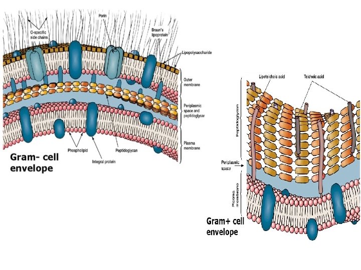
GRAM STAIN • There are two main types of bacterial cell walls, Gram positive and Gram negative, which are differentiated by their Gram staining characteristics. • Gram stain Procedure: 1. 2. 3. 4. Crystal violet Iodine Alcohol Saffranine

Gram stain

Gram +ve and Gram –ve Cell Wall • The Gram positive cell • The Gram negative cell wall is characterized by wall contains a thin the presence of a very peptidoglycan layer, thick peptidoglycan which is responsible for layer, which is the cell wall's inability responsible for the to retain the crystal retention of the crystal violet stain upon violet dyes during the decolourisation with Gram staining procedure. ethanol during Gram staining. Color of Bacteria: Blue-violet Color of Bacteria: RED

C-Capsule • Some species of bacteria have a third protective covering, a capsule made up of polysaccharides (complex carbohydrates). • Functions of the Capsule: 1. To keep the bacterium from drying out. 2. To protect bacterium from phagocytosis (engulfing) by larger microorganisms. 3. The capsule is a major virulence factor in the major diseasecausing bacteria, such as Escherichia coli and Streptococcus pneumoniae. (Noncapsulated mutants of these organisms are avirulent, i. e. they don't cause disease).
 are long hair-like structures that can be found")
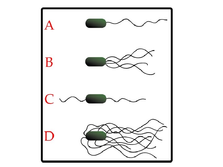
II- Flagella • Flagella (singular, flagellum) are long hair-like structures that can be found at either or both ends of a bacterium or all over its surface. • Function of the Flagella: the flagella beat in a propeller-like motion to help the bacterium move toward nutrients; away from toxic chemicals; or, in the case of the photosynthetic cyanobacteria; toward the light.
, short hairlike structures")
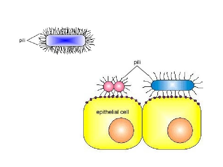
III- Pili • Many species of bacteria have pili (singular, pilus), short hairlike structures emerging from all around the outside cell surface. • Function of the Pili: the pili assist the bacteria in attaching to other cells and surfaces, such as teeth, intestines, and rocks. • Without pili, many disease-causing bacteria lose their ability to infect because they are unable to attach to host tissue.

IV- The Cytoplasm • The cytoplasm, or protoplasm, of bacterial cells is where the functions for cell growth, metabolism, and replication are carried out. • It is a gel-like matrix composed of water, enzymes, nutrients, wastes, and gases and contains cell structures such as ribosomes, a chromosome, and plasmids. • The cell envelope encases the cytoplasm and all its components. • Unlike the eukaryotic (true) cells, bacteria do not have a membrane enclosed nucleus. The chromosome, a single, continuous strand of DNA, is localized, but not contained, in a region of the cell called the nucleoid. All the other cellular components are scattered throughout the cytoplasm.

The Nucleoid • The nucleoid is a region of cytoplasm where the chromosomal DNA is located. It is not a membrane bound nucleus, but simply an area of the cytoplasm where the strands of DNA are found. • Most bacteria have a single, circular chromosome that is responsible for replication, although a few species do have two or more.

Ribosomes • They translate the genetic code from the molecular language of nucleic acid to that of amino acids (building blocks of proteins). • Differences between Bacteria and Eukaryotes 1. Bacterial ribosomes are similar to those of eukaryotes, but are smaller and have a slightly different composition and molecular structure. 2. Bacterial ribosomes are never bound to other organelles as they sometimes are (bound to the endoplasmic reticulum) in eukaryotes, but are free-standing structures distributed throughout the cytoplasm. 3. Some antibiotics will inhibit the functioning of bacterial ribosomes, but not a eukaryote's, thus killing bacteria but not the eukaryotic organisms they are infecting.

Plasmids • Plasmids are small extrachromosomal genetic structures carried by many strains of bacteria. • Like the chromosome, plasmids are made of a circular piece of DNA. Unlike the chromosome, they are NOT involved in reproduction. • Plasmids replicate independently of the chromosome and, while not essential for survival, appear to give bacteria a selective advantage. • Plasmids are passed-on to other bacteria through Two ways: 1. For most plasmid types, copies in the cytoplasm are passed on to daughter cells during binary fission.

Plasmids 2. Other types of plasmids form a tubelike structure at the surface called a pilus that passes copies of the plasmid to other bacteria during conjugation, a process by which bacteria exchange genetic information. • Plasmids have been shown to be instrumental in the transmission of special properties, such as antibiotic drug resistance, resistance to heavy metals, and virulence factors necessary for infection of animal or plant hosts. The ability to insert specific genes into plasmids have made them extremely useful tools in the fields of molecular biology and genetics, specifically in the area of genetic engineering.

Endospores • Endospores are bacterial survival structures that are highly resistant to many different types of chemical and environmental stresses and therefore enable the survival of bacteria in environments that would be lethal for these cells in their normal vegetative form.
: Streptococci, Staphylococci 2. Bacillus (rod-like):")
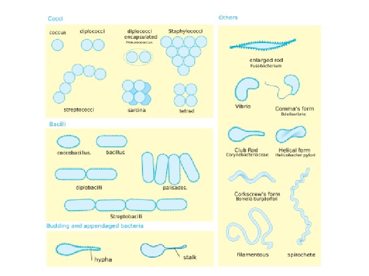
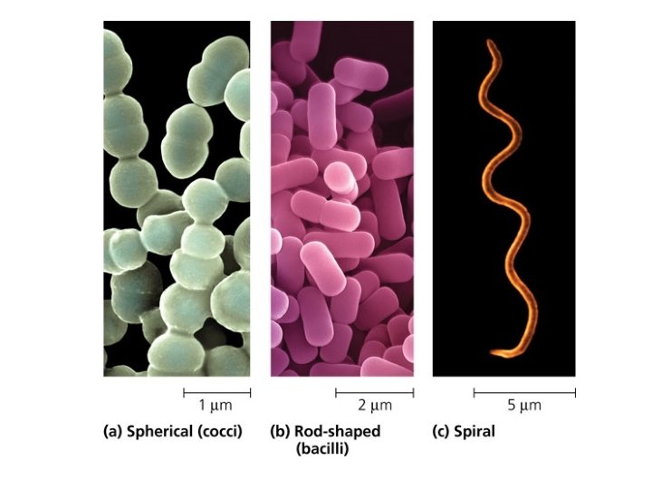
Cell Morphology & Shape of Bacteria 1. Coccus (spherical): Streptococci, Staphylococci 2. Bacillus (rod-like): Enterobacteriacea spp. 3. Spirillum (spiral): Treponema spp.

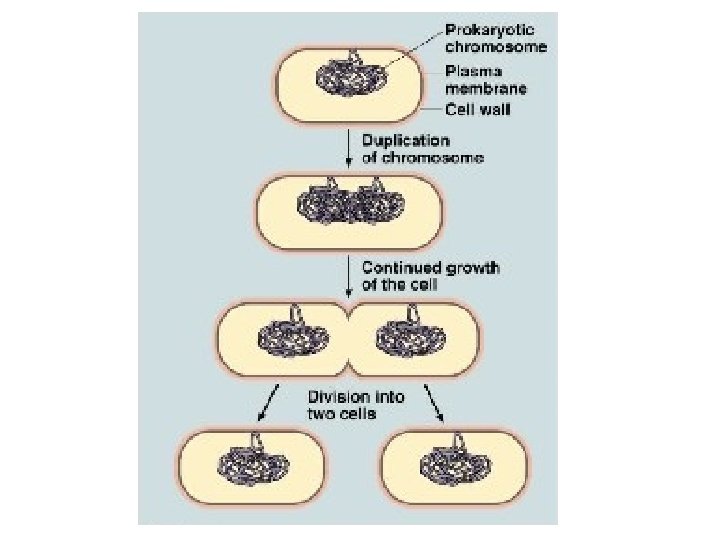
Replication of Bacteria • Bacterial cells replicate asexually by a process called: Binary Fission. • One cell doubles in size and splits in half to produce two identical daughter cells. These daughter cells can then double in size again to produce four sibling cells and these to produce eight, and so on. • Doubling Time: the time it takes for a bacterial cell to grow and divide in two.
 DNA (and proteins) • The cytoplasm")
Replication of Bacteria DNA transcription RNA translation (Ribosomes) DNA (and proteins) • The cytoplasm of a bacterial cell contains the DNA molecules that make up the bacterial genome, the transcriptional machinery that copies DNA into ribonucleic acid (RNA), and the ribosomes that translate the messenger RNA information into protein sequence. • Since there is no nucleus, all of these processes occur simultaneously. The rapid growth rate of the bacterial cell requires constant DNA replication and ways to segregate the two new chromosomes into the two daughter cells without tangling them.

Microbial Growth CLS 212: Medical Microbiology

Factors Affecting Microbial Growth • There are some factors that affect and control the growth of microorganisms around us, in hospitals, in the laboratory, and in industrial settings. These factors are: 1. 2. 3. 4. 5. 6. 7. Availability of Nutrients Moisture Temperature p. H Osmotic Pressure and Salinity Atmospheric Pressure Gaseous Atmosphere

Availability of Nutrients • Nutrients are crucial for microorganisms to survive in the environment. • These nutrients are chemicals that can be broken into essential elements like: carbon, oxygen, hydrogen, nitrogen, sodium, potassium, calcium, iron, ext. . . which are required for growth.

Moisture • All organisms on planet need water for their metabolic processes and most will die if moisture is too little. • Some bacteria and parasites can stay dormant in endospores and cysts until moisture is available for their growth.

Temperature • Microorganisms have optimum temperature required for growth, this temperature depends on their enzymes. • The temperature (which ranges from minimum to maximum growth temp. ) is different from one organism to another. • Microorganisms can be classified according to their preferred temp. into: : ( chapter 8 , page 122) 1. Thermophiles 2. Mesophiles: 3. Psychrophiles

p. H • Most microorganisms prefer a neutral or slightly alkaline growth medium p. H 7 -7. 4. • Some microorganisms like acidic or alkaline environments so are classified into: 1. Acidophiles: microorganisms that grow best in acidic media p. H 2 -5 e. g. Fungi. 2. Alkaliphiles: microorganisms that grow best in alkaline media p. H 8. 5 -11 e. g. Vibrio cholera (the only alkaliphilic human pathogen).

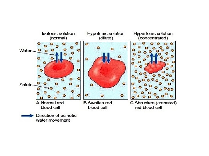
Osmotic Pressure Isotonic solutions : • solutions where the concentration of the solute is equal to that of normal cells found in it; thus no osmotic pressure is exerted. Most organisms prefer isotonic solutions. Hypotonic solutions: • Solutions where solute concentration outside the cell is less than that inside the cell. This cause microbial cells to swell then burst (die). Hypertonic solutions : • Solutions where solute concentration outside the cell is more than that inside the cell. This cause microbial cells to shrink (inhibiting growth).
: Are organisms that prefer salt environment to grow.")
Osmotic Pressure Halophilic organisms (salt lovers): Are organisms that prefer salt environment to grow. e. g. microorganisms living in the Dead Sea.
 and")
Atmospheric Pressure • Most bacteria live at normal atmospheric pressure (14. 7 psi) and are not affected by minor changes in it. • Some like very high atmospheric pressure (Barophiles) like in oil wells and deep oceans.

Gaseous Atmosphere • Microorganisms can be classified according to the requirement of oxygen to survive into: • Aerobes: require 20 -22% O 2. • Anaerobes: will die in the presence of O 2. • Microaerophiles: require 5% only of O 2. • Some microorganisms are Capnophiles i. e. require 5 -10% of CO 2 for their growth.

Bacterial Growth In Vitro • In order for bacteria to grow in the laboratory it need appropriate growth medium and special environmental conditions like temperature, p. H, O 2, . . to multiply. • Bacteria can be cultured on many different culture media according to its nutritional needs such as Nutrient Agar, Blood Agar, Mac Conkey Agar, CLED, . . • After inoculation of media, they should be incubated in chambers to maintain appropriate environment. Temperature and time of incubation differ for each type of bacteria to grow.

Bacterial Count • Microbiologists tend to measure the number of bacteria present in a liquid for quality control purposes in FDA (Food and Drug Administration) monitored fields e. g. dairy farms, drinking water supply, drug industry. . . • This is done by measuring: 1. The total number of bacteria present in the sample (dead and alive bacteria) by using a spectrophotometer. The amount of light transmitted by the machine is proportional to the number of bacterial cells present. 2. The number of viable bacteria present in the sample by a method called the viable plate count.

Bacterial Count Spectrophotometer

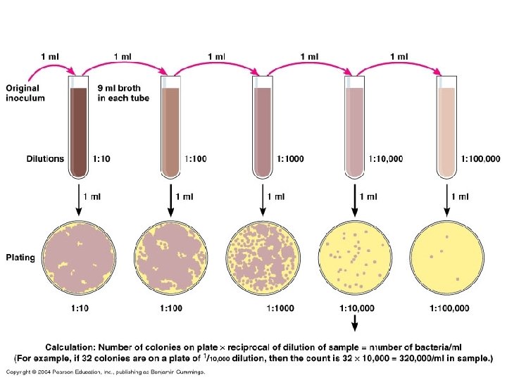
The Viable Plate Count Method 1. Serial dilutions of the sample are prepared. 2. From each dilution, 1 ml or 0. 1 ml is inoculated on Nutrient Agar media. 3. All the plates are incubated for 24 hours at 37°C. 4. After incubation, the bacterial colonies are counted from the plates. Then the number is multiplied by the dilution factor to get the number of bacteria in the original sample.

The Viable Plate Count Method

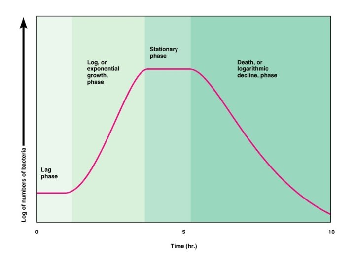
Bacterial Growth Curve • In order to plot a growth curve for a certain bacteria, a pure bacterial broth is to be prepared and incubated. Then a sample is collected from the broth every 30 minutes and a viable plate count is done on each sample. • The data is then plotted on a logarithmic graph paper where the x-axis represent the incubation time and the y-axis represent the log 10 of the number of viable bacteria.

4 Phases in a Bacterial Growth Curve 1. Lag Phase: where the bacteria absorb nutrients, synthesize enzymes, and prepare for division. There is no increase in bacterial number in this phase. 2. Log Phase (logarithmic growth phase): where rapid multiplication occur causing very high increase in the number of bacteria. 3. Stationary Phase: where the nutrients in the media decrease and the toxic waste resulting from bacterial metabolism increase. As a result, the multiplication is slowing down. The number of dividing bacteria equals the number of dead bacteria. 4. Death Phase: where overcrowding occurs and the bacteria are dying very rapidly because of lack of nutrients and accumulation of toxic waste. Very few bacteria will remain alive in this stage.
- Slides: 52