Avian Radiology Birds are very well suited to

Avian Radiology • Birds are very well suited to radiographic study. • The air sac system provides excellent contrast with soft tissues. • Their small size reduces exposure required to produce an image. • Positioning is easily accomplished if proper equipment is available

• An acrylic plate with restraint mechanisms for the head and legs will facilitate positioning. • The wings are restrained with masking tape or other means. • Technique will vary with each machine and an avian technique chart will eliminate many retakes.

• Radiographs are indicated in any bird with clinical signs of weight loss (wasting), vomiting, regurgitation, polyuria/polydipsia, or abdominal swelling (although details may be obscured in these cases). • Orthopedic conditions such as fractures, of course, are indications for radiographs.

Positioning of avian patient for radiographs

Ventrodorsal

Lateral

Lateral

• The focal film distance will sometimes be reduced to give some magnification and to increase radiographic density without increasing k. VP. • Fast exposure times (1/60 sec or faster) are necessary to avoid excessive motion on the films. • Lowest k. Vp, high m. A and short exposure • k. Vp of 70, m. A of 300 at 1/120 second

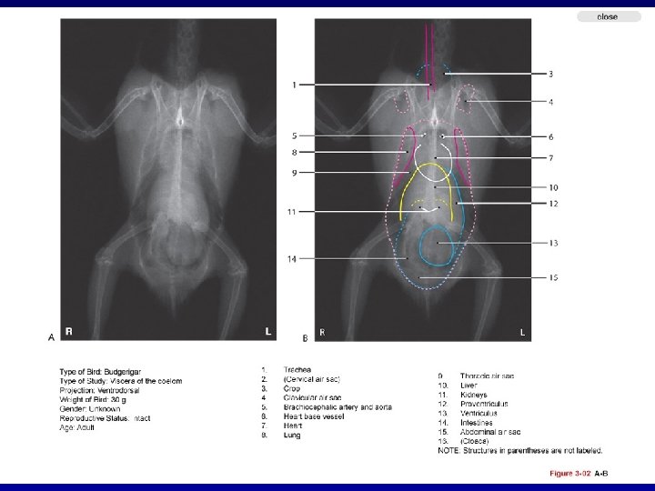
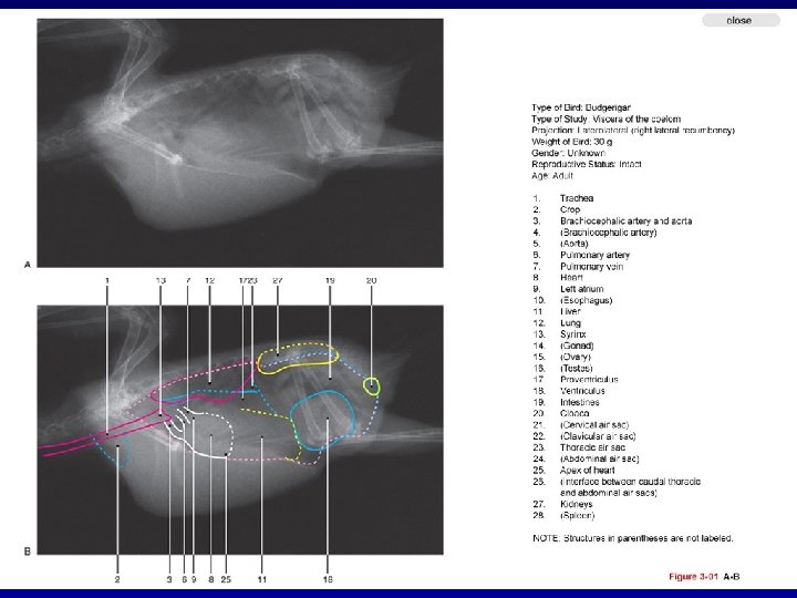
• Radiographs should be examined systematically. • The radiograph should be evaluated for technique and positioning • The radiograph is then examined starting with the skin and working inward to the deepest tissues. • Then each system is followed from cranial to caudal.


Radiographic anatomy of a normal cockatiel.

Avian • Tibiotarsus • Intertarsal joint • Tarsometatarsus

Avian tibiotarsus tarsometatarsus

Avian # phalanges minus 1 equals digit

Avian • Furcula • Corocoid • Scapula

Avian - Furcula

Avian - Corocoid

Avian - Scapula

Avian

Avian • Alular digit

Questions
- Slides: 25