AVIAN INFECTIOUS BRONCHITIS DR SANJIV KUMAR ASSISTANT PROFESSOR

AVIAN INFECTIOUS BRONCHITIS DR. SANJIV KUMAR ASSISTANT PROFESSOR, DEPTT. OF PATHOLOGY, BVC, PATNA

INTRODUCTION • Highly infectious and contagious respiratory disease of chicks • Also affect the oviduct, intestine and kidneys • Great economic importance due to its adverse effect on egg production in layers, and on production in broilers • Other pathogens such as mycoplasma or E. coli increases the severity and duration of the disease
, a coronavirus. § Affect only chickens §")
ETIOLOGY • Avian infectious bronchitis virus (IBV), a coronavirus. § Affect only chickens § Direct spread by Airborne transmission § Egg transmission is exceptional.

PATHOGENESIS • Different serotypes o Respiratory tract - massachusetts and connecticut o Nephrotoxic - T, Gray and Holte • • New virus appear 3 -4 hours after infection Maximum output per cell within 10 -12 hrs. Incubation period: 1 -3 days. The virus first attack tracheal cells, multiply and then reaches different organs like kidneys, oviduct etc.

CLINICAL SIGNS • Spread very fast. • Morbidity is 100%, Mortality varies according to the virus strain. Respiratory form In young chicks IB, causes asphyxia, preceded by severe respiratory distress.
 • Egg")
Reproductive form • Damage oviduct. • Egg internal quality changes (watery albumin) • Egg deformed. Renal (Nephritic) form • Young growing birds • Mortality upto 30 %.
 in trachea, froth in")
Macroscopic lesions Respiratory tract • Mucus and redness (no haemorrhage) in trachea, froth in air sacs in older chickens. • In young chicks a yellow cheesy plug at the tracheal bifurcation is indicative of IB infection. Reproductive tract • Fluid yolk material may be found in the abdominal • Abnormal ovary having the misshapen follicles • Egg- Outer surface - Ridges or concretions Watery albumin Nephrotic form • Swollen, pale kidneys, with tubules and ureters distended with urates • In layers, urolithiasis is associated with certain dietary factors
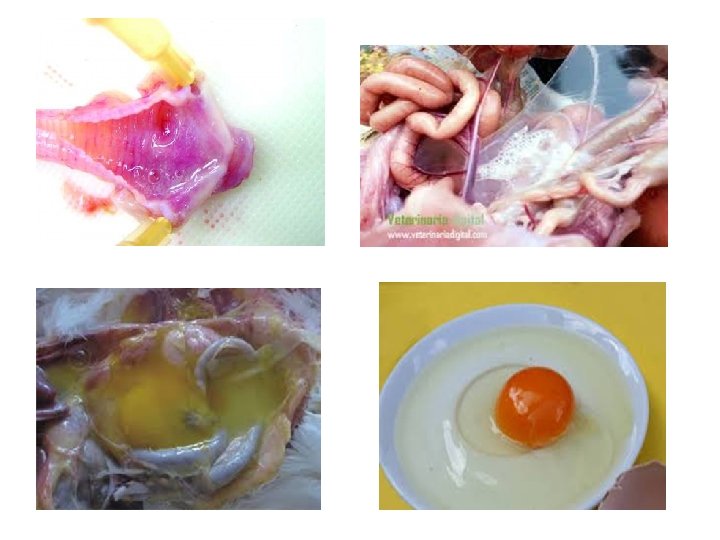
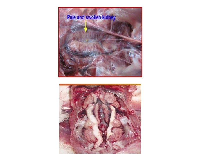

Microscopic lesions • Trachea & Bronchi: Inflammatory and degenerative changes • Kidney: Interstitial nephritis, Degenerative changes, Urolithiasis, urates in ureter • Oviduct: Epithelial damage, dilatation of tubular glands, infiltration of mononuclear cells, proliferation of lymphoid follicles.

DIAGNOSIS • Based on clinical signs • Gross and microscopic lesions • Serological tests: o Virus neutralisaton (VN) o Immunodiffusion (ID) o Haemagglutination inhibition (HI) o Immunofluorescence (IF) and ELISA - more sensitive

THANK YOU
- Slides: 12