AVIAN ENCEPHALOMYELITIS EPIDEMIC TREMOR AE It is an


- Slides: 29

AVIAN ENCEPHALOMYELITIS “EPIDEMIC TREMOR” (AE)

It is an infectious viral disease primarily affecting young chickens. It is characterized by: Ataxia Rapid tremors (specially of the head and neck) The disease was of great economic importance to the poultry industry before using of the commercial vaccines. No public health significance has been attached to the disease.

Causative agent AE virus is belonging to genius enterovirus of the family picorna-viridea. o Field isolates (Natural strains): Varies in pathogenecity All are enterotropic They infect chicks via oral route and they shed in the feces. Some tend to be more neurotropic than the others and produce sever CNS lesions. Non lethal for the embryos by rapid passage.

• Embryo-adapted (Van roekel strain ) It is highly neurotropic It causes signs of the disease in all ages by parentral inoculation. It dose not infect via the oral route (except with very high dosage) It dos not spread from bird to bird. It is patogenic to embryos from non immune flocks. All strains of AE are chemically, physically, and serologically uniform.

Susceptibility The natural host range is limited to chicken, turkeys, Japanese quail and pheasents Experimental hosts are ducklings, squabs, and guinea fowl.

Transmission Oral route is the most probable route after hatching. Vertical transmission is very important means of virus transmission. Virus transmission can occur in the incubator.

Incubation period 11 days among chicks that infected by contact or oral administration. 1 -7 days among chicks infected by embryo transmission.

Signs Depression, ataxia due to muscular incoordination. Chicks like to sit on their hocks and when disturbed they move with out control for speed and gait. Some may refuse to move or may walk on their hocks. Fine tremor of the head and neck. Temporary decrease in egg production in layer flocks. Usually morbidity rate is 40 – 60 % Mortality averages 25 % may reaches 50 %.

Clinical signs consist of progressive ataxia, weakness, dullness and sometimes a fine tremor of the head and neck. Slight incoordination can be elicited by forcing the chicks to move. Reluctance or inability to stand (as seen in these chicks) is common.

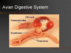
PM Lesions The only gross lesions associated with AE in chicks are whitish areas in the muscularis of the proventriculus.

Gizzard musculature may have grossly visible pale areas resulting from massive infiltration by mononuclear cells.

Cataract formation as seen in the affected eye on the left may occur in a high percentage of the survivors of an early outbreak of AE. The lesion is usually seen as an opacity or bluish discoloration of one or both eyes.

Histopathology Principal changes present in CNS and some viscera. Peripheral nervous system is not involved. In CNS: - non purulent encephalomyelitis and ganglionitis of the dorsal root ganglia Perivascular cuffs in all portion of the brain and spinal cord except the cerebellum in which gliosis occurs. Neural degeneration of the Purkinje cells.

Compact and diffuse microgliosis is a characteristic lesion in cerebellum, particularly in the white matter (as shown) or molecular layer

Compact and diffuse microgliosis is a characteristic lesion in cerebellum, particularly in the white matter or molecular layer

Perivascular cuffs of small lymphocytes, often several layers thick are found in nearly all parts of the CNS.

Neuronal degeneration is considered one of the most striking features of AE. The normal neurons seen in the left contrast with an affected one undergoing central chromatolysis in the write

Similarly, Purkinje cells may undergo necrosis, and some areas of the cerebellum are often found to be essentially devoid of these cells. The normal cerebellum (shown in left silde) has a uniform row of Purkinje cells separating the granular and molecular layers. In contrast, Purkinje cells may undergo necrosis, and some areas of the cerebellum

The muscular layer in this proventriculus is infiltrated by dense aggregates of lymphocytes. A lesion like this, perhaps coupled with abnormal aggregates in the gizzard and pancreas, is of diagnostic significance in AE only when accompanied by characteristic microscopic lesions in the CNS.

Gizzard musculature is frequently infiltrated in the same manner as the proventricular musculature.

Infiltration of the myocardium is frequent in AE but because some lymphoid cell aggregates are found in "normal" hearts, caution is required in interpretation. In this specimen, abnormal numbers of cells infiltrate between the muscle fibers.

A lymphoid focus in the liver of an infected chick. The comments regarding the heart and pancreas also pertain to the liver.

Diagnosis Clinical signs. Absence of gross lesions. Histopathlogical lesions. Absence of Nutritional deficiencies

Diagnosis Viral isolation • By inoculation of suspension of brain from an infected chicks into the yolk sac of 5 -7 - day old susceptible checken embryos. • Examination of smears from the brain or cryostat sections stained by direct immunofluorscence for virus demonstration.

Diagnosis Serological tests Virus nutralization (VN): - inoculation of dilutions of the virus alone and virus together with specific ant-sera into susceptible chick embryos. And determine the nutralization index. (index of log 10 1. 1 consider positive).

Embryo susceptibility test 36 – 48 eggs from a flock are incubated for 6 days and then each live embryo is inoculated into the yolk sac with 100 EDS 50 of egg adapted virus. The embryos are incubated for further 12 days and examined. If less than 50 % of the embryos show lesions that means the flock is consider immune. If 100 % of the embryos show lesions that means the flock is consider susceptible

Susceptible (antibody free) embryos, inoculated with embryo-adapted AE virus via the yolk sac at 5 or 6 days of incubation, develop lesions illustrated by the 2 embryos on the left. Severe stunting results primarily from marked muscular dystrophy, as seen in the embryo from which the skin has been removed. In addition, affected embryos are paralyzed and the legs are rigidly extended rather than hanging loosely from the body. The 2 embryos on the right are normal. Because embryos from immune flocks are resistant to infection, an "embryo susceptibility" test can be used to determine if a breeding flock has been exposed to AE.

Differential diagnosis • The disease must be differentiated from ND, Marek’s disease, nutritional encephalomalicia.

Control Drug therapy is of no value. Control is depend on the vaccination of the parent flocks. Live and inactivated vaccine are available. Live vaccine can be given by mass administration usually in drinking water. They protect against egg transmission and drop egg production and decrease hatchability.
 Intention tremor vs essential tremor usmle
 Myalgic encephalomyelitis
 Epidemic dropsy
 Aids epidemic
 Epidemic dropsy
 Endemic epidemic
 Doctors attributed the epidemic to the rampant
 Epidemic transition model
 Epidemic broadcast trees
 Endemic epidemic
 Liskantin biverkningar
 Action tremor
 Esential tremor
 Flapping tremor youtube
 Psikojenik tremor belirtileri
 Flapping tremor
 Somatic tremor artifact
 Trihexyphenidyl tremor
 Low pathogenic avian influenza
 Paraphrasing proverbs
 Definition of avian
 Polygastric examples
 Ahas bird condition
 Aves kingdom
 Avian taxonomy
 Bash ahas
 Avian phylogeny
 Avian ku ethnicity
 Bird digestive system functions
