AV Blocks FirstDegree PR AV Block interval 0

�AV Blocks �First-Degree � PR AV Block interval >0. 21 seconds �Second-Degree � Progressive delay until conduction blocked �Second-Degree � Conduction AV Block (Mobitz II) of selected impulses through the AV node �Third-Degree � No AV Block (Mobitz I) AV Block atrial impulses are conducted through the AV node � Atria and ventricles fire at their intrinsic rates

�Bundle Branch Blocks Right Bundle Branch Block

� The Turn-Signal Rule � QRS >0. 12 seconds throughout the ECG � Look at the QRS in V 1 � Identify the J point � Draw a horizontal line � Triangle pointing up indicates RBBB � Triangle pointing down indicates LBBB

�Hemiblocks �Left Posterior Hemiblock �Principal finding is a rightward shift in the QRS axis

�Hemiblocks �Left Anterior Hemiblock �Principal abnormality is a shift in the QRS axis far to the left

�Left Bundle Branch Block

�Right Bundle Branch Block

�Chamber Enlargement �Atrial Enlargement �Ventricular Hypertrophy �Causes Right-sided enlargement and hypertrophy � Secondary to long-term pulmonary disease Left-sided enlargement and hypertrophy � Secondary to long-term hypertension

�Right Atrial Enlargement

�Left Atrial Enlargement

�Right Ventricular Hypertrophy

�Left Ventricular Hypertrophy
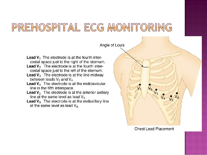

�Cardiac Conductive System �ECG Recording �ECG Leads �Mean QRS Axis Determination �The Normal 12 -Lead ECG �Disease Findings �Conduction Abnormalities �Prehospital ECG Monitoring
- Slides: 15