Autonomic Nervous System The Autonomic Nervous Assess Prof


 neuron leaves CNS to synapse with 12 the")
 ganglia near vertebral bodies")


 Postganglionic neurons:")


. All parasympathetic postganglionic release")
 as its neurotransmitter. q")


- Slides: 42
Autonomic Nervous System The Autonomic Nervous Assess Prof. Fawzia Al-Rouq System Department of Physiology College of Medicine King Saud University
INTRODUCTION
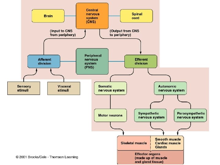
THE NERVOUS SYSTEM • INTRODUCTION • The nervous system monitors and controls almost every organ / system through a series of positive and negative feedback loops. • The Central Nervous System (CNS): Includes the brain and spinal cord. • The Peripheral Nervous System (PNS): Formed by neurons & their process present in all the regions of the body. • It consists of cranial nerves arises from the brain & spinal nerves arising from the spinal cord. • The peripheral NS is divided into • Somatic Nervous system • Autonomic nervous system
Functional Anatomy & Physiology of Autonomic NS
OBJECTIVES
Anatomy and physiology of Autonomic Nervous System At the end of this lectutre the student should be able to: -appreciate the anatomy of sympathetic& parasympathetic nervous system. -explain physiological functions of Sympathetic ¶sympathetic nerves in head&neck, chest, abdomen and pelvis
FUNCTIONAL ANATOMY OF THE AUTONOMIC NERVOUS SYSTEM
Basic anatomical difference between the motor pathways of the voluntary somatic nervous system (to skeletal muscles) and those of the autonomic nervous system
Basic anatomical difference between the motor pathways of the voluntary somatic nervous system (to skeletal muscles) and those 10 of the autonomic nervous system Somatic division: Cell bodies of motor neurons reside in CNS (brain or spinal cord) Their axons (sheathed in spinal nerves) extend all the way to their skeletal muscles Autonomic system: chains of two motor neurons 1 st = preganglionic neuron (in brain or cord) 2 nd = gangionic neuron (cell body in ganglion outside CNS) Slower because lightly or unmyelinated
ANS is the subdivision of the peripheral nervous system that regulates body activities that are generally not under conscious control Visceral motor innervates non-skeletal (non-somatic) muscles Composed of a special group of neurons serving: Cardiac muscle (the heart) Smooth muscle (walls of viscera and blood vessels) Internal organs Skin 11
Axon of 1 st (preganglionic) neuron leaves CNS to synapse with 12 the 2 nd (ganglionic) neuron Axon of 2 nd (ganglionic) neuron extends to the organ it serves this dorsal root ganglion is sensory
LOCATIONS OF AUTONOMIC GANGLIA Sympathetic Ganglia Location q Trunk (chain) ganglia near vertebral bodies q Prevertebral ganglia near large blood vessel in gut : celiac , superior mesenteric & inferior mesenteric
Parasympathetic Ganglia Location : Terminal ganglia in the wall of organ
Sympathetic Innervation of Visceral Targets • Short, lightly myelinated preganglionic neurons • Long, unmyelinated postganglionic neurons Ganglia close to spinal cord Spina l
Parasympathetic Innervation of Visceral Targets Ganglia close to or on target organs • Preganglionic neurons - long • Post ganglionic neurons - short
SYMPATHETIC & PARASYMPATHETIC NERVOUS SYSTEM ORIGIN Blue= Para symp; Red symp
Sympathetic - Origin Thoracolumbar lateral horns of the spinal segments T 1 -L 2. Nerve fibers originate between T 1 & L 2
Parasympathetic - Origin q Craniosacral Cell bodies of the motor nuclei of the cranial nerves III, VII, IX and X in the brain stem Second, third and fourth [S 2 -S 4] sacral segments of the spinal cord Nerve fibers emerge from brain & sacrum cranio-sacral outflow q
PARASYMPATHETIC NERVOUS SYSTEM q The cranial nerves III, VII and IX affect the pupil and salivary gland secretion q Vagus nerve (X) carries fibres to the heart, lungs, stomach, upper intestine and ureter q The sacral fibres form pelvic plexuses which innervate the distal colon, rectum, bladder and reproductive organs.
Autonomic Nervous System 2 divisions: Sympathetic “Fight or flight” “E” division Exercise, excitement, emergency, and embarrassment Parasympathetic “Rest and digest” “D” division Digestion, defecation, and diuresis
SYMPATHETIC NERVOUS SYSTEM FUNCTIONS FEAR, FLIGHT OR FIGHT q The sympathetic system enables the body to be prepared for fear, flight or fight q Sympathetic responses include an increase in heart rate, blood pressure and cardiac output q Diversion of blood flow from the skin and splanchnic vessels to those supplying skeletal muscle q Increased pupil size, bronchiolar dilation, contraction of sphincters and metabolic changes such as the mobilisation of fat and glycogen.
FUNCTIONS OF SYMPATHETIC NERVOUS SYSTEM Bronchioles dilate, which allows for greater alveolar oxygen exchange. It increases heart rate and the contractility of cardiac cells (myocytes), thereby providing a mechanism for the enhanced blood flow to skeletal muscles. Sympathetic nerves dilate the pupil and relax the lens, allowing more light to enter the eye.
PARASYMPATHETIC FUNCTIONS NERVOUS SYSTEM q The parasympathetic nervous system has "rest and digest" activity. q In physiological terms, the parasympathetic system is concerned with conservation and restoration of energy, as it causes a reduction in heart rate and blood pressure, and facilitates digestion and absorption of nutrients, and consequently the excretion of waste products q The chemical transmitter at both pre and postganglionic synapses in the parasympathetic system is Acetylcholine (Ach).
THE AUTONOMIC NERVOUS SYSTEM Subdivis Nerves ion Employed Location of Ganglia Chemical Messenger General Function Sympath Thoracolum Alongside etic bar vertebral column Norepineph Fight or rine flight Parasym Craniosacral On or near pathetic an effector organ Acetylcholi Conservati ne on of body energy
PHYSIOLOGICAL FUNCTIONS OF THE AUTONOMIC NERVOUS SYSTEM
The Autonomic Nervous System Structu re Sympathetic Stimulation Parasympathetic Stimulation Iris (eye Pupil dilation muscle) Pupil constriction Salivary Saliva production Glands reduced Saliva production increased Oral/Na Mucus production sal reduced Mucosa Mucus production increased Heart rate and force decreased Lung Bronchial muscle relaxed Bronchial muscle contracted
The Autonomic Nervous System Structure Sympathetic Stimulation Parasympathetic Stimulation Stoma ch Peristalsis reduced Gastric juice secreted; motility increased Small Intes Motility reduced Digestion increased Large Intes Motility reduced Secretions and motility increased Liver Increased conversion of glycogen to glucose Kidney Decreased urine secretion Adrenal medulla Norepinephrine and epinephrine secreted Bladder Wall relaxed Sphincter closed Increased urine secretion Wall contracted Sphincter relaxed
MECHANISM OF ACTIONS The neurotransmitters & receptors of Autonomic NS
OBJECTIVES describe neurotransmitters that can release at pre and post ganglionic of Autonomic NS. Describe Autonomic NS receptors.
Sympathetic Neurotransmitters Preganglionic neurons - Cholinergic = ( release acetylcholine ) Postganglionic neurons: release norepinepherine at target organs ie. Adrenergic
Parasympathetic Neurotransmitters • Pre & Postganglionic neurons release acetylcholine = Cholinergic
ANS Neurotransmitters: Classified as either cholinergic or adrenergic neurons based upon the neurotransmitter released Adrenergic Cholinergic
Chemical or neural transmitter All preganglionic fibers release acetylcholin (Ach). All parasympathetic postganglionic release Ach. All sympathetic postganglionic release noradrenalin except sweat glands & bl vessels to skeletal muscles
RECEPTORS q The parasympathetic nervous system uses only acetylcholine (ACh) as its neurotransmitter. q The ACh acts on two types of receptors, the muscarinic and nicotonic choloinergic receptors. q Most transmissions occur in two stages: When stimulated, the preganglionic nerve releases ACh at the ganglion, which acts on nicotinic receptors of the postganglionic nerve. q The postganglionic nerve then releases ACh to stimulate the muscarinic receptors of the target organ.
ANS Receptors : Classified as either parasympathetic or sympathetic Adrenergic Cholinergic
The Sympathetic NS Acts on tow types of receptors : α and β. What do the receptors do? Activation of receptors leads to smooth muscle contraction Activation of 2 receptors leads to smooth muscle relaxation Activation of 1 receptors leads to smooth muscle contraction (especially in heart)
THE STRESS REACTION When stress occurs, the sympathetic nervous system is triggered. Norepinephrine is released by nerves, and epinephrine is secreted by the adrenal glands. By activating receptors in blood vessels and other structures, these substances ready the heart and working muscles for action. Acetylcholine is released in the parasympathetic nervous system, producing calming effects. The digestive tract is stimulated to digest a meal, the heart rate slows, and the pupils of the eyes become smaller. The neuroendocrine system also maintains the body’s normal internal functioning.
Chronic stress When glucocorticoids or adrenaline are secreted in response to the prolonged psychological stress commonly encountered by humans, the results are not ideal. Normally, bodily systems gear up under stress and release hormones to improve memory, increase immune function, enhance muscular activity, and restore homeostasis. If you are not fighting or fleeing, but standing frustrated in a supermarket checkout line or sitting in a traffc jam, you are not engaging in muscular exercise. Yet these systems continue to be stimulated, and when they are stimulated chronically, there are different consequences: Memory is impaired, immune function is suppressed, and energy is stored as fat.
Response to stress