
AUTONOMIC NERVOUS SYSTEM Rimbun Desy Purwidyastuti Meutia Maulina
AUTONOMIC NERVOUS SYSTEM Rimbun Desy Purwidyastuti Meutia Maulina Ilmu Kedokteran Dasar Anatomi-Histologi Fakultas Kedokteran Universitas Airlangga
Definition Nerve cells located within both the CNS and PNS Innervation and control of visceral organs, smooth muscle and secretory gland
Function Homeostasis of the internal environment Regulation of the cardiovascular, respiratory, digestive, excretory and thermoregulatory mechanism
The pathways Visceral afferent pathways in many ways resemble somatic afferent pathways. The peripheral processes of visceral afferents run through autonomic ganglia or plexuses and possibly through somatic nerves. The central processes accompany somatic afferent fibres through CN or dorsal spinal roots in the CNS establishing connections to mediate
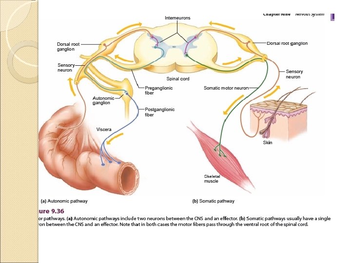
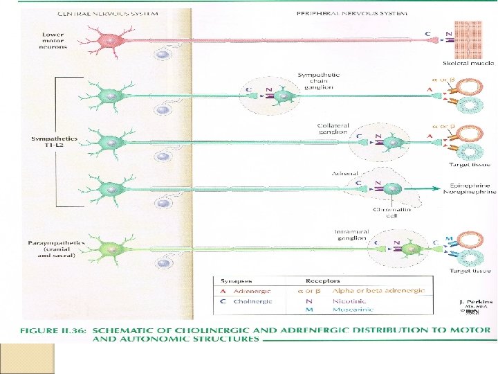
The pathways cont’d… Visceral efferent pathways differ from somatic equivalents. In the ANS there is a sequence of two neurones between the CNS and the innervated structure. The first neurone preganglionic neurone, its cell body is located in the spinal cord or brain stem. The second neurone postganglionic neurone, its cell body is located peripherally in an autonomic ganglion (the sinaptic connection between the two neurones)
Sympathetic and Parasympathetic Division The efferent neurones of the ANS are divided according to its anatomical and functional sympathetic and parasympathetic. The sympathetic and parasympathetic components are both structurally and functionally segregated. They have antagonistic effects upon the innervated structure.
Sympathetic and Parasympathetic Division cont’d… Changes in internal and external environment and emotional factors, profoundly influence autonomic activity, most notably via descending connections from the hypothalamus
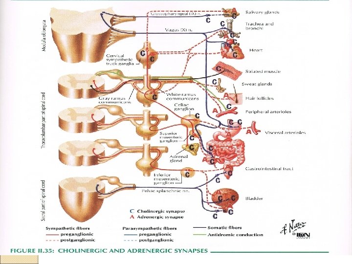
Neurotransmitter of ANS Sympathetic neurotransmitter: Preganglionic: acetylcholine Postganglionic: norepinephrine (noradrenaline), except in the sweat glands. Parasympathetic neurotransmitter: Preganglionic: acetylcholine Postganglionic: acetylcholine
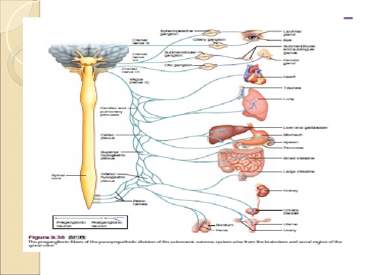
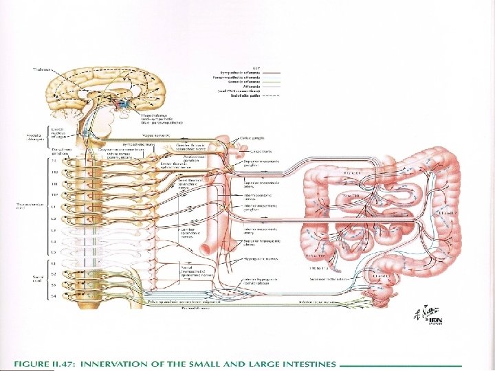
Spinal nuclei • Sympathetic spinal neurons lie in the lateral horn (intermediolateral and intermediomedial cell columns) of the thoracolumbarspinal cord (T 1–L 2) thoracolumbar system • Parasympathetic spinal neurons lie in the brain stem (projections along CN III, VII, IX, X) and the sacral spinal cord (S 2–S 4) craniosacral system • The intestine has its own autonomic ganglia myenteric and submucous plexuses
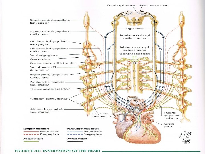
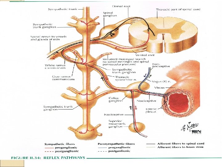
Sympathetic Division Preganglionic neurones lie in lateral horn of the spinal cord (T 1 until L 2 -L 3), leave the cord in the ventral nerve roots and join the spinal nerve Postganglionic neuron have their cell bodies in one of two locations : ◦ Sympathetic chain of ganglia (alongside of vertebral column) ◦ The plexuses (coeliac, superior or inferior mesenteric)
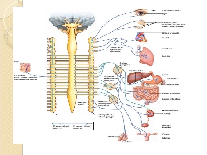
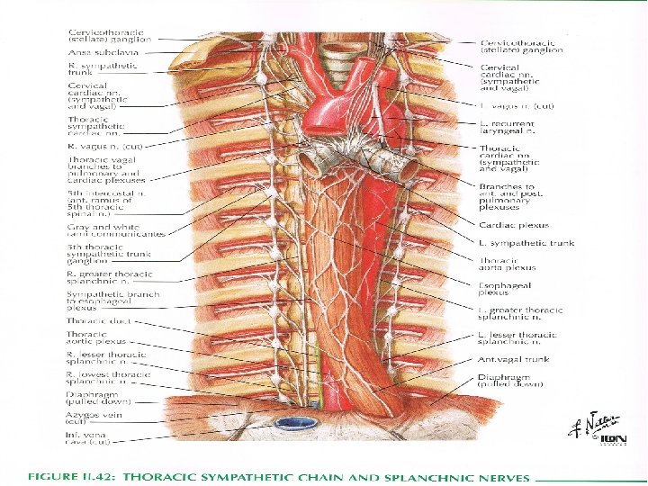
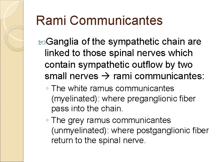
Rami Communicantes Ganglia of the sympathetic chain are linked to those spinal nerves which contain sympathetic outflow by two small nerves rami communicantes: ◦ The white ramus communicantes (myelinated): where preganglionic fiber pass into the chain. ◦ The grey ramus communicantes (unmyelinated): where postganglionic fiber return to the spinal nerve.
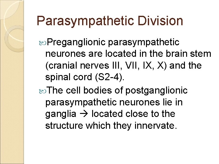
Parasympathetic Division Preganglionic parasympathetic neurones are located in the brain stem (cranial nerves III, VII, IX, X) and the spinal cord (S 2 -4). The cell bodies of postganglionic parasympathetic neurones lie in ganglia located close to the structure which they innervate.
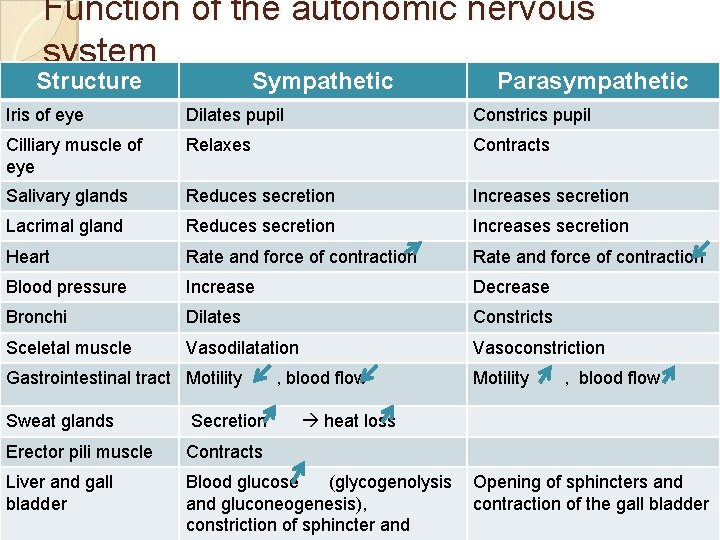
Function of the autonomic nervous system Structure Sympathetic Parasympathetic Iris of eye Dilates pupil Constrics pupil Cilliary muscle of eye Relaxes Contracts Salivary glands Reduces secretion Increases secretion Lacrimal gland Reduces secretion Increases secretion Heart Rate and force of contraction Blood pressure Increase Decrease Bronchi Dilates Constricts Sceletal muscle Vasodilatation Vasoconstriction Gastrointestinal tract Motility , blood flow Sweat glands Secretion Erector pili muscle Contracts Liver and gall bladder Blood glucose (glycogenolysis and gluconeogenesis), constriction of sphincter and Motility , blood flow heat loss Opening of sphincters and contraction of the gall bladder
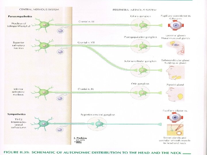


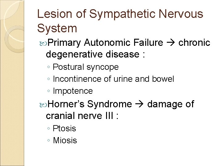
Lesion of Sympathetic Nervous System Primary Autonomic Failure chronic degenerative disease : ◦ Postural syncope ◦ Incontinence of urine and bowel ◦ Impotence Horner’s Syndrome damage of cranial nerve III : ◦ Ptosis ◦ Miosis
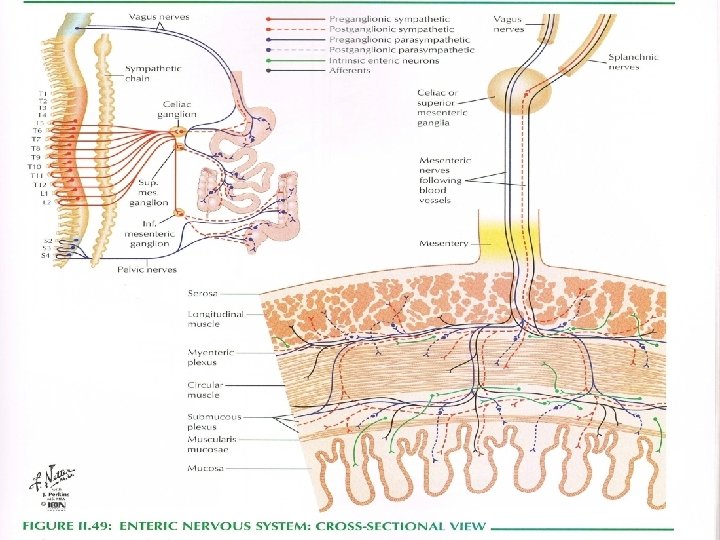
Enteric nervous system Within the alimentary canal, postganglionic parasympathetic neurones contribute to the myenteric (Auerbach’s) and submucosal (Meissner’s) plexuses. Additionally contain afferent neurones and interneurones. Capable of sustaining the motility of the gastrointestinal tract in the absence of input from the CNS.


Thank you

Central portion of ANS (Afferent connections) Afferent impulses enter the ANS from : q spinal tracts (anterolateral fasciculus = spinothalamic + spinocerebellar + spinoreticular tracts) q brain stem tracts (arising in the reticular formation), q corticothalamic tracts q the circumventricular organs
Central portion of ANS (Efferent connections) Projections from the hypothalamus and brain stem lateral horn of the thoracolumbar spinal cord synapses into the sympathetic neurons of the spinal cord project preganglionic fibers to the sympathetic ganglia • The parasympathetic neurons receive input from higher centers project in turn to parasympathetic ganglia located near the end organs •
Peripheral Portion Afferent and efferent connections Afferent: impulses to the ANS enter the spinal cord via the dorsal roots, and the brain stem via CN III, VII, IX and X. • Efferent: preganglionic fibers exit the spinal cord in the ventral roots and travel to the paravertebral ganglia, synapse onto the next neuron of the pathway. • The sympathetic preganglionic fibers travel a short distance to the paravertebral sympathetic chain, and the postganglionic fibers a relatively long distance to the effector organs. • The parasympathetic preganglionic fibers are long; they project to ganglia near the effector organs, which give off short postganglionic processes.
- Slides: 34