AUTONOMIC NERVOUS SYSTEM Functions Contraction and relaxation of


")


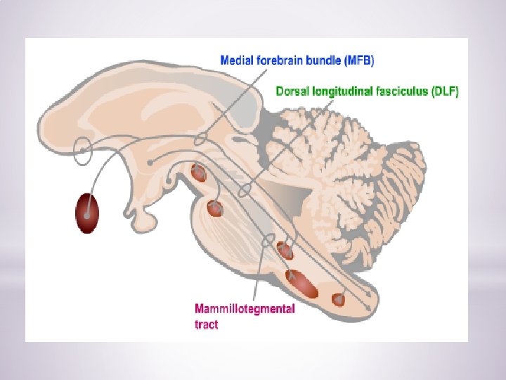
 q fasciculus telencephalicus medialis (medial forebrain")


- Slides: 25
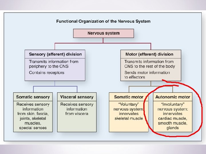
AUTONOMIC NERVOUS SYSTEM Functions: Contraction and relaxation of smooth muscles Function of all exocrine glands Heart rate Some metabolic processes
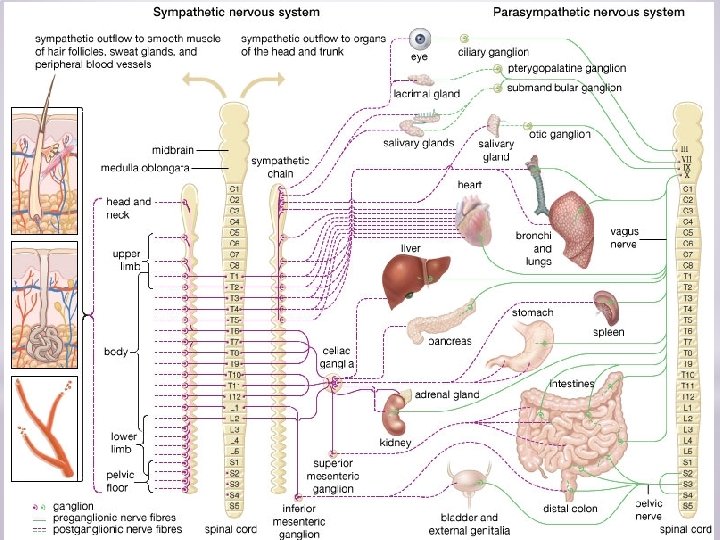
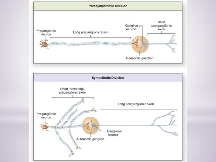
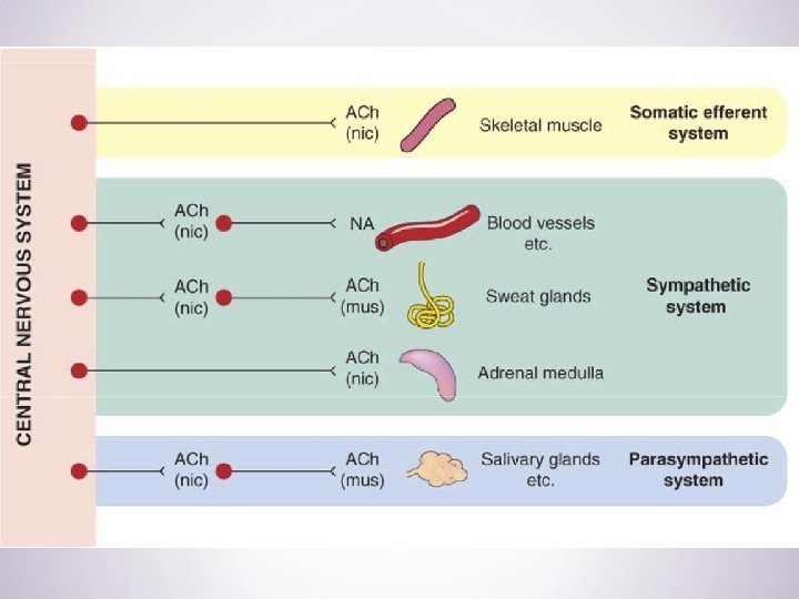
AUTONOMIC NERVOUS SYSTEM q sympathetic NS § ncl. intermediolateralis in T 1 – L 2 segments of spinal cord = thoracolumbar system § paravertebral ganglia (tr. sympathicus) and prevertebral ganglia § neurotransmitters Ø pregangl. – acetylcholine Ø postgangl. – norepinephrin (ex. sweat glands and piloerector muscle) q parasympathetic NS § parasympathetic nuclei of CN III, VII, IX, X § segments S 2 – S 4 = craniosacral system § ganglia near the target organ § neurotransmitter acetylcholine
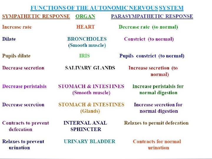
Sympathetic system Catabolic reaction (activities that are mobilized during emergency and stress situations, “fight, fright and flight” responses) Parasympathetic system Anabolic reactions (activities associated with conservation and restoration of body resources, “rest and digest” responses) y anatom
Central autonomic network (CAN)
Paraventricular nucleus
PVN in CAN DN of CN X s. IMLN r. VLM c. SN of CN V NTS
Other hypothalamic nuclei in CAN: Dorsomedial nucleus Posterior hypothalamic nucleus Mammillary nucleus Lateral hypothalamic area - cardiovascular control, control of feeding, satiety and insulin release
Extrahypothalamic structures in CAN: Control of symp. outflow Locus coeruleus Rostral and caudal VLM Raphe nuclei Control of parasymp. outflow Central ncl. of amygdala Dorsal ncl. of CN X Raphe nuclei PAG Parabrachial ncl.
Limbic cortex – control of both autonomic outflows Cingulate c. Orbitofrontal c. Insula Rhinal c. Hippocampus
Descendent modulatory pathways q fasciculus longitudinalis dorsalis (FLD) q fasciculus telencephalicus medialis (medial forebrain bundle MFB) q tr. mammillotegmentalis
Hypothalamus Nuclei of the anterior part q ncl. paraventricularis q stimulation of parasympathetic system Stimulation of the anterior part of hypothalamus q miosis q decrease in heart rate and blood pressure q dilation of cutaneous arteries q increase in peristalsis and secretion in the GIT
Hypothalamus Nuclei of the posterior part q ncl. mammillaris and hypothalamicus post. q stimulation of sympathetic system Stimulation of the posterior part of hypothalamus q mydriasis q increase in heart rate and blood pressure q constriction of cutaneous arteries q decrease in peristalsis and secretion in the GIT q erection of hairs
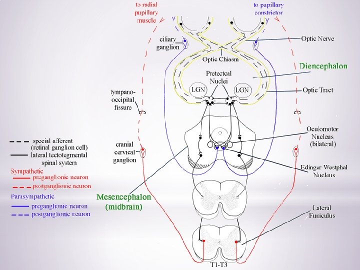
PUPILARY LIGHT REFLEX q a reflex that controls the diameter of the pupil, in response to the intensity of light (luminance) that falls on the retina of the eye q mydriasis: dilation of the pupil q miosis: constriction of the pupil
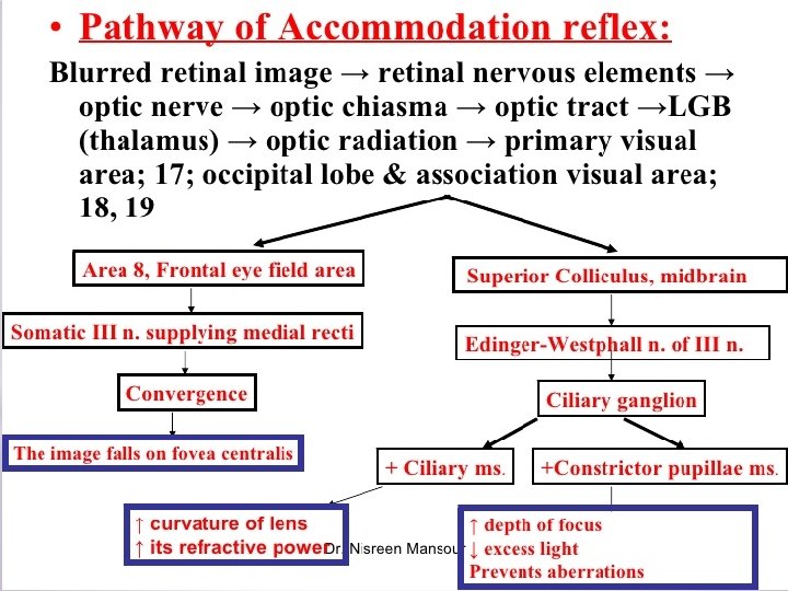
ACCOMMODATION
Near reflex triad: convergence accommodation of the lens pupillary constriction
Illustrations were copied from: Neuroscience Online, the Open-Access Neuroscience Electronic Textbook Department of Neurobiology and Anatomy University of Texas Medical School at Houston