Automated Hematology Dr P Dahim Health Reference Laboratory

Automated Hematology Dr. P. Dahim Health Reference Laboratory 1

• • Principles of Automation Calibration Quality Control Flagging 2
 § Light scattering")
Hematology Automation • Two General Principles § Electronic resistance ( impedance) § Light scattering 3
 • Utilizes non-conductive properties of blood cells § as blood cell")
Electronic Resistance (Impedance) • Utilizes non-conductive properties of blood cells § as blood cell passes through orifice of aperture it displaces its own volume § increased resistance between electrodes results in an electrical pulse § RBCs and Platelets counted together, separated by pulse heights § hydrodynamic focusing forces cells to pass single file through sensing zone 4

Cell counting 5
 •")
Light scattering • Cells counted as passed through focused beam of light( LASER) • Sum of diffraction(bending around corners), refraction (bending due to change in speed) and reflection (light rays turned back by obstruction). • Multi angle polarized scatter separation (M. A. P. S. S) § § 0° : indicator of cell size 10° : indicator of cell structure and complexity 90° polarized: indicates nuclear lobularity 90° depolarized: differentiate eosinophils 6

Principles of counting &sizing • Counting and sizing red cells , WBCs , Plts by counting the number of pulses and height of pulses • Measuring Hb by modified Hi. CN method • Measuring MCV by computing the mean of height of pulses • Hct (PCV ) derived from MCV & RBC • MCH derived from Hb & RBC 7 • MCHC derived from Hb , RBC &

• RDW : SD or CV of individual measurement of red cell volume • HDW : The degree of variation in red cell hemoglobinization, CV of measurements of hemoglobin concentration of individual cells • NRBC : included in the WBC counting 8

WBC Differential Count : • Impedance technology with current of various frequencies • Light scattering & light absorbance 9

Reticulocyte counting : • Flourescence – based methods 10

Histograms • RBC, PLT, and WBC plotted on histogram • X-Axis § Cell size in femtoliters (f. L) • Y-Axis § # of cells 11

Histogram § WBC: Distribution with three individual peaks and valleys at specific regions representing the lymphocytes, monocytes, and granulocytes. § All curves normally start and end at baseline 12


Calibration • Calibration provides the most accurate results possible. • For best performance, calibrate all the CBC parameters. • The WBC differential is calibrated at the factory. They do not require calibration in the laboratory. 20

When to Calibrate • At installation • After the replacement of any component that involves dilution characteristics or the primary measurements (such as the apertures) • When advised to do so by your service representative 21

Calibration Normal fresh blood samples Calibrators 22
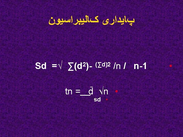

Precision check • Purpose: check reproducibility of instrument • Precision quick method § Run 5 replicates of one patient’s sample free of elevated bilirubin, lipemia, and hemoloysis • Calculate % CV or SD • Refer to the methodology in procedure manual for expected % CV or SD 25

Quality Control • Purpose of QC § Assure proper functionality of instrumentation § Monitoring the Integrity of the Calibration 26
 • Previously analyzed patient samples §")
Quality Control Methods • Assayed stabilized material (Commercial) • Previously analyzed patient samples § Easily obtained § Cost effective § Results and samples readily available 27
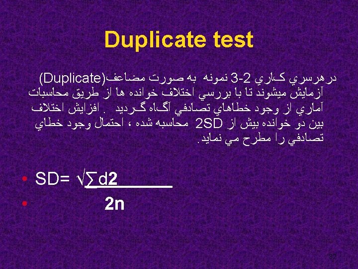
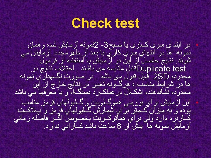
 Control charts Duplicate & Check tests")
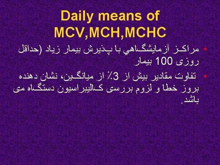
Quality control • • Replicate tests (control samples) Control charts Duplicate & Check tests Correlation system Delta check Daily means of MCV, MCHC Comparison the results with reference methods 28

Quality Control • OUT OF CONTROL!!! § Repeat the assay ( One time occurrence ) § Check integrity of material § Troubleshoot § Verify instrumentation 31

Delta check Hb 2 g/l PCV 0. 05 L/L MCV >6 f. L MCH > 5 pg WBC Normal to abnormal • Platelets Reduced or increased by more than 50% • • • 34

ERRORS INACCURACY 37

Reliability of Electronic Counters • High Precision • Accuracy? - Recirculation of cells - Red cell agglutination - Lipid droplets, Microorganisms, - Extraneous particles - Faulty maintenance - Incorrectly calibration 38

Interfering substances • • Cold Agglutinins Cryoglobulins Lipemia Platelet Clumps RBC fragments NRBCs Clot 39

WBC Count Interferences Unusual RBC abnormalities that resist lysing malarial parasites giant platelets, platelet clumps, NRBCs fragmented white cells, agglutinated white cells, § lyse-resistant red cells, § cryoglobulin, some extremely elevate proteins, § unlysed particles greater than 35 f. L in size § § 40

RBC Count Interferences • Very high WBC count, • high concentration of very large platelets, • Auto-agglutination 41

Hgb • Very high WBC count, • Severe lipemia, • certain unusual RBC abnormalities that resist lysing 42

MCV • Very high WBC count, • high concentration of very large platelets, • Auto-agglutination 43

Flagging • WBC Suspect flags § Blasts § Imm Grans § Variant lymphs 44

More Flagging • RBC Suspect flags § NRBCs § Macrocytic RBCs § Dimorphic RBC population § Micro RBCs/RBC fragments § RBC agglutination 45

Troubleshooting Flagged Results: • Refer to the Instrument Manual to troubleshoot codes, flags, and messages displayed with patient results 46

47
- Slides: 47