Autoimmunity and Systemic Autoimmune Diseases Prof Dr mit


 induces")
 surplus CTLA-4 receptors blockade Autoimmune Diseases")


 antigens. •")


 alleles and autoimmune diseases in")


- Slides: 45
Autoimmunity and Systemic Autoimmune Diseases Prof. Dr. Ümit Ölmez
• Immune system , can react to an enormous variety of microbes but does not react against the individual’s own (self) antigens. This unresponsiveness to self antigens, also called immunological tolerance. • If these mechanisms fail, the immune system may attack the individual’s own cells and tissues. Such reactions are called autoimmunity, and the diseases they cause are called autoimmune diseases.
• How does the immune system maintain unresponsiveness to self antigens? The important principles and features of self- tolerance • What are the factors that may contribute to the loss of self-tolerance and the development of autoimmunity?
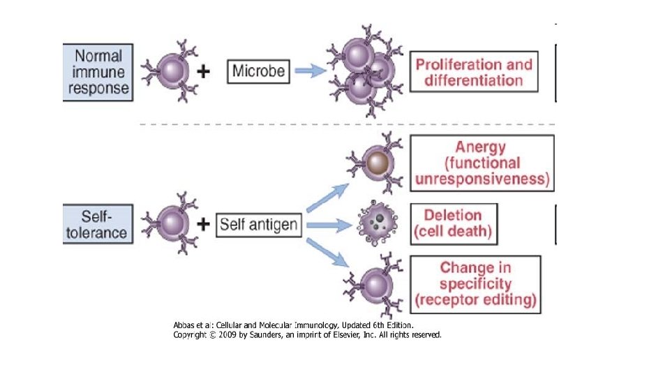
IMMUNOLOGICAL TOLERANCE: SIGNIFICANCE AND MECHANISMS • Immunological tolerance is a lack of response to antigens that is induced by exposure of lymphocytes to these antigens. • Normally, microbes are immunogenic and self antigens are tolerogenic. • In fact, the same antigen may be administered in different ways to induce an immune response or tolerance. • Immunological tolerance to different self antigens : • Central tolerance ( central lymphoid organs) • Peripheral tolerance ( secondary lymphoid organs or peripheral tissues)
Fates of lymphocytes after encounter with antigens
FIGURE 9 -1 Central and peripheral tolerance to self antigens. Central tolerance: Immature lymphocytes specific for self antigens may encounter these antigens in the generative (central) lymphoid organs and are deleted; B lymphocytes change their specificity (receptor editing); and some T lymphocytes develop into regulatory T cells. Peripheral tolerance: Mature self-reactive lymphocytes may be inactivated or deleted by encounter with self antigens in peripheral tissues or suppressed by regulatory T cells. Basic Immunology (Functions and Disorders of the Immune System) , AK Abbas, AH Lichtman, S Pillai , 5 th edition. 2016
• CD 4+ helper T cells, orchestrate virtually all immune responses to protein antigens, so tolerance in these cells may be enough to prevent both cell-mediated and humoral immune responses against self proteins. • Conversely, failure of tolerance in helper T cells may result in autoimmunity manifested by T cell–mediated attack against self antigens or by the production of autoantibodies against self proteins.
CENTRAL T LYMPHOCYTE TOLERANCE • The principal mechanisms of central tolerance in T cells are death of immature T cells and the generation of CD 4+ regulatory T cells. • Strong recognition of self antigens by immature T cells in the thymus may lead to death of the cells (negative selection) is a major mechanism of central tolerance. • The process of negative selection affects self-reactive CD 4+ T cells and CD 8+ T cells, which recognize self peptides displayed by class II MHC and class I MHC molecules, respectively. • Some immature CD 4+ T cells that recognize self antigens in the thymus with high affinity do not die but develop into regulatory T cells and enter peripheral tissues.
FIGURE 9 -2 Central T cell tolerance. Strong recognition of self antigens by immature T cells in the thymus may lead to death of the cells (negative selection, or deletion), or the development of regulatory T cells that enter peripheral tissues. Basic Immunology (Functions and Disorders of the Immune System) , AK Abbas, AH Lichtman, S Pillai , 5 th edition. 2016
• Many self proteins that are normally present only in certain peripheral tissues are also expressed in some of the epithelial cells of the thymus. A protein called AIRE (autoimmune regulator) is responsible for the thymic expression of these peripheral tissue antigens. • Mutations in the AIRE gene are the cause of a rare disorder called autoimmune polyendocrine syndrome. • The lymphocytes that survive negative selection in the thymus go on mature and are depleted of potentially dangerous autoreactive T cells. Peripheral mechanisms may prevent the activation of these lymphocytes.
PERIPHERAL T LYMPHOCYTE TOLERANCE • Peripheral tolerance is induced when mature T cells recognize self antigens in peripheral tissues, leading to functional inactivation (anergy) or death, or when the self-reactive lymphocytes are suppressed by regulatory T cells. • Peripheral tolerance , is clearly important for preventing T cell responses to self antigens that are present mainly in peripheral tissues and not in the thymus. • It also may provide backup mechanisms for preventing autoimmunity in situations where central tolerance is incomplete.
Peripheral T cell Tolerance Mechanisms • Anergy • Suppression by regulatory T cells • Deletion (apoptotic cell death)
FIGURE 9 -3 Peripheral T cell tolerance. A, Normal T cell responses require antigen recognition and costimulation. B, Three major mechanisms of peripheral T cell tolerance are illustrated: cell-intrinsic anergy, suppression by regulatory T cells, and deletion (apoptotic cell death). Basic Immunology (Functions and Disorders of the Immune System) , AK Abbas, AH Lichtman, S Pillai , 5 th edition. 2016
• Antigen recognition without adequate costimulation results in T cell anergy or death, or makes T cells sensitive to suppression by regulatory T cells. • T lymphocytes need at least two signals to induce their proliferation and differentiation into effector and memory cells: • Signal 1 is always antigen, and signal 2 is provided by costimulators that are expressed on antigen-presenting cells (APCs), typically as part of the innate immune response to microbes. • The presence or absence of costimulation is a major factor determining whether T cells are activated or tolerized.
Anergy • Anergy in T cells refers to long-lived functional unresponsiveness that is induced when these cells recognize self antigens. • Antigen recognition without adequate costimulation is thought to be the basis of anergy induction. Anergic cells survive but are incapable of responding to the antigen. • When T cells recognize antigens without costimulation, the TCR complex may lose its ability to transmit activating signals. • If a T cell recognizes antigen without strong costimulation, the T cell receptors may lose their ability to deliver activating signals or the T cell may engage inhibitory receptors, such as cytotoxic T lymphocyte–associated protein 4 (CTLA 4), that block activation. • Cytotoxic T lymphocyte–associated protein 4(CTLA-4) , is a regulatory molecule; • Inhibit T Cell reseptors of costimulator B 7 -1 and B 7 -2. On recognition of B 7 -1 and B 7 -2 by CTLA-4 receptors, supresses T cell responce and strengthen anergy. • As a result; CTLA-4 inhibit immune response to self antigens and lacking this function cause autoimmunity.
Figure : 9. 5 T cell anergy. An antigen presented by costimulator-expressing cells(APCs) induces a normal T cell response. If the T cell recognizes antigen without costimulation, or in the presence of CTLA-4 -B 7 interactions, the T cell fails to respond and is rendered incapable of responding even if the antigen is subsequently presented by costimulator-expressing APCs.
or Costimulator (B 7) surplus CTLA-4 receptors blockade Autoimmune Diseases
Regulation of T Cell Responses by Inhibitory Receptors • In T cells, the best-defined inhibitory receptors are CTLA-4 and PD-1; • CTLA-4 , is expressed transiently on activated CD 4+ T cells and constitutively on regulatory T cells. It functions to terminate activation of responding T cells and also mediates the suppressive function of regulatory T cells. • CTLA-4 works by blocking and removing B 7 molecules from the surface of APCs, thus reducing costimulation and preventing the activation of T cells; CTLA-4 might also deliver inhibitory signals to T cells. • When B 7 levels are low (as would be expected normally when APCs are displaying self antigens), the receptor that is preferentially engaged is the high-affinity CTLA-4, but when B 7 levels are high (as in infections), the low affinity activating receptor CD 28 is engaged to a greater extent. • PD-1, is expressed on CD 4+ and CD 8+ T cells after antigen stimulation. It has an immunoreceptor tyrosine-based inhibitory motif (ITIM) typical of receptors that deliver inhibitory signals. PD-1 terminates responses of T cells to self antigens and also to chronic infections, notably virus infections. • Several other receptors on T cells other than CTLA-4 and PD-1 have been shown to inhibit immune responses and are currently the targets of checkpoint blockade therapy. Such treatment leads to enhanced antitumor immune responses and tumor regression in a significant fraction of the patients.
Immune Suppression by Regulatory T Cells • Regulatory T cells develop in the thymus or peripheral tissues on recognition of self antigens and suppress the activation of potentially harmful lymphocytes specific for these self antigens. • The majority of self-reactive regulatory T cells probably develop in the thymus, but they may also arise in peripheral lymphoid organs. • Most regulatory T cells are CD 4+ and express high levels of CD 25, the α chain of the interleukin-2 (IL-2) receptor. • They also express a transcription factor called Fox. P 3, which is required for the development and function of the cells. • Mutations of the gene encoding Fox. P 3 in humans or in mice cause a systemic, multiorgan autoimmune disease, demonstrating the importance of Fox. P 3+ regulatory T cells for the maintenance of selftolerance.
• The survival and function of regulatory T cells are dependent on the cytokine IL-2. • IL-2 is an example of a cytokine that serves two opposite roles: • It promotes immune responses by stimulating T cell proliferation, and it inhibits immune responses by maintaining functional regulatory T cells. • The cytokine transforming growth factor β (TGF-β) also plays a role in the generation of regulatory T cells, perhaps by stimulating expression of the Fox. P 3 transcription factor. • However, the importance of defects in regulatory T cells in common human autoimmune diseases is not established.
Deletion: Apoptosis of Mature Lymphocytes • Recognition of self antigens may trigger pathways of apoptosis that result in elimination (deletion) of the self-reactive lymphocytes. • There are two likely mechanisms of death of mature T lymphocytes induced by self antigens: q Recognition of self antigens may lead to the coexpression of death receptors and their ligands. • This ligand-receptor interaction generates signals through the death receptor that culminate in the activation of caspases and apoptosis. • The best-defined death receptor– ligand pair involved in self-tolerance is a protein called Fas (CD 95), which is expressed on many cell types, and Fas ligand (Fas. L), which is expressed mainly on activated T cells. q Antigen recognition induces the production of pro-apoptotic proteins in T cells that induce cell death by causing mitochondrial proteins to leak out and activate caspases, cytosolic enzymes that induce apoptosis. • Self antigens, which are recognized without strong costimulation, do not stimulate production of anti-apoptotic proteins, and the relative deficiency of survival signals induces death of the cells that recognize these antigens.
FIGURE 9 -6 Mechanisms of apoptosis of T lymphocytes. T cells respond to antigen presented by normal antigen-presenting cells (APCs) by secreting interleukin-2 (IL 2), expressing anti-apoptotic (pro-survival) proteins, and undergoing proliferation and differentiation. Restimulation of recently activated T cells by antigen leads to coexpression of Fas and Fasligand (Fas. L), engagement of the death receptor Fas leads to apoptosis of the T cells by the death receptor (extrinsic) pathway. Self antigen recognition by T cells without costimulation may lead to relative deficiency of intracellular anti-apoptotic proteins, and the excess of pro-apoptotic proteins causes cell death by inducing release of mediators of apoptosis from mitochondria (death by the mitochondrial [intrinsic] pathway of apoptosis).
B LYMPHOCYTE TOLERANCE • Self polysaccharides, lipids, and nucleic acids are T-independent antigens that are not recognized by T cells. These antigens must induce tolerance in B lymphocytes to prevent autoantibody production. Central B Cell Tolerance • When immature B lymphocytes interact strongly with self antigens in the bone marrow, the B cells either change their receptor specificity (receptor editing) or are killed (deletion). • Receptor editing • Deletion • Anergy
FIGURE 9 -8 Central tolerance in immature B lymphocytes. An immature B cell that recognizes self antigen in the bone marrow changes its antigen receptor (receptor editing), dies by apoptosis (negative selection, or deletion), or reduces antigen receptor expression and becomes functionally unresponsive. Basic Immunology (Functions and Disorders of the Immune System) , AK Abbas, AH Lichtman, S Pillai , 5 th edition. 2016
Peripheral B Cell Tolerance • Peripheral tolerance is induced when mature B cells recognize self antigens without T cell help, which results in anergy and death of the B cells, or engagement of inhibitory receptors. Mature B lymphocytes that encounter self antigens in peripheral lymphoid tissues become incapable of responding to that antigen. • B cells that recognize self antigens in the periphery may also undergo apoptosis, or inhibitory receptors on the B cells may be engaged, thus preventing activation, regulatory T cells may also contribute to B cell tolerance. • During somatic hypermutation of Ig genes in germinal centers , some antigen receptors may be generated that are capable of recognizing self antigens. • B cells expressing these autoreactive receptors die either because there are no follicular helper T cells to rescue them or because germinal center B cells express high levels of Fas and are killed by Fas. L-expressing T cells. • The autoimmune disease that results from FAS mutations may in part be caused by survival of these self-reactive germinal center B cells.
FIGURE 9 -9 Peripheral tolerance in B lymphocytes. A mature B cell that recognizes a self antigen without T cell help is functionally inactivated and becomes incapable of responding to that antigen (anergy), or it dies by apoptosis (deletion), or its activation is suppressed by engagement of inhibitory receptors. Basic Immunology (Functions and Disorders of the Immune System) , AK Abbas, AH Lichtman, S Pillai , 5 th edition. 2016
AUTOIMMUNITY • Autoimmunity is defined as an immune response against self (autologous) antigens. • It is an important cause of disease, estimated to affect 2% to 5% of the population in developed countries, and the prevalence of several autoimmune diseases is increasing. • Different autoimmune diseases may be organ-specific, affecting only one or a few organs, or systemic, with widespread tissue injury and clinical manifestations. • Tissue injury in autoimmune diseases may be caused by antibodies against self antigens or by T cells reactive with self antigens.
Although the non-organ-specific diseases characteristically produce symptoms in the skin, joints, kidney, and muscle, individual organs are more markedly affected by particular diseases, for example the kidney in SLE and the joints in rheumatoid arthritis.
Autoimmune diseases may be organ specific, affecting only one or a few organs, or non-organ specific (systemic) diseases
PATHOGENESIS • The principal factors in the development of autoimmunity are the inheritance of susceptibility genes and environmental triggers, such as infections. • It is postulated that susceptibility genes interfere with pathways of self-tolerance and lead to the persistence of self-reactive T and B lymphocytes. • Environmental stimuli may cause cell and tissue injury and inflammation and activate these self-reactive lymphocytes, resulting in the generation of effector T cells and autoantibodies that are responsible for the autoimmune disease.
FIGURE 9 -10 Postulated mechanisms of autoimmunity. In this proposed model of organ-specific T cell– mediated autoimmunity, various genetic loci may confer susceptibility to autoimmunity, probably by influencing the maintenance of self-tolerance. Environmental triggers, such as infections and other inflammatory stimuli, promote the influx of lymphocytes into tissues and the activation of antigen-presenting cells (APCs) and subsequently of self-reactive T cells, resulting in tissue injury. Basic Immunology (Functions and Disorders of the Immune System) , AK Abbas, AH Lichtman, S Pillai , 5 th edition. 2016
• Despite our growing knowledge of the immunological abnormalities that may result in autoimmunity, we still do not know the etiology of common human autoimmune diseases. • Several factors, for the lack of understanding the etiology of common human autoimmune diseases; q Autoimmune diseases in humans usually are heterogeneous and multifactorial q The self antigens that are the inducers and targets of the autoimmune reactions often are unknown q Diseases may manifest clinically long after the autoimmune reactions have been initiated
Genetic Factors • Inherited risk for most autoimmune diseases is attributable to multiple gene loci, of which the largest contribution is made by MHC genes. • In rare cases, autoimmunity- associated genes are variants (mutations) that are essentially nonexistent in healthy individuals, rather than commonly detected polymorphisms. • Such rare variants can have a large impact on the development of autoimmunity.
• The association between human leukocyte antigen (HLA) alleles and autoimmune diseases in humans was recognized many years ago. • It was one of the first indications that T cells played an important role in these disorders (because the only known function of MHC molecules is to present peptide antigens to T cells). • The incidence of a particular autoimmune disease often is greater among individuals who inherit a particular HLA allele(s) than in the general population.
FIGURE 9 -11 Association of autoimmune diseases with alleles of the major histocompatibility complex (MHC) locus. Family and linkage studies show a greater likelihood of developing certain autoimmune diseases in persons who inherit particular human leukocyte antigen (HLA) alleles than in persons who lack these alleles (odds ratio or relative risk). Selected examples of HLA-disease associations are listed. For instance, in people who have the HLA-B 27 allele, the risk of development of ankylosing spondylitis, an autoimmune diseases of the spine, is 90 to 100 times higher than in B 27 -negative people; other diseases show various degrees of association with other HLA alleles. Breeding studies in animals have also shown that the incidence of some autoimmune diseases correlates strongly with the inheritance of particular MHC alleles (e. g. , type 1 diabetes mellitus with a mouse class II allele called I-Ag 7). Basic Immunology (Functions and Disorders of the Immune System) , AK Abbas, AH Lichtman, S Pillai , 5 th edition. 2016
• The HLA allele is not, by itself, the cause of the disease. In fact, the disease never develops in the vast majority of people who inherit an HLA allele that does confer increased risk of the disease. • Despite the clear association of MHC alleles with several autoimmune diseases, how these alleles contribute to the development of the diseases remains unknown. • Some hypotheses are that particular MHC alleles may be especially effective at presenting pathogenic self peptides to autoreactive T cells, or they are inefficient at displaying certain self antigens in the thymus, leading to defective negative selection of T cells.
• Polymorphisms , in non-HLA genes are associated with various autoimmune diseases and may contribute to failure of self-tolerance or abnormal activation of lymphocytes.
• Some rare autoimmune disorders are Mendelian in origin, caused by mutations in single genes that have high penetrance and lead to autoimmunity in most or all individuals who inherit these mutations. • These genes, alluded to earlier, include AIRE, FOXP 3, FAS, and CTLA 4. Mutations in these genes have been valuable for identifying key molecules and pathways involved in self-tolerance. • These Mendelian forms of autoimmunity are exceedingly rare, however, and common autoimmune diseases are not caused by mutations in any of these known genes.
Role of Infections and Other Environmental Influences • Infections may activate self-reactive lymphocytes, thereby triggering the development of autoimmune diseases. • Infections may contribute to autoimmunity in several ways : q An infection of a tissue may induce a local innate immune response, which may lead to increased production of costimulators and cytokines by tissue APCs. • These activated tissue APCs may be able to stimulate self-reactive T cells that encounter self antigens in the tissue. • In other words, infection may break T cell tolerance and promote the activation of self reactive lymphocytes. q Some infectious microbes may produce peptide antigens that are similar to, and cross- react with, self antigens. Immune responses to these microbial peptides may result in an immune attack against self antigens. • Such cross reactions between microbial and self antigens are termed molecular mimicry. • In some rare disorders, antibodies produced against a microbial protein bind to self proteins.
FIGURE 9 -13 Mechanisms by which microbes may promote autoimmunity. A, Normally, an encounter of mature T cells with self antigens presented by resting tissue antigen-presenting cells (APCs) results in peripheral tolerance. B, Microbes may activate the APCs to express costimulators, and when these APCs present self antigens, the specific T cells are activated, rather than being rendered tolerant. C, Some microbial antigens may cross-react with self antigens (mimicry). Therefore, immune responses initiated by the microbes may become directed at self cells and tissues. This figure illustrates concepts as they apply to T cells; molecular mimicry also may apply to self-reactive B lymphocytes. Basic Immunology (Functions and Disorders of the Immune System) , AK Abbas, AH Lichtman, S Pillai , 5 th edition. 2016
• Infections, also may injure tissues and release antigens that normally are sequestered from the immune system. • For example, some sequestered antigens (e. g. , in testis and eye) normally are not seen by the immune system and are ignored. • Release of these antigens (e. g. , by trauma or infection) may initiate an autoimmune reaction against the tissue. • The abundance and composition of normal commensal microbes in the gut, skin, and other sites (the microbiome) may also influence the health of the immune system and the maintenance of self-tolerance.
• Several other environmental and host factors may contribute to autoimmunity. • Many autoimmune diseases are more common in women than in men, but how gender might affect immunological tolerance or lymphocyte activation remains unknown. • Exposure to sunlight is a trigger for the development of the autoimmune disease systemic lupus erythematosus (SLE), in which autoantibodies are produced against self nucleic acids and nucleoproteins.